Investigating the Uncertainty of Cellular Microenvironment Parameter Estimations via Diffusion MRI Cytometry
Pith reviewed 2026-06-27 17:14 UTC · model grok-4.3
The pith
Diffusion MRI using IMPULSED sequences can robustly estimate cell diameter, intracellular volume fraction, and extracellular diffusion coefficient.
A machine-rendered reading of the paper's core claim, the machinery that carries it, and where it could break.
Core claim
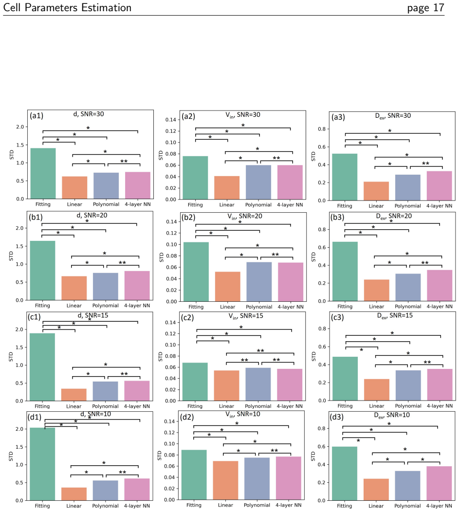
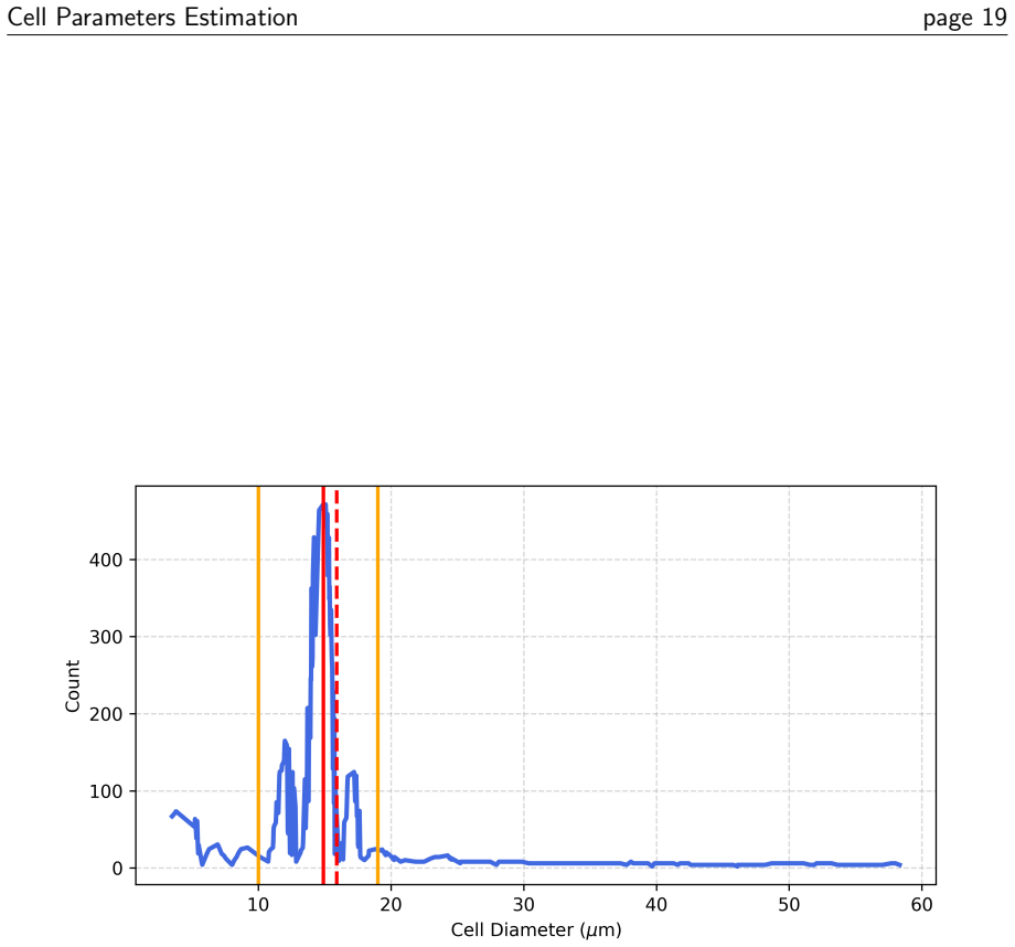
Simulations with one pulsed gradient spin echo and two oscillating gradient spin echo sequences show that cell diameter d, intracellular volume fraction Vin, and extracellular diffusion coefficient Dex exhibit relative uncertainties below 1.0 under the IMPULSED model at SNR of 30. A four-layer neural network achieves the lowest errors when mapping signals to these parameters, with mean absolute errors of 1.7 μm, 5.06%, and 0.28 μm²/ms respectively. In vitro experiments with MC38 cells yield a 6.7% error in diameter estimation, supporting the use of IMPULSED dMRI for robust parameter estimation in tumor microenvironments.
What carries the argument
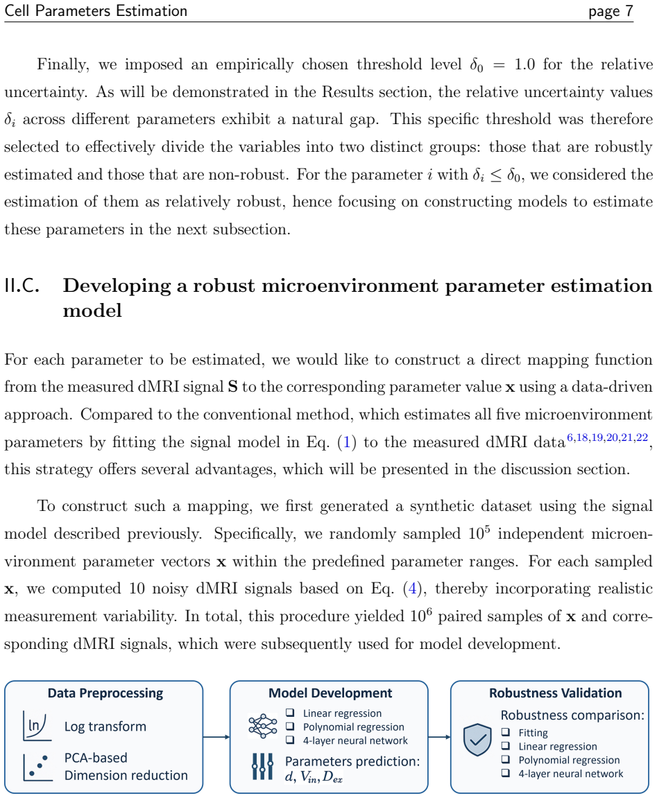
Jacobian-based sensitivity analysis for parameter uncertainty quantification in the IMPULSED diffusion model, paired with principal component analysis and neural network regression for signal-to-parameter mapping.
If this is right
- d, Vin, and Dex are robustly derivable with relative uncertainty below 1.0.
- The four-layer neural network outperforms linear and polynomial regression for parameter estimation.
- In vitro validation achieves 6.7% error in cell diameter estimation.
- This provides a framework for noninvasive assessment of tumor microenvironment changes.
Where Pith is reading between the lines
- Extending the analysis to lower SNR or in vivo conditions could reveal additional robust parameters.
- The neural network approach might be adapted for real-time clinical mapping if trained on more diverse data.
- Combining these estimates with other imaging modalities could improve tumor response monitoring accuracy.
Load-bearing premise
The IMPULSED model accurately represents diffusion in actual cellular environments under the tested sequence parameters and noise levels.
What would settle it
Direct comparison of estimated cell diameters from IMPULSED dMRI against independent microscopy measurements in the same MC38 cell samples at matched SNR would test if the reported uncertainties hold.
Figures
read the original abstract
This study aims to identify cell microenvironment parameters that can be robustly estimated from IMPULSED diffusion MRI signals and to develop a reliable mapping-based estimation framework. Diffusion MRI signals were simulated using the established IMPULSED model with one pulsed gradient spin echo sequence and two oscillating gradient spin echo sequences at different frequencies. Five cellular parameters were considered: cell diameter ($d$), intracellular diffusion coefficient ($D_{in}$), intracellular volume fraction ($V_{in}$), extracellular diffusion coefficient ($D_{ex}$), and the frequency-dependent slope of $D_{ex}$ ($\beta_{ex}$). Parameter uncertainty was quantified using Jacobian-based sensitivity analysis at an SNR of 30, representing clinically achievable conditions on a 1.5T MRI scanner. To enable direct parameter mapping, signals were logarithmically transformed, reduced in dimension using principal component analysis, and then used to estimate parameters with linear regression, fourth-order polynomial regression, and a fully connected four-layer neural network. Model validation was performed in vitro using MC38 cell lines. Uncertainty analysis identified $d$, $V_{in}$, and $D_{ex}$ as robustly derivable parameters, each with relative uncertainty below 1.0. Among the tested models, the four-layer neural network performed best, with mean absolute errors of 1.7 $\mu$m for $d$, 5.06% for $V_{in}$, and 0.28 $\mu$m$^2$/ms for $D_{ex}$. In vitro validation showed a 6.7% error in cell diameter estimation. These results demonstrate that IMPULSED dMRI can support robust estimation of key cell microenvironment parameters and provide a practical framework for noninvasive assessment of tumor microenvironment changes during radiation therapy response monitoring.
Editorial analysis
A structured set of objections, weighed in public.
Referee Report
Summary. The manuscript simulates IMPULSED dMRI signals (one PGSE + two OGSE sequences) using the forward model to estimate five cellular parameters (d, Din, Vin, Dex, beta_ex). Jacobian-based sensitivity analysis at fixed SNR=30 is used to quantify uncertainty and identify d, Vin, and Dex as robust (relative uncertainty <1). Signals are log-transformed, dimension-reduced via PCA, and mapped to parameters via linear regression, polynomial regression, and a four-layer NN; the NN yields the lowest MAEs (1.7 μm for d, 5.06% for Vin, 0.28 μm²/ms for Dex). In-vitro validation on MC38 cells reports 6.7% error for d. The central claim is that IMPULSED dMRI supports robust estimation of these key microenvironment parameters for tumor monitoring.
Significance. If the Jacobian-derived uncertainties prove representative of the actual NN estimator and the IMPULSED forward model holds under realistic cellular conditions, the work supplies a concrete, simulation-validated pipeline for extracting d, Vin, and Dex from clinically feasible 1.5 T acquisitions. The explicit comparison of three mapping architectures and the in-vitro check for d are positive features that could be extended to multi-cell-line or in-vivo settings.
major comments (3)
- [Abstract / Uncertainty analysis] Abstract and Methods (uncertainty analysis): the claim that d, Vin, and Dex are 'robustly derivable' with relative uncertainty below 1.0 rests exclusively on first-order Jacobian sensitivity evaluated at a single nominal parameter vector and fixed SNR=30. This local linear proxy does not incorporate (i) correlations among the five parameters, (ii) the PCA reduction step, or (iii) the nonlinear NN mapping that is ultimately used for estimation; consequently the reported uncertainties may not bound the actual mapping errors.
- [Validation] Validation section: experimental ground truth is provided only for cell diameter d (6.7% error in MC38 cells). No corresponding in-vitro or phantom measurements are reported for Vin or Dex, so the assertion that these two parameters are 'robustly derivable' in practice rests solely on simulation MAE values.
- [Methods] Methods (simulation and model assumptions): all results presuppose that the IMPULSED analytic expressions accurately describe diffusion inside and outside cells across the simulated parameter ranges and sequence timings. No test of forward-model misspecification (e.g., non-Gaussian intracellular diffusion, membrane permeability) is performed, yet this assumption is load-bearing for translating the SNR=30 Jacobian results to clinical 1.5 T data.
minor comments (2)
- [Abstract] The abstract states 'relative uncertainty below 1.0' without specifying whether this is the coefficient of variation or a normalized standard deviation; a brief definition or reference to the exact formula used would improve clarity.
- [Abstract] The ranges and sampling strategy for the five simulated parameters are not stated in the abstract or summary; adding this information would aid reproducibility assessment.
Simulated Author's Rebuttal
We thank the referee for their insightful comments, which have helped us improve the clarity and scope of our manuscript. We address each major comment below and indicate the revisions made.
read point-by-point responses
-
Referee: [Abstract / Uncertainty analysis] Abstract and Methods (uncertainty analysis): the claim that d, Vin, and Dex are 'robustly derivable' with relative uncertainty below 1.0 rests exclusively on first-order Jacobian sensitivity evaluated at a single nominal parameter vector and fixed SNR=30. This local linear proxy does not incorporate (i) correlations among the five parameters, (ii) the PCA reduction step, or (iii) the nonlinear NN mapping that is ultimately used for estimation; consequently the reported uncertainties may not bound the actual mapping errors.
Authors: We agree that the Jacobian-based sensitivity analysis provides a local, first-order approximation and does not fully account for parameter correlations, the PCA dimensionality reduction, or the nonlinear nature of the neural network estimator. This analysis was intended as an initial step to screen for parameters with sufficient sensitivity under the given SNR conditions. The subsequent simulation results with the NN, showing low MAEs, offer supporting evidence for the estimability of d, Vin, and Dex. To address this, we will revise the abstract and methods sections to explicitly state that the robustness claim is based on the Jacobian sensitivity combined with simulation mapping errors, rather than a comprehensive uncertainty propagation through the full pipeline. revision: partial
-
Referee: [Validation] Validation section: experimental ground truth is provided only for cell diameter d (6.7% error in MC38 cells). No corresponding in-vitro or phantom measurements are reported for Vin or Dex, so the assertion that these two parameters are 'robustly derivable' in practice rests solely on simulation MAE values.
Authors: We acknowledge this limitation in the experimental validation. The in-vitro experiments provided direct comparison only for cell diameter d via microscopy. For Vin and Dex, we rely on the simulation-based MAEs. We will revise the abstract, results, and discussion to clarify that only d has been validated in vitro, while Vin and Dex are supported by simulations, and highlight this as an area for future experimental validation. revision: yes
-
Referee: [Methods] Methods (simulation and model assumptions): all results presuppose that the IMPULSED analytic expressions accurately describe diffusion inside and outside cells across the simulated parameter ranges and sequence timings. No test of forward-model misspecification (e.g., non-Gaussian intracellular diffusion, membrane permeability) is performed, yet this assumption is load-bearing for translating the SNR=30 Jacobian results to clinical 1.5 T data.
Authors: The IMPULSED model is a well-established framework in the diffusion MRI literature for modeling restricted diffusion in cellular microenvironments. Our study assumes its validity as per prior validations in the field. We did not conduct additional misspecification tests in this work. We will add a dedicated limitations paragraph in the discussion section to acknowledge the dependence on the forward model assumptions and to suggest that future studies could explore model robustness under conditions such as membrane permeability or non-Gaussian effects. revision: partial
Circularity Check
No circularity: uncertainty and mapping results are computed outputs, not redefinitions of inputs
full rationale
The paper simulates signals from the IMPULSED forward model, applies Jacobian sensitivity analysis directly to that model at fixed SNR=30 to compute relative uncertainties, trains regression/NN mappings on the same simulated dataset (with PCA preprocessing), and reports MAE on held-out simulations plus one in-vitro check for diameter. These quantities are derived quantities from the described procedures rather than tautological re-statements of the model equations or fitted parameters renamed as predictions. No self-citation chain, ansatz smuggling, or uniqueness theorem is invoked to force the central claims; the derivation chain remains self-contained against the external in-vitro benchmark.
Axiom & Free-Parameter Ledger
free parameters (1)
- SNR=30
axioms (1)
- domain assumption The IMPULSED model correctly describes the diffusion signals from cellular microenvironments with the chosen sequences.
Reference graph
Works this paper leans on
-
[1]
Therasse et al., New guidelines to evaluate the response to treatment in solid tumors, Journal of the national cancer institute 92 , 205--216 (2000)
P. Therasse et al., New guidelines to evaluate the response to treatment in solid tumors, Journal of the national cancer institute 92 , 205--216 (2000)
2000
-
[2]
R. C. Newton, S. V. Kemp, P. L. Shah, D. Elson, A. Darzi, K. Shibuya, S. Mulgrew, and G.-Z. Yang, Progress toward optical biopsy: bringing the microscope to the patient, Lung 189 , 111--119 (2011)
2011
-
[3]
J. E. Swartz, J. P. Driessen, P. M. van Kempen, R. de Bree, L. M. Janssen, F. A. Pameijer, C. H. Terhaard, M. E. Philippens, and S. Willems, Influence of tumor and microenvironment characteristics on diffusion-weighted imaging in oropharyngeal carcinoma: A pilot study, Oral oncology 77 , 9--15 (2018)
2018
-
[4]
Bai et al., Study of diffusion weighted imaging derived diffusion parameters as biomarkers for the microenvironment in gliomas, Frontiers in Oncology 11 , 672265 (2021)
Y. Bai et al., Study of diffusion weighted imaging derived diffusion parameters as biomarkers for the microenvironment in gliomas, Frontiers in Oncology 11 , 672265 (2021)
2021
-
[5]
B. F. Jordan, M. Runquist, N. Raghunand, A. Baker, R. Williams, L. Kirkpatrick, G. Powis, and R. J. Gillies, Dynamic contrast-enhanced and diffusion MRI show rapid and dramatic changes in tumor microenvironment in response to inhibition of HIF-1 using PX-478, Neoplasia 7 , 475--485 (2005)
2005
-
[6]
Xu et al., MRI-cytometry: mapping nonparametric cell size distributions using diffusion MRI, Magnetic resonance in medicine 85 , 748--761 (2021)
J. Xu et al., MRI-cytometry: mapping nonparametric cell size distributions using diffusion MRI, Magnetic resonance in medicine 85 , 748--761 (2021)
2021
-
[7]
E. E. Sigmund, G. Y. Cho, S. Kim, M. Finn, M. Moccaldi, J. H. Jensen, D. K. Sodickson, J. D. Goldberg, S. Formenti, and L. Moy, Intravoxel incoherent motion imaging of tumor microenvironment in locally advanced breast cancer, Magnetic resonance in medicine 65 , 1437--1447 (2011)
2011
-
[8]
T. L. Chenevert, P. E. McKeever, and B. D. Ross, Monitoring early response of experimental brain tumors to therapy using diffusion magnetic resonance imaging., Clinical cancer research: an official journal of the American Association for Cancer Research 3 , 1457--1466 (1997)
1997
-
[9]
J. Xu, M. D. Does, and J. C. Gore, Quantitative characterization of tissue microstructure with temporal diffusion spectroscopy, Journal of magnetic resonance 200 , 189--197 (2009)
2009
-
[10]
Jiang, H
X. Jiang, H. Li, J. Xie, P. Zhao, J. C. Gore, and J. Xu, Quantification of cell size using temporal diffusion spectroscopy, Magnetic resonance in medicine 75 , 1076--1085 (2016)
2016
-
[11]
E. O. Stejskal, Use of spin echoes in a pulsed magnetic-field gradient to study anisotropic, restricted diffusion and flow, The Journal of Chemical Physics 43 , 3597--3603 (1965)
1965
-
[12]
P. T. Callaghan, Principles of nuclear magnetic resonance microscopy , Clarendon press, 1993
1993
-
[13]
Cory, Measurement of translational displacement probabilities by NMR: an indicator of compartmentation, Magnetic resonance in medicine 14 , 435--444 (1990)
D. Cory, Measurement of translational displacement probabilities by NMR: an indicator of compartmentation, Magnetic resonance in medicine 14 , 435--444 (1990)
1990
-
[14]
K. R. Brownstein and C. Tarr, Importance of classical diffusion in NMR studies of water in biological cells, Physical review A 19 , 2446 (1979)
1979
-
[15]
J. E. Tanner and E. O. Stejskal, Restricted self-diffusion of protons in colloidal systems by the pulsed-gradient, spin-echo method, The Journal of Chemical Physics 49 , 1768--1777 (1968)
1968
-
[16]
D. A. Yablonskiy, G. L. Bretthorst, and J. J. Ackerman, Statistical model for diffusion attenuated MR signal, Magnetic Resonance in Medicine: An Official Journal of the International Society for Magnetic Resonance in Medicine 50 , 664--669 (2003)
2003
-
[17]
L. Zhao, A. Sukstanskii, C. Kroenke, J. Song, D. Piwnica-Worms, J. Ackerman, and J. J. Neil, Intracellular water specific MR of microbead-adherent cells: HeLa cell intracellular water diffusion, Magnetic Resonance in Medicine: An Official Journal of the International Society for Magnetic Resonance in Medicine 59 , 79--84 (2008)
2008
-
[18]
M. Iima, O. Reynaud, T. Tsurugizawa, L. Ciobanu, J.-R. Li, F. Geffroy, B. Djemai, M. Umehana, and D. Le Bihan, Characterization of glioma microcirculation and tissue features using intravoxel incoherent motion magnetic resonance imaging in a rat brain model, Investigative radiology 49 , 485--490 (2014)
2014
-
[19]
Ichikawa, U
S. Ichikawa, U. Motosugi, D. Hernando, H. Morisaka, N. Enomoto, M. Matsuda, and H. Onishi, Histological grading of hepatocellular carcinomas with intravoxel incoherent motion diffusion-weighted imaging: inconsistent results depending on the fitting method, Magnetic Resonance in Medical Sciences 17 , 168--173 (2018)
2018
-
[20]
Ades-Aron, J
B. Ades-Aron, J. Veraart, P. Kochunov, S. McGuire, P. Sherman, E. Kellner, D. S. Novikov, and E. Fieremans, Evaluation of the accuracy and precision of the diffusion parameter EStImation with Gibbs and NoisE removal pipeline, Neuroimage 183 , 532--543 (2018)
2018
-
[21]
J. M. Oeschger, K. Tabelow, and S. Mohammadi, Axisymmetric diffusion kurtosis imaging with Rician bias correction: A simulation study, Magnetic Resonance in Medicine 89 , 787--799 (2023)
2023
-
[22]
Iima, Perfusion-driven intravoxel incoherent motion (IVIM) MRI in oncology: applications, challenges, and future trends, Magnetic Resonance in Medical Sciences 20 , 125--138 (2021)
M. Iima, Perfusion-driven intravoxel incoherent motion (IVIM) MRI in oncology: applications, challenges, and future trends, Magnetic Resonance in Medical Sciences 20 , 125--138 (2021)
2021
-
[23]
Xu et al., Magnetic resonance imaging of mean cell size in human breast tumors, Magnetic resonance in medicine 83 , 2002--2014 (2020)
J. Xu et al., Magnetic resonance imaging of mean cell size in human breast tumors, Magnetic resonance in medicine 83 , 2002--2014 (2020)
2002
-
[24]
C. Shen, D. Nguyen, Z. Zhou, S. B. Jiang, B. Dong, and X. Jia, An introduction to deep learning in medical physics: advantages, potential, and challenges, Physics in Medicine & Biology 65 , 05TR01 (2020)
2020
-
[25]
W. Li et al., Multi-institutional investigation of model generalizability for virtual contrast-enhanced MRI synthesis, in International Conference on Medical Image Computing and Computer-Assisted Intervention , pages 765--773, Springer, 2022
2022
-
[26]
Assaf, R
Y. Assaf, R. Z. Freidlin, G. K. Rohde, and P. J. Basser, New modeling and experimental framework to characterize hindered and restricted water diffusion in brain white matter, Magnetic Resonance in Medicine: An Official Journal of the International Society for Magnetic Resonance in Medicine 52 , 965--978 (2004)
2004
-
[27]
D. C. Alexander, P. L. Hubbard, M. G. Hall, E. A. Moore, M. Ptito, G. J. Parker, and T. B. Dyrby, Orientationally invariant indices of axon diameter and density from diffusion MRI, Neuroimage 52 , 1374--1389 (2010)
2010
-
[28]
J. Xu, H. Li, K. D. Harkins, X. Jiang, J. Xie, H. Kang, M. D. Does, and J. C. Gore, Mapping mean axon diameter and axonal volume fraction by MRI using temporal diffusion spectroscopy, Neuroimage 103 , 10--19 (2014)
2014
-
[29]
H. Li, J. C. Gore, and J. Xu, Fast and robust measurement of microstructural dimensions using temporal diffusion spectroscopy, Journal of magnetic resonance 242 , 4--9 (2014)
2014
-
[30]
Jiang, H
X. Jiang, H. Li, S. P. Devan, J. C. Gore, and J. Xu, MR cell size imaging with temporal diffusion spectroscopy, Magnetic resonance imaging 77 , 109--123 (2021)
2021
-
[31]
B. J. Hos et al., Identification of a neo-epitope dominating endogenous CD8 T cell responses to MC-38 colorectal cancer, Oncoimmunology 9 , 1673125 (2020)
2020
-
[32]
Panagiotaki, S
E. Panagiotaki, S. Walker-Samuel, B. Siow, S. P. Johnson, V. Rajkumar, R. B. Pedley, M. F. Lythgoe, and D. C. Alexander, Noninvasive quantification of solid tumor microstructure using VERDICT MRI, Cancer research 74 , 1902--1912 (2014)
1902
-
[33]
Klassen, P
N. Klassen, P. Walker, C. Ross, J. Cygler, and B. Lach, Two-stage cell shrinkage and the OER for radiation-induced apoptosis of rat thymocytes, International journal of radiation biology 64 , 571--581 (1993)
1993
-
[34]
Jiang, E
X. Jiang, E. T. McKinley, J. Xie, H. Li, J. Xu, and J. C. Gore, In vivo magnetic resonance imaging of treatment-induced apoptosis, Scientific reports 9 , 9540 (2019)
2019
-
[35]
Schlemmer, P
H.-P. Schlemmer, P. Bachert, M. Henze, R. Buslei, K. Herfarth, J. Debus, and G. Van Kaick, Differentiation of radiation necrosis from tumor progression using proton magnetic resonance spectroscopy, Neuroradiology 44 , 216--222 (2002)
2002
-
[36]
S. Y. Lee, M. K. Ju, H. M. Jeon, E. K. Jeong, Y. J. Lee, C. H. Kim, H. G. Park, S. I. Han, and H. S. Kang, Regulation of tumor progression by programmed necrosis, Oxidative medicine and cellular longevity 2018 , 3537471 (2018)
2018
-
[37]
S. Zhu, Y. Wang, J. Tang, and M. Cao, Radiotherapy induced immunogenic cell death by remodeling tumor immune microenvironment, Frontiers in immunology 13 , 1074477 (2022)
2022
-
[38]
M. M. Matuszak, R. Kashani, M. Green, C. Lee, Y. Cao, D. Owen, S. Jolly, and M. Mierzwa, Functional adaptation in radiation therapy, in Seminars in radiation oncology , volume 29, pages 236--244, Elsevier, 2019
2019
-
[39]
Nguyen, M.-H
D. Nguyen, M.-H. Lin, D. Sher, W. Lu, X. Jia, and S. Jiang, Advances in automated treatment planning, in Seminars in radiation oncology , volume 32, pages 343--350, Elsevier, 2022
2022
-
[40]
Li et al., Automatic treatment plan re-optimization for adaptive radiotherapy guided with the initial plan DVHs, Physics in medicine and biology 58 , 8725--8738 (2013)
N. Li et al., Automatic treatment plan re-optimization for adaptive radiotherapy guided with the initial plan DVHs, Physics in medicine and biology 58 , 8725--8738 (2013)
2013
-
[41]
Gudbjartsson and S
H. Gudbjartsson and S. Patz, The Rician distribution of noisy MRI data, Magnetic resonance in medicine 34 , 910--914 (1995)
1995
-
[42]
D. K. Sharma, M. Chatterjee, G. Kaur, and S. Vavilala, Deep learning applications for disease diagnosis, in Deep learning for medical applications with unique data , pages 31--51, Elsevier, 2022
2022
-
[43]
Yinghui et al., Artificial intelligence in four-dimensional imaging for motion management in radiation therapy, Artificial Intelligence Review 58 , 103 (2025)
W. Yinghui et al., Artificial intelligence in four-dimensional imaging for motion management in radiation therapy, Artificial Intelligence Review 58 , 103 (2025)
2025
-
[44]
W. Li et al., Evaluating Virtual Contrast-Enhanced Magnetic Resonance Imaging in Nasopharyngeal Carcinoma Radiation Therapy: A Retrospective Analysis for Primary Gross Tumor Delineation, International Journal of Radiation Oncology* Biology* Physics 120 , 1448--1457 (2024)
2024
-
[45]
Karimi, C
D. Karimi, C. Jaimes, F. Machado-Rivas, L. Vasung, S. Khan, S. K. Warfield, and A. Gholipour, Deep learning-based parameter estimation in fetal diffusion-weighted MRI, Neuroimage 243 , 118482 (2021)
2021
-
[46]
Y. Dai, X. Jia, Y.-p. Liao, and D. Jie, R-index: A robust metric for IVIM parameter estimation on clinical MRI scanners, Magnetic Resonance Imaging , 110560 (2025)
2025
-
[47]
Y. Dai, X. Jia, T. Aguilera, K. Jiang, A. P. Rodriguez, I. Vanhaezebrouck, and J. Deng, Optimizing IMPULSED Acquisition Protocols for Clinical 3T Scanners Through Bayesian Experimental Design, 2026
2026
discussion (0)
Sign in with ORCID, Apple, or X to comment. Anyone can read and Pith papers without signing in.