REVIEW 4 major objections 5 minor 60 references
Contrast-Free Ultrasound Microvascular Imaging via Radiality and Similarity Weighting
T0 review · 4 major / 5 minor · reviewed 2026-08-15 · deepseek-v4-flash
Pith's one-line read The paper argues that a new ultrasound processing method, HR-XPD, can sharpen contrast-free microvascular images two- to threefold by combining radiality weighting with cross-subset coherence, while suppressing background noise.
desk verdict Clever combination of radiality and coherence weighting that visibly cleans up contrast-free Doppler, but the 2–3x resolution gain is an artifact-prone FWHM claim until a point-target phantom is run. read the letter →
The pith
A machine-rendered reading of the paper's core claim, the machinery that carries it, and where it could break.
The reading
What carries the argument
The load-bearing object is the radiality map $G_q$, defined at each pixel as the average over eight points on a surrounding annulus of the normalized dot product between the local image gradient and the outward radial vector. A value near 1 means gradients converge symmetrically, marking the center of a vessel's point-spread function. The companion machinery is the similarity map $M_0(x,z)$, the normalized cross-correlation between two clutter-filtered in-phase and quadrature (IQ) subsets over the frame ensemble. Radiality sharpens the spatial peak, while cross-correlation and similarity weighting suppress the uncorrelated noise that radiality alone amplifies; multiplying the radiality-weighted cross-correlation (HR-XPD*) by $M_0$ produces the final HR-XPD image.
What would settle it
Place a wire or microsphere phantom at several depths and noise levels, reconstruct with both conventional PD and HR-XPD, and measure the full width of the point-spread function at multiple thresholds; if HR-XPD leaves the point-spread width unchanged while lowering the background floor, the reported resolution improvement is a contrast effect, whereas a narrowing of the point-spread itself would confirm true resolution enhancement.
Extended reading notes
Core claim
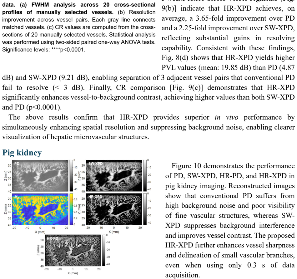
The central claim is that HR-XPD (high-resolution cross-correlation power Doppler) makes contrast-free microvascular ultrasound images both sharper and cleaner than conventional power Doppler. The method works by splitting ultrafast plane-wave transmissions into two subsets, clutter-filtering each subset separately, and combining two complementary weightings: a radiality map, which rewards pixels whose surrounding intensity gradients point inward like a point-spread-function center, and a similarity map, the normalized cross-correlation between the two subsets over time, which keeps coherent blood flow and rejects random noise. The product is a power Doppler image in which vessel centers are emphasized while sidelobes and background are suppressed. The paper supports this with simulations across signal-to-noise levels and in vivo demonstrations, reporting roughly 2.5- to 3.7-fold full-width-at-half-maximum resolution improvements over conventional PD, higher peak-to-valley separation of adjacent vessels, and background intensity reductions of about 20 dB.
Load-bearing premise
The load-bearing premise is that the -3 dB width of manually selected vessel profiles measures true spatial resolution rather than the edge of a suppressed background, because the paper does not calibrate against a point target or resolution phantom.
Editorial extensions
If this is right
- If the reported gains hold, microvascular anatomy can be imaged without contrast agents in under a second, making high-resolution vascular imaging practical in breath-held liver and kidney scans.
- Adjacent vessels that conventional PD leaves unresolved (valley intensity below the 3 dB threshold) become separable, enabling more reliable vessel counting and microvascular density quantification.
- The short acquisition time and high frame rate open a path to functional ultrasound activation mapping, where spatial sharpness and temporal resolution both matter.
- The same radiality-plus-coherence weighting could be applied to microbubble contrast-enhanced data, potentially stabilizing radiality-based super-resolution in lower-signal-to-noise clinical settings, a possibility the paper notes.
- Because the method relies on flow-signal coherence, it may be extended from power Doppler to color Doppler to add velocity information without losing the resolution gain.
Reading between the lines
- A direct experimental separation of true resolution from contrast would be to run HR-XPD on a wire or point-scatterer phantom: if the point-spread function width is unchanged while background drops, the two- to threefold FWHM figure is mainly contrast enhancement rather than actual resolution gain.
- The 2-3x resolution claim likely depends on where the -3 dB contour sits on a vessel profile; measuring widths at a lower threshold, such as -10 dB, would test whether the apparent sharpening persists across the full point-spread function.
- Splitting transmissions into two angle subsets reduces the signal available to each subset, so the method's advantage may depend on having enough frames for the similarity map to stabilize; this can be tested by sweeping frame count.
- If confirmed, HR-XPD could be combined with spectral decomposition or deconvolution approaches, since radiality and coherence suppression are complementary to model-based resolution restoration.
Signed reviews
Editorial analysis
A structured set of objections, weighed in public.
Referee Report
Summary. The paper proposes HR-XPD, a contrast-free microvascular ultrasound imaging method that combines radiality weighting (derived from local gradient convergence) with cross-correlation between two interleaved angular Doppler subsets and a similarity map. The claimed contributions are improved spatial resolution, lower background intensity, and enhanced vascular contrast compared with conventional power Doppler (PD), SW-XPD, and HR-PD. Validation is provided through Field II simulations of two crossing 1-mm tubes under varying noise levels and in vivo imaging of a transplanted human kidney, a healthy human liver, and a pig kidney. The central quantitative claim is a 2- to 3-fold spatial-resolution improvement over PD (up to 3.65-fold in the liver) and contrast increases of up to 20 dB, achieved with 0.3-1.2 s of acquisition without contrast agents.
Significance. If the resolution improvement is real, HR-XPD would be a practically valuable addition to microvascular ultrasound because it is contrast-free, uses short acquisition times, and is evaluated across multiple in vivo settings. The method is a transparent nonlinear processing recipe: the radiality annulus radius, sampling-point count, and SVD block/rank settings are explicit, and no parameters are fitted to the reported outcomes, which is a strength. The paper also provides useful comparisons against three baselines (PD, SW-XPD, HR-PD) and quantifies robustness to noise. However, the headline resolution claim rests on metrics that are inherently contrast-dependent, and no point-target or wire-phantom experiment is reported to separate genuine spatial-resolution gain from nonlinear background suppression. The simulation results themselves show only a modest FWHM gain (about 1.2-1.4x), which is inconsistent with the much larger in vivo gains and underscores the need for a direct resolution test.
major comments (4)
- [Quantitative Evaluation; Figs. 7, 9, 11] The central 2-3x spatial-resolution claim is measured exclusively through the -3 dB FWHM of manually selected vessel cross-sections (Eq. 7 definition; Figs. 7, 9, 11). This metric is contrast-dependent: HR-XPD strongly suppresses background and sidelobe intensity, so the -3 dB contour of any bright structure narrows even if the underlying point spread function (PSF) is unchanged. The simulation FWHM gains are only about 1.2-1.4x (Fig. 2e, Fig. 4c), while the in vivo gains are 2.46x, 3.65x, and 1.5-2.8x in Figs. 7b, 9b, and 11e, respectively. This inconsistency suggests that most of the reported in vivo gain is a contrast-enhancement artifact rather than a true spatial-resolution improvement. A point-target or wire-phantom experiment, or an isolated sub-resolution scatterer simulation, is needed to measure the PSF width directly and separate resolution gain from contrast enhancement.
- [Eq. (9); Figs. 7c, 8d, 11c] The peak-to-valley level (PVL) metric is used to claim that HR-XPD resolves adjacent vessels that PD and SW-XPD fail to resolve (PVL < 3 dB). PVL compares the valley intensity to the peak intensity, but a valley can rise above the 3 dB threshold purely because the noise floor between two unresolved PSF peaks has been suppressed by the similarity weighting. PVL therefore does not constitute a two-point resolution test unless the center-to-center separation of a phantom target is known. A resolution phantom with echo-free point scatterers or wires at known separations is required to support the resolvability claim.
- [Simulation (Materials and Methods)] The simulation geometry consists of two crossing tubes with a diameter of 1 mm, not point targets. The FWHM of a 1-mm tube profile is determined by the convolution of the PSF with the tube's finite spatial extent and the flow velocity profile, so the measured FWHM and resolution-improvement ratio in Fig. 2(e) do not directly quantify PSF narrowing. A simulation with a point scatterer or a sub-resolution wire, or a deconvolution-based resolution estimator, would provide a cleaner measure of the method's true resolution capability.
- [Eq. (1); Materials and Methods, 'Principle of HR-XPD'] The radiality G_q in Eq. (1) is the average of normalized dot products of gradient vectors with radial vectors and can take negative values for outward-pointing gradients. The text states that the radiality maps R1 and R2 are 'log-compressed and normalized' before being applied as weights, but no mapping is specified for negative values. Since radiality weighting is the core mechanism for resolution enhancement, this ambiguity makes the method irreproducible as written and should be clarified with the exact transformation (e.g., absolute value, positive-part, or offset before log compression).
minor comments (5)
- [Abstract] The abstract claims 'up to a 2 to 3-fold enhancement' in spatial resolution, but the liver results in Fig. 9(b) report a 3.65-fold mean improvement over PD; the claim should be updated to reflect the full range or the abstract should be qualified.
- [Fig. 2 caption and main text] There is a mismatch between the caption of Fig. 2 and the main text: the caption labels (d) as 'Estimated resolution improvement' and (e) as 'PVL', while the main text refers to PVL in Fig. 2(d) and resolution improvement in Fig. 2(e). The labeling should be corrected.
- [Materials and Methods, 'Principle of HR-XPD'] In the sentence after Eq. (2), 'Details in [5]' is too vague for a described mechanism; a brief derivation or intuitive explanation of why the cross-terms involving noise are suppressed would make the method more self-contained.
- [Statistics] The text says that FWHM, CR, and resolution improvement ratios were compared using 'one-way analysis of variance (RM one-way ANOVA)'; the abbreviation RM should be defined as repeated-measures, and the specific post-hoc correction should be stated consistently.
- [Fig. 6, Fig. 9] The text and figure captions repeatedly use 'randomly selected' to describe vessel cross-sections (e.g., Fig. 7c, Fig. 8d, Fig. 11c), but the selection is manual and therefore subjective; the term 'randomly' should be replaced with 'manually selected' to avoid implying an objective sampling procedure.
Circularity Check
No significant circularity: HR-XPD is a fixed nonlinear processing recipe evaluated against independent baselines; no fitted parameter or self-citation chain forces the reported outcome.
full rationale
The paper's derivation chain is self-contained: HR-XPD is explicitly defined by Eqs. (1)-(6) from the two angular IQ subsets, radiality maps, and normalized similarity map, and its outputs are then compared with conventional PD, SW-XPD, and HR-PD in simulations and in vivo. No parameter is fitted to the reported FWHM, PVL, CR, or background-intensity outcomes, and the central claim is not a renamed input. The cited prior work on radiality ([47], [48]) and on cross-correlation/similarity processing ([5], [42]) supplies methodological motivation, but the present algorithm and its quantitative evaluation do not reduce to those citations. The FWHM-based resolution claim could be challenged as a measurement-validity issue, because nonlinear weighting can narrow -3 dB vessel profiles by suppressing background and sidelobes even if the underlying point spread function is unchanged; however, that is a correctness or experimental-design concern, not a circularity. Since the paper does not define its predictions in terms of its inputs or rely on a load-bearing self-citation chain, the appropriate circularity finding is no significant circularity.
Assumptions & free parameters
free parameters (3)
- radiality annulus inner radius r =
0.5 pixel
- number of radiality sampling points =
8
- SVD block size and rank threshold =
not reported
assumptions (4)
- domain assumption Blood flow signals are coherent between odd and even steering-angle subsets, while noise is uncorrelated across subsets.
- domain assumption Radiality (gradient convergence) computed from envelope IQ data indicates vessel/PSF centers for red-blood-cell echoes without contrast agents.
- domain assumption Local block-wise SVD clutter filtering cleanly separates blood signal from tissue clutter in large field-of-view human imaging.
- standard math Normalized cross-correlation as defined in Eq. (3) is a valid similarity metric for flow coherence.
Cite this review
Pith. "Pith review of Contrast-Free Ultrasound Microvascular Imaging via Radiality and Similarity Weighting." pith.science (2026). https://pith.science/paper/WM63EAXK
@misc{pith2026250907128,
  author       = {Pith},
  title        = {Pith review of: Contrast-Free Ultrasound Microvascular Imaging via Radiality and Similarity Weighting},
  year         = {2026},
  howpublished = {\url{https://pith.science/paper/WM63EAXK}},
  note         = {Machine review of arXiv:2509.07128}
}
read the original abstract
Microvascular imaging has advanced significantly with ultrafast data acquisition and improved clutter filtering, enhancing the sensitivity of power Doppler imaging to small vessels. However, the image quality remains limited by spatial resolution and elevated background noise, both of which impede visualization and accurate quantification. To address these limitations, this study proposes a high-resolution cross-correlation Power Doppler (HR-XPD) method that integrates spatial radiality weighting with Doppler signal coherence analysis, thereby enhancing spatial resolution while suppressing artifacts and background noise. Quantitative evaluations in simulation and in vivo experiments on healthy human liver, transplanted human kidney, and pig kidney demonstrated that HR-XPD significantly improves microvascular resolvability and contrast compared to conventional PD. In vivo results showed up to a 2 to 3-fold enhancement in spatial resolution and an increase in contrast by up to 20 dB. High-resolution vascular details were clearly depicted within a short acquisition time of only 0.3 s-1.2 s without the use of contrast agents. These findings indicate that HR-XPD provides an effective, contrast-free, and high-resolution microvascular imaging approach with broad applicability in both preclinical and clinical research.
Figures


Figures from the paper (6 more)



Reference graph
Works this paper leans on
-
[1]
R. A. Nishimura, F. A. J. Miller, M. J. Callahan et al., Doppler echocardiography: theory, instrumentation, technique, and application: Elsevier, 1985
work page 1985
-
[2]
K. J. Taylor, P. N. Burns, and P. N. Well, Clinical applications of Doppler ultrasound, 1987
work page 1987
-
[3]
Coherent plane-wave compounding for very high frame rate ultrasonography and transient elastography,
G. Montaldo, M. Tanter, J. Bercoff et al. , “Coherent plane-wave compounding for very high frame rate ultrasonography and transient elastography,” IEEE Trans. Ultrason. Ferroelectr. Freq. Control, vol. 56, no. 3, pp. 489-506, 2009
work page 2009
-
[4]
Ultrafast ultrasound vector Doppler for small vasculature imaging,
S. Yan, J. Shou, J. Y u et al., “Ultrafast ultrasound vector Doppler for small vasculature imaging,” IEEE Trans. Ultrason. Ferroelectr. Freq. Control, vol. 70, no. 7, pp. 613-624, 2023
work page 2023
-
[5]
C. Huang, P. So ng, J. D. Trzasko et al., “Simultaneous noise suppression and incoherent artifact reduction in ultrafast ultrasound vascular imaging,” IEEE Trans. Ultrason. Ferroelectr. Freq. Control, vol. 68, no. 6, pp. 2075-2085, 2021
work page 2021
-
[6]
Ultrafast compound Doppler imaging: Providing full blood flow characterization,
J. Bercoff, G. Montaldo, T. Loupas et al., “Ultrafast compound Doppler imaging: Providing full blood flow characterization,” IEEE Trans. Ultrason. Ferroelectr. Freq. Control, vol. 58, no. 1, pp. 134-147, 2011
work page 2011
-
[7]
C. Demené, T. Deffieux, M. Pernot et al., “Spatiotemporal clutter filtering of ultrafast ultrasound data highly increases Doppler and fUltrasound sensitivity,” IEEE Trans. Med. Imaging, vol. 34, no. 11, pp. 2271-2285, 2015
work page 2015
-
[8]
Functional ultrasound imaging of the brain,
E. Macé, G. Montaldo, I. Cohen et al., “Functional ultrasound imaging of the brain,” Nat. Methods, vol. 8, no. 8, pp. 662-664, 2011
work page 2011
Show all 60 references
-
[9]
Ultrasound small vessel imaging with block-wise adaptive local clutter filtering,
P. Song, A. Manduca, J. D. Trzasko et al., “Ultrasound small vessel imaging with block-wise adaptive local clutter filtering,” IEEE Trans. Med. Imaging, vol. 36, no. 1, pp. 251-262, 2016
2016
-
[10]
Ultrafas t imaging in biomedical ultrasound,
M. Tanter, and M. Fink, “Ultrafas t imaging in biomedical ultrasound,” IEEE Trans. Ultrason. Ferroelectr. Freq. Control, vol. 61, no. 1, pp. 102-119, 2014
2014
-
[11]
Eigen-based clutter f ilter design for ultrasound color flow imaging: A review,
C. Alfred, and L. Lovstakken, “Eigen-based clutter f ilter design for ultrasound color flow imaging: A review,” IEEE Trans. Ultrason. Ferroelectr. Freq. Control, vol. 57, no. 5, pp. 1096-1111, 2010. [ 1 2 ] M . K i m , Y . Z h u , J . H e d h l i et al. , “Multidimensional clu...
2010
-
[13]
Wide field-of-view ultrafast curved array imaging using diverging waves,
J. Kang, D. Go, I. Song et al., “Wide field-of-view ultrafast curved array imaging using diverging waves,” IEEE Trans. Biomed. Eng., vol. 67, no. 6, pp. 1638-1649, 2019
2019
-
[14]
Independent component-based spatiotemporal clutter filtering for slow flow ultrasound,
J. Tierney, J. Baker, D. Brown et al., “Independent component-based spatiotemporal clutter filtering for slow flow ultrasound,” IEEE Trans. Med. Imaging, vol. 39, no. 5, pp. 1472-1482, 2019
2019
-
[15]
In vivo visualization of vasculature in adult zebrafish by using high-frequency ultrafast ultrasound imaging,
C.-C. Chang, P .-Y . Chen, H. Huang et al., “In vivo visualization of vasculature in adult zebrafish by using high-frequency ultrafast ultrasound imaging,” IEEE Trans. Biomed. Eng., vol. 66, no. 6, pp. 1742-1751, 2018
2018
-
[16]
Whole-brain functional ultrasound imaging in awake head-fixed mice,
C. Brunner, M. Grillet, A. Urban et al. , “Whole-brain functional ultrasound imaging in awake head-fixed mice,” Nat. Protoc., vol. 16, no. 7, pp. 3547-3571, 2021
2021
-
[17]
Super-resolution ultrasound imaging,
K. Christensen-Jeffries, O. Couture, P. A. Dayton et al., “Super-resolution ultrasound imaging,” Ultrasound Med. Biol., vol. 46, no. 4, pp. 865-891, 2020
2020
-
[18]
Retinal functional ultrasound imaging (rfUS) for assessing neurovascular alterations: a pilot study on a rat model of dementia,
C. Morisset, A. Dizeux, B. Larrat et al. , “Retinal functional ultrasound imaging (rfUS) for assessing neurovascular alterations: a pilot study on a rat model of dementia,” Sci. Rep., vol. 12, no. 1, pp. 19515, 2022
2022
-
[19]
Cerebral blood flow threshold of ischemic penumbra and infarct core in acute ischemic stroke: a systematic review,
E. Bandera, M. Botteri, C. Minelli et al., “Cerebral blood flow threshold of ischemic penumbra and infarct core in acute ischemic stroke: a systematic review,” Stroke, vol. 37, no. 5, pp. 1334-1339, 2006
2006
-
[20]
Quantitative assessment of ultrasound microvessel imaging in Crohn’s disease: correlation with pathological inflammation,
U.-W. Lok, S. Tang, P. Gong et al., “Quantitative assessment of ultrasound microvessel imaging in Crohn’s disease: correlation with pathological inflammation,” Eur. Radiol., vol. 35, no. 5, pp. 2806-2817, 2024
2024
-
[21]
Ultrasensitive ultrasound microvessel imaging for characterizing benign and malignant breast tumors,
P. Gong, P. Song, C. Huang et al., “Ultrasensitive ultrasound microvessel imaging for characterizing benign and malignant breast tumors,” Ultrasound Med. Biol., vol. 45, no. 12, pp. 3128-3136, 2019
2019
-
[22]
Doppler slicing for ultrasound super-resolution without contrast agents,
A. Bar-Zion, O. Solomon, C. Rabut et al., "Doppler slicing for ultrasound super-resolution without contrast agents," BioRxiv, 2021]
2021
-
[23]
Improved ultrasound microvessel imaging using deconvolution with total variation regularization,
U.-W. Lok, J. D. Trzasko, C. Huang et al., “Improved ultrasound microvessel imaging using deconvolution with total variation regularization,” Ultrasound Med. Biol., vol. 47, no. 4, pp. 1089-1098, 2021
2021
-
[24]
Resolution and contra st improved ultrafast power Doppler microvessel imaging with null subtraction imaging,
M. L. Oelze, and Z. Kou, “Resolution and contra st improved ultrafast power Doppler microvessel imaging with null subtraction imaging,” J. Acoust. Soc. Am., vol. 153, no. 3, pp. A30-A30, 2023
2023
-
[25]
Contrast -free microvessel imaging using null subtraction imaging combined with harmonic imaging,
Z. Kou, R. Miller, and M. L. Oelze, “Contrast -free microvessel imaging using null subtraction imaging combined with harmonic imaging,” J. Acoust. Soc. Am., vol. 155, no. 3, pp. A55-A55, 2024
2024
-
[26]
Acoustic Angiography: Superharmonic Contrast-Enhanced Ultrasound Imaging for Noninvasive Visualization of Microvasculature,
I. G. Newsome, and P. A. Dayton, "Acoustic Angiography: Superharmonic Contrast-Enhanced Ultrasound Imaging for Noninvasive Visualization of Microvasculature," Biomedical Engineering Technologies: Volume 1, pp. 641-655: Springer, 2021
2021
-
[27]
Mapping microvasculature with acoustic angiography yields quantifiable differences between healthy and tumor-bearing tissue volumes in a rodent model,
R. C. Gessner, S. R. Aylward, and P. A. Dayton, “Mapping microvasculature with acoustic angiography yields quantifiable differences between healthy and tumor-bearing tissue volumes in a rodent model,” Radiology, vol. 264, no. 3, pp. 733-740, 2012
2012
-
[28]
Contrast-free super-resolution power Doppler (CS-PD) based on deep neural networks,
Q. You, M. R. Lowerison, Y . Shin et al., “Contrast-free super-resolution power Doppler (CS-PD) based on deep neural networks,” IEEE Trans. Ultrason. Ferroelectr. Freq. Control, vol. 70, no. 10, pp. 1355-1368, 2023
2023
-
[29]
Super-resolution ultrasound imaging using the erythrocytes— Part I: Density images,
J. A. Jensen, M. A. Naji, S. K. Præsius et al., “Super-resolution ultrasound imaging using the erythrocytes— Part I: Density images,” IEEE Trans. Ultrason. Ferroelectr. Freq. Control, vol. 71, no. 8, pp. 925-944, 2024
2024
-
[30]
Super-resolution ultrasound imaging using the erythrocytes—Part II: Velocity images,
M. A. Naji, I. Taghavi, M. Schou et al., “Super-resolution ultrasound imaging using the erythrocytes—Part II: Velocity images,” IEEE Trans. Ultrason. Ferroelectr. Freq. Control, vol. 71, no. 8, pp. 945-959, 2024
2024
-
[31]
Deep learning-based super-resolution ultrasound speckle tracking velocimetry,
J. H. Park, W. Choi, G. Y . Yoon et al. , “Deep learning-based super-resolution ultrasound speckle tracking velocimetry,” Ultrasound Med. Biol., vol. 46, no. 3, pp. 598-609, 2020
2020
-
[32]
Coherent flow imaging: A power Doppler imaging technique based on backscatter spatial coherence
J. J. Dahl, N. Bottenus, M. A. L. Bell et al., "Coherent flow imaging: A power Doppler imaging technique based on backscatter spatial coherence." pp. 639-642
-
[33]
Visualization of small-diameter vessels by reduction of incoherent reverberation with coherent flow power doppler,
Y . L. Li, D. Hyun, L. Abou-Elkacem et al. , “Visualization of small-diameter vessels by reduction of incoherent reverberation with coherent flow power doppler,” IEEE Trans. Ultrason. Ferroelectr. Freq. Control, vol. 63, no. 11, pp. 1878-1889, 2016
2016
-
[34]
High-quality ultrafast power Doppler imaging based on spatial angular coherence factor,
L. Huang, Y . Wang, R. Wang et al., “High-quality ultrafast power Doppler imaging based on spatial angular coherence factor,” IEEE Trans. Ultrason. Ferroelectr. Freq. Control, vol. 70, no. 5, pp. 378-392, 2023
2023
-
[35]
A spatial coherence beamformer design for power Doppler imaging,
K. Ozgun, J. Tierney, and B. Byram, “A spatial coherence beamformer design for power Doppler imaging,” IEEE Trans. Med. Imaging, vol. 39, no. 5, pp. 1558-1570, 2019
2019
-
[36]
ASAP: Super-contrast vasculature imaging using coherence analysis and high frame-rate c ontrast enhanced ultrasound,
A. Stanziola, C. H. Leow, E. Bazigou et al. , “ASAP: Super-contrast vasculature imaging using coherence analysis and high frame-rate c ontrast enhanced ultrasound,” IEEE Trans. Med. Imaging, vol. 37, no. 8, pp. 1847-1856, 2018
2018
-
[37]
3-D microvascular imaging using high frame rate ultrasound and ASAP without contrast agents: Development and initial in vivo evaluation on nontumor and tumor models,
C. H. Leow , N. L. Bush, A. Stanziola et al., “3-D microvascular imaging using high frame rate ultrasound and ASAP without contrast agents: Development and initial in vivo evaluation on nontumor and tumor models,” IEEE Trans. Ultrason. Ferroelectr. Freq. Control, vol. 66, no. ...
2019
-
[38]
Adaptive noise reduction for power Doppler imaging using SVD filtering in the channel domain and coherence weighting of pixels,
B. Pialot, C. Lach ambre, A. Lorente Mur et al., “Adaptive noise reduction for power Doppler imaging using SVD filtering in the channel domain and coherence weighting of pixels,” Phys. Med. Biol., vol. 68, no. 2, pp. 025001, 2023
2023
-
[39]
Improved contrast-enhanced power Doppler using a coherence-based estimator,
C. Tremblay-Darveau, A. Bar-Zion, R. Williams et al., “Improved contrast-enhanced power Doppler using a coherence-based estimator,” IEEE Trans. Med. Imaging, vol. 36, no. 9, pp. 1901-1911, 2017
1901
-
[40]
Improved ultrafast power Doppler imaging by using spatiotemporal non- local means filtering,
L. Huang, J. Zhang, X. Wei et al., “Improved ultrafast power Doppler imaging by using spatiotemporal non- local means filtering,” IEEE Trans. Ultrason. Ferroelectr. Freq. Control, vol. 69, no. 5, pp. 1610-1624, 2022
2022
-
[41]
Blood flow imaging in the neonatal brain using angular coherence power Doppler,
M. Jakovljevic, B. C. Yoon, L. Abou-Elkacem et al., “Blood flow imaging in the neonatal brain using angular coherence power Doppler,” IEEE Trans. Ultrason. Ferroelectr. Freq. Control, vol. 68, no. 1, pp. 92-106, 2020
2020
-
[42]
Enhancement of Ultrasound Microbubble and Blood Flow Imaging using Similarity Measurement,
C. Huang, “Enhancement of Ultrasound Microbubble and Blood Flow Imaging using Similarity Measurement,” Authorea Preprints, 2023
2023
-
[43]
Ultrafast power Doppler imaging using frame-multiply-and-sum-based nonlinear compounding,
J. Kang, D. Go, I. Song et al. , “Ultrafast power Doppler imaging using frame-multiply-and-sum-based nonlinear compounding,” IEEE Trans. Ultrason. Ferroelectr. Freq. Control, vol. 68, no. 3, pp. 453-464, 2020
2020
-
[44]
DMAS beamforming with complementary subset transmit for ultrasound coherence-based power Doppler detection in multi-angle plane-wave imaging,
C.-C. Shen, and Y .-C. Chu, “DMAS beamforming with complementary subset transmit for ultrasound coherence-based power Doppler detection in multi-angle plane-wave imaging,” Sensors, vol. 21, no. 14, pp. 4856, 2021
2021
-
[45]
Improved ultrafast power Doppler imaging using united spatial–angular adaptive scaling Wiener postfilter,
Y . Wang, L. Huang, R. Wang et al., “Improved ultrafast power Doppler imaging using united spatial–angular adaptive scaling Wiener postfilter,” IEEE Trans. Ultrason. Ferroelectr. Freq. Control, vol. 70, no. 9, pp. 1118-1134, 2023
2023
-
[46]
Real-time adaptive and localized spatiotemporal clutter filtering for ultrasound small vessel imaging,
C. Huang, U.-W. Lok, J. Zhang et al., “Real-time adaptive and localized spatiotemporal clutter filtering for ultrasound small vessel imaging,” arXiv preprint arXiv:2405.11105, 2024
2024 arXiv
-
[47]
Ultrasound microvascular imaging based on super -resolution radial fluctuations,
J. Zhang, N. Li, F. Dong et al. , “Ultrasound microvascular imaging based on super -resolution radial fluctuations,” J. Ultrasound Med., vol. 39, no. 8, pp. 1507-1516, 2020
2020
-
[48]
Ultrasound microvasculature imaging with entropy-based radiality super- resolution (ERSR),
J. Yin, J. Zhang, Y . Zhu et al. , “Ultrasound microvasculature imaging with entropy-based radiality super- resolution (ERSR),” Phys. Med. Biol., vol. 66, no. 21, pp. 215012, 2021
2021
-
[49]
In Vivo Dynamic Coronary Arteries Blood Flow Imaging Based on Multi- Cycle Phase Clustering Ultrafast Ultrasound,
H. Y u, J. Zhang, F. Feng et al., “In Vivo Dynamic Coronary Arteries Blood Flow Imaging Based on Multi- Cycle Phase Clustering Ultrafast Ultrasound,” Adv. Sci., vol. 12, no. 31, pp. e05485, 2025
2025
-
[50]
Fast live-cell conventional fluorophore nanoscopy with ImageJ through super-resolution radial fluctuations,
N. Gustafsson, S. Culley, G. Ashdown et al., “Fast live-cell conventional fluorophore nanoscopy with ImageJ through super-resolution radial fluctuations,” Nat. Commun., vol. 7, no. 1, pp. 12471, 2016
2016
-
[51]
Calculation of pressure fields from arbitrarily shaped, apodized, and excited ultrasound transducers,
J. A. Jensen, and N. B. Svendsen, “Calculation of pressure fields from arbitrarily shaped, apodized, and excited ultrasound transducers,” IEEE Trans. Ultrason. Ferroelectr. Freq. Control, vol. 39, no. 2, pp. 262- 267, 1992
1992
-
[52]
Field: A progra m for simulating ultrasound systems,
J. A. Jensen, “Field: A progra m for simulating ultrasound systems,” Med. Biol. Eng. Comput., vol. 34, no. 1, pp. 351-353, 1997
1997
-
[53]
Localization free super-resolution microbubble velocimetry using a long short-term memory neural network,
X. Chen, M. R. Lowerison, Z. Dong et al., “Localization free super-resolution microbubble velocimetry using a long short-term memory neural network,” IEEE Trans. Med. Imaging, vol. 42, no. 8, pp. 2374-2385, 2023
2023
-
[54]
Optimizing in vivo data acquisition for robust clinical microvascular imaging using ultrasound localization microscopy,
C. Huang, U.-W. Lok, J. Zhang et al., “Optimizing in vivo data acquisition for robust clinical microvascular imaging using ultrasound localization microscopy,” Physics in Medicine & Biology, vol. 70, no. 7, pp. 075017, 2025
2025
-
[55]
Eigenspace based minimum variance beamforming applied to ultrasound imaging of acoustically hard tissues,
S. Mehdizadeh, A. Austeng, T. F. Johansen et al., “Eigenspace based minimum variance beamforming applied to ultrasound imaging of acoustically hard tissues,” IEEE Trans. Med. Imaging, vol. 31, no. 10, pp. 1912- 1921, 2012
1912
-
[56]
Ultrafast ultrasound localization microscopy for deep super-resolution vascular imaging,
C. Errico, J. Pierre, S. Pezet et al., “Ultrafast ultrasound localization microscopy for deep super-resolution vascular imaging,” Nature, vol. 527, no. 7579, pp. 499-502, 2015
2015
-
[57]
Assessment of Takayasu's ar teritis activity by ultrasound localization microscopy,
G. Goudot, A. Jimenez, N. Mohamedi et al. , “Assessment of Takayasu's ar teritis activity by ultrasound localization microscopy,” EBioMedicine, vol. 90, pp. 104502, 2023
2023
-
[58]
Pattern recognition of microcirculation with super-resolution ultrasound imaging provides markers for early tumor response to anti-angiogenic therapy,
J. Yin, F. Dong, J. An et al., “Pattern recognition of microcirculation with super-resolution ultrasound imaging provides markers for early tumor response to anti-angiogenic therapy,” Theranostics, vol. 14, no. 3, pp. 1312- 1314, 2024
2024
-
[59]
Enhancing Row-column array (RCA)-based 3D ultrasound vascular imaging with spatial-temporal similarity weighting,
J. Zhang, C. Huang, U.-W. Lok et al., “Enhancing Row-column array (RCA)-based 3D ultrasound vascular imaging with spatial-temporal similarity weighting,” IEEE Trans. Med. Imaging, vol. 44, no. 1, pp. 297-309, 2024
2024
-
[60]
Poisson statistical model of ultrasound super-resolution imaging acquisition time,
K. Christensen-Jeffries, J. Brown, S. Harput et al., “Poisson statistical model of ultrasound super-resolution imaging acquisition time,” IEEE Trans. Ultrason. Ferroelectr. Freq. Control, vol. 66, no. 7, pp. 1246-1254, 2019
2019
-
[61]
Microvascular flow dictates the compromise between spatial resolution and acquisition time in ultrasound localization microscopy,
V . Hingot, C. Errico, B. Heiles et al., “Microvascular flow dictates the compromise between spatial resolution and acquisition time in ultrasound localization microscopy,” Sci. Rep., vol. 9, no. 1, pp. 2456, 2019. Supplementary information TABLE S1 PARMETER SETTINGS OF IN VIV...
2019
Reviewed August 15, 2026 · model on record in the stance chip above.
Discussion (0). Continue with ORCID to comment.