REVIEW 4 major objections 5 minor 41 references
Dataset and Benchmark for Enhancing Critical Retained Foreign Object Detection
T0 review · 4 major / 5 minor · reviewed 2026-08-06 · deepseek-v4-flash
Pith's one-line read The paper introduces Hopkins RFOs Bench, the first publicly available chest X-ray dataset dedicated to critical retained foreign objects, and shows that physics-based synthetic X-ray augmentation consistently improves detector…
desk verdict The dataset is the real contribution; the synthetic augmentation comparison is useful but rests on point estimates from a tiny test set, so the headline claim needs statistical grounding before this is final. read the letter →
The pith
A machine-rendered reading of the paper's core claim, the machinery that carries it, and where it could break.
The reading
What carries the argument
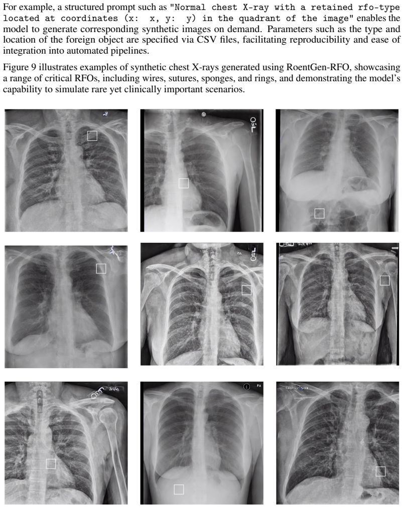
The load-bearing machinery is the dataset itself plus two synthetic-generation pipelines that are directly compared. DeepDRR-RFO is a physics-based pipeline that segments CT volumes into air, soft tissue, and bone, reconstructs 3D models of real surgical items from single photographs, embeds them into the CT volumes, and simulates X-ray formation with material-specific attenuation to produce automatically annotated radiographs. RoentGen-RFO is a diffusion-based pipeline that adapts a pretrained DDPM through carefully designed text prompts, without fine-tuning on RFO images, to generate synthetic chest X-rays containing critical foreign objects. The comparison of these two pipelines, evaluated with the same detectors and metrics on the same held-out test set, carries the paper's central argument about which synthetic-data strategy is effective for rare critical findings.
What would settle it
Train the four detectors with and without +2,000 physics-based synthetic images exactly as described, but evaluate on a separate external test set of 100 or more critical-RFO chest X-rays from another institution; if the synthetic-augmented models do not beat the real-data baseline on AUC and FROC by a margin larger than the run-to-run variance across several random seeds, the paper's central empirical claim would be undermined.
Extended reading notes
Core claim
Hopkins RFOs Bench is claimed to be the first publicly available dataset dedicated explicitly to critical RFO cases, containing 144 critical-RFO chest X-ray images from distinct patients, annotated at both image level and object level with bounding boxes or polygons and a critical/non-critical label. On this benchmark, all four tested detectors (Faster R-CNN, FCOS, RetinaNet, YOLO) improve substantially when pretrained on the non-critical Object-CXR dataset and fine-tuned on Hopkins RFOs Bench, e.g., Faster R-CNN's AUC rises from 0.62 to 0.80. When additional physics-based synthetic radiographs are added to the training set, every model improves across ACC, FNR, AUC, and FROC, peaking at 2,000 synthetic images (e.g., RetinaNet reaches 79.5% ACC, 0.23 FNR, 0.78 AUC, 63.5 FROC) and declining slightly at 4,000. Diffusion-based synthetic images, by contrast, generally reduce performance, illustrating the authors' conclusion that physically grounded simulation currently offers more useful training data for critical RFO detection than zero-shot diffusion generation.
Load-bearing premise
The quantitative conclusions rest on a small held-out test set (about 29 critical-RFO images) with no reported confidence intervals or multiple training runs, so a few hard images could change the ranking of training strategies, and the synthetic RFOs themselves are acknowledged to be overly contrasted.
Editorial extensions
If this is right
- Researchers gain a public, IRB-approved benchmark with image- and object-level annotations, enabling direct comparison of future critical-RFO detection models.
- Physics-based synthetic augmentation at moderate scale (around 2,000 images) is a viable strategy to mitigate extreme data scarcity for rare radiological findings.
- Diffusion-based synthetic data, at least in zero-shot form, is not yet useful for training critical-RFO detectors and may harm performance by introducing distribution shift.
- Pretraining on a large non-critical RFO dataset (Object-CXR) followed by fine-tuning on a small critical-RFO dataset is an effective transfer recipe, at least on this benchmark.
- The same benchmark can be used to fine-tune diffusion models on real critical-RFO examples, potentially improving their ability to generate useful synthetic cases.
Reading between the lines
- An implication the paper leaves implicit is that the reported performance gains rest on a small test set: with a 70/10/20 split of 144 critical cases, the critical test partition contains roughly 29 images, so the superiority of +2,000 physics-based images over +1,000 or +4,000 could shift with a few difficult cases; the authors report no confidence intervals or multi-seed variance.
- A testable extension is to evaluate the trained detectors on external chest X-rays from another institution to see whether the physics-based synthetic gains transfer beyond the single health system used to build the benchmark.
- The physics-based pipeline's acknowledged limitation that synthetic RFOs appear overly contrasted suggests a concrete improvement: applying appearance randomization or domain adaptation to the synthetic images before training, then measuring whether gains persist on subtle real-world objects.
- Because the dataset includes non-critical RFOs and no-RFO images alongside critical cases, it could support multi-class training and calibrated screening workflows, not just binary critical-versus-absent detection.
Editorial analysis
A structured set of objections, weighed in public.
Referee Report
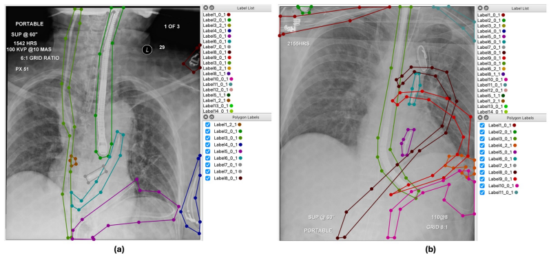
Summary. The paper introduces Hopkins RFOs Bench, a retrospectively collected dataset of 144 chest X-ray images containing critical retained foreign objects (RFOs) from the Johns Hopkins Health System, together with image- and object-level annotations. The authors benchmark four object detectors and compare two synthetic-data augmentation strategies: DeepDRR-RFO, a physics-based pipeline, and RoentGen-RFO, a diffusion-based pipeline. The central claims are that the dataset is the first publicly available resource dedicated to critical RFOs and that physics-based synthetic augmentation consistently improves detection performance across all reported metrics, while DDPM-based augmentation does not.
Significance. If the dataset is released as described, it would fill a real gap: the public RFO datasets listed in Table 1 contain only non-critical objects. The curation process is described carefully, including IRB approval, radiologist second reads, and patient-level data splits, and the authors commit to open release of code and (upon acceptance) data. The paper also provides a useful side-by-side comparison of physics-based and diffusion-based synthetic augmentation. However, the central quantitative claim is not yet statistically grounded: the held-out critical test set is small, Table 2 reports no confidence intervals or seed variability, and the same baseline condition is reported with different values in Table 2 and Table 3. These issues must be resolved before the benchmark results can be relied upon.
major comments (4)
- [Section 3 and Section 6.2, Table 2] The held-out critical test set contains roughly 29 images (20% of 144), and Table 2 reports only single point estimates with no confidence intervals, bootstrap resampling, or multiple-seed variability. The observed improvements are small in absolute terms (e.g., FCOS ACC 75.1 to 76.4 and FROC 52.0 to 55.1; Faster-RCNN FROC 50.5 to 54.3), so a change of one or two test cases could alter the rankings. The Section 6.2 conclusion that physics-based augmentation 'consistently improves model performance across all metrics' is therefore not statistically supported as stated.
- [Table 2 vs Appendix G, Table 3] The baseline condition is reported inconsistently across the manuscript: Table 2 'Base' for Faster-RCNN gives ACC 74.0, AUC 0.62, FROC 50.5, while Table 3 'Hopkins RFOs Bench' gives ACC 74.3, AUC 0.73, FROC 49.8; FCOS likewise differs (ACC 75.1 vs 71.4, AUC 0.61 vs 0.67). Since these appear to describe the same training condition, the discrepancy must be reconciled before any augmentation comparison is interpretable.
- [Section 4.1 and Appendix D] The task definition in Section 4.1 requires object-level predictions with a category ci ∈ {non-critical, critical}, but Appendix D describes binary training that assigns 'class label 1 for all RFOs' and a classification head that distinguishes images with and without RFOs. It is therefore unclear whether any baseline model is trained or evaluated to separate critical from non-critical objects. Because the paper's stated focus is critical RFO detection, the label space used in the benchmark and the metrics reported for critical-object localization must be clarified, or the benchmark should be described as any-RFO detection.
- [Section 7 and Limitations paragraph] The authors acknowledge in Section 7 that synthetic RFOs 'often appear with lower image resolution and are overly contrasted against the surrounding anatomical background,' and the Limitations paragraph states that synthetic images 'may still lack certain clinical subtleties found in real data.' These statements qualify the generalizability of the Table 2 gains, since the improvement may be driven by high-contrast synthetic objects rather than by features that transfer to subtle real-world critical RFOs. The discussion should temper the 'consistently improves' claim, or provide per-type or difficulty-stratified evidence that the gains are not an artifact of synthetic object appearance.
minor comments (5)
- [Section 3 and Figure 2] The text and figure captions use 'No-critical' where 'non-critical' is intended; please correct this terminology consistently.
- [Abstract and Appendix B.1] The Abstract and Section 3 state that the dataset is publicly available/open at GitHub, while Appendix B.1 says the dataset 'will be made fully and publicly available upon acceptance of the corresponding manuscript'; please reconcile these availability statements.
- [Table 3 caption] Table 3 is captioned as results on 'different synthetic datasets,' but the rows describe real-data training settings (Object-CXR, Hopkins RFOs Bench, and pretrain-plus-finetune); the caption should be corrected.
- [Appendix E] The last line of Appendix E, 'The rndring volume of each RFO are open access on our Hopkins RFO Bench,' contains a typo and unclear wording; please revise it.
- [Section 2.3] The definition of DDPMs is given twice in Section 2.3 ('iteratively denoising random inputs' and 'iteratively denoising random noise'); please remove the redundancy.
Circularity Check
No circular derivation: the benchmark and synthetic-augmentation conclusions rest on a held-out real test set, not on the paper's own equations or self-citations.
full rationale
Walked the full derivation chain. Dataset construction: 144 critical RFO cases come from radiologist review of 6,371 keyword-flagged reports (Section 3, Figure 1); labels are human annotations, not outputs of the proposed models. Benchmark: models trained on Object-CXR, on Hopkins RFOs Bench, or on synthetic-augmented training sets are evaluated on a patient-disjoint held-out split (70/10/20, Section 3); none of the reported ACC/AUC/FROC values are used to fit parameters, and no training configuration is derived by inverting the test metric. Synthetic augmentation: DeepDRR-RFO generates automatic annotations by projecting known 3D RFO placements through the same X-ray simulator (Appendix E), so synthetic ground truth is exact by construction; however, the claim that physics-based augmentation 'consistently improves model performance across all metrics' (Section 6.2) is supported by evaluation on real held-out chest X-rays, so the synthetic labels do not force the result. RoentGen-RFO's prompt-based coordinates are also training-only. No equation defines a predicted quantity in terms of the measured quantity being predicted, and no load-bearing premise is justified solely by a self-citation; refs. [30,35,36] are contextual self-citations, not uniqueness theorems or ansatz sources. The paper's own limitations—single health system, small dataset, 'overly contrasted' synthetic RFOs (Section 7 and Limitation paragraph)—are generalizability and statistical-power caveats, not circularity. The lack of confidence intervals and reliance on a single split is a statistical robustness concern, not a circular-derivation concern.
Assumptions & free parameters
assumptions (4)
- domain assumption Radiologist annotations are accurate ground truth for RFO location and type.
- domain assumption Chest X-ray is a sufficient modality for detecting the labeled retained foreign objects.
- domain assumption TotalSegmentator CT segmentation and TripoSR 3D reconstruction yield physically valid inputs for DeepDRR simulation.
- domain assumption RoentGen zero-shot prompts place RFOs at the requested coordinates with realistic appearance.
Cite this review
Pith. "Pith review of Dataset and Benchmark for Enhancing Critical Retained Foreign Object Detection." pith.science (2026). https://pith.science/paper/Y5OJVBUK
@misc{pith2026250706937,
  author       = {Pith},
  title        = {Pith review of: Dataset and Benchmark for Enhancing Critical Retained Foreign Object Detection},
  year         = {2026},
  howpublished = {\url{https://pith.science/paper/Y5OJVBUK}},
  note         = {Machine review of arXiv:2507.06937}
}
read the original abstract
Critical retained foreign objects (RFOs), including surgical instruments like sponges and needles, pose serious patient safety risks and carry significant financial and legal implications for healthcare institutions. Detecting critical RFOs using artificial intelligence remains challenging due to their rarity and the limited availability of chest X-ray datasets that specifically feature critical RFOs cases. Existing datasets only contain non-critical RFOs, like necklace or zipper, further limiting their utility for developing clinically impactful detection algorithms. To address these limitations, we introduce "Hopkins RFOs Bench", the first and largest dataset of its kind, containing 144 chest X-ray images of critical RFO cases collected over 18 years from the Johns Hopkins Health System. Using this dataset, we benchmark several state-of-the-art object detection models, highlighting the need for enhanced detection methodologies for critical RFO cases. Recognizing data scarcity challenges, we further explore image synthetic methods to bridge this gap. We evaluate two advanced synthetic image methods, DeepDRR-RFO, a physics-based method, and RoentGen-RFO, a diffusion-based method, for creating realistic radiographs featuring critical RFOs. Our comprehensive analysis identifies the strengths and limitations of each synthetic method, providing insights into effectively utilizing synthetic data to enhance model training. The Hopkins RFOs Bench and our findings significantly advance the development of reliable, generalizable AI-driven solutions for detecting critical RFOs in clinical chest X-rays.
Figures


Figures from the paper (8 more)





Reference graph
Works this paper leans on
-
[1]
Ct-based data generation for foreign object detection on a single x-ray projection
V Andriiashen, R Liere, T Leeuwen, and KJ Batenburg. Ct-based data generation for foreign object detection on a single x-ray projection. sci. rep. 13 (1), 1881 (2023)
work page 2023
-
[2]
Bolanle Asiyanbola, Chao Cheng-Wu, Jonathan S Lewin, and Ralph Etienne-Cummings. Modified map-seeking circuit: Use of computer-aided detection in locating postoperative retained foreign bodies. Journal of Surgical Research, 175(2):e47–e52, 2012
work page 2012
-
[3]
Diagnostic assessment of deep learning algorithms for detection of lymph node metastases in women with breast cancer
Babak Ehteshami Bejnordi, Mitko Veta, Paul Johannes Van Diest, Bram Van Ginneken, Nico Karssemeijer, Geert Litjens, Jeroen AWM Van Der Laak, Meyke Hermsen, Quirine F Manson, Maschenka Balkenhol, et al. Diagnostic assessment of deep learning algorithms for detection of lymph node metastases in women with breast cancer. Jama, 318(22):2199–2210, 2017
2017
-
[4]
A vision–language foundation model for the generation of realistic chest x-ray images
Christian Bluethgen, Pierre Chambon, Jean-Benoit Delbrouck, Rogier van der Sluijs, Małgorzata Połacin, Juan Manuel Zambrano Chaves, Tanishq Mathew Abraham, Shivanshu Purohit, Curtis P Langlotz, and Akshay S Chaudhari. A vision–language foundation model for the generation of realistic chest x-ray images. Nature Biomedical Engineering, pages 1–13, 2024
2024
-
[5]
Free response approach to measurement and characterization of radiographic observer performance
PC Bunch. Free response approach to measurement and characterization of radiographic observer performance. AJR Am J Roentgenol, 130(2):382, 1978
work page 1978
-
[6]
Robert R Cima, Anantha Kollengode, Janice Garnatz, Amy Storsveen, Cheryl Weisbrod, and Claude Deschamps. Incidence and characteristics of potential and actual retained foreign object events in surgical patients. Journal of the American College of Surgeons, 207(1):80–87, 2008
work page 2008
-
[7]
Operating characteristics, signal detectability, and the method of free response
James P Egan, Gordon Z Greenberg, and Arthur I Schulman. Operating characteristics, signal detectability, and the method of free response. The Journal of the Acoustical Society of America, 33(8):993–1007, 1961
work page 1961
-
[8]
Preventable errors in the operating room: retained foreign bodies after surgery—part i
Verna C Gibbs, Fergus D Coakley, and H David Reines. Preventable errors in the operating room: retained foreign bodies after surgery—part i. Current problems in surgery, 44(5):281–337, 2007
work page 2007
Show all 41 references
-
[9]
Fast r-cnn
Ross Girshick. Fast r-cnn. In Proceedings of the IEEE international conference on computer vision, pages 1440–1448, 2015
2015
-
[10]
Foreign object detection and removal to improve automated analysis of chest radiographs
Laurens Hogeweg, Clara I Sánchez, Jaime Melendez, Pragnya Maduskar, Alistair Story, Andrew Hayward, and Bram Van Ginneken. Foreign object detection and removal to improve automated analysis of chest radiographs. Medical physics, 40(7):071901, 2013
2013
-
[11]
Is synthetic data generation effective in maintaining clinical biomarkers? investigating diffusion models across diverse imaging modalities
Abdullah Hosseini and Ahmed Serag. Is synthetic data generation effective in maintaining clinical biomarkers? investigating diffusion models across diverse imaging modalities. Frontiers in Artificial Intelligence, 7:1454441, 2025
2025
-
[12]
Denoising diffusion probabilistic models for addressing data limitations in chest x-ray classification
Evi MC Huijben, Josien PW Pluim, and Maureen AJM van Eijnatten. Denoising diffusion probabilistic models for addressing data limitations in chest x-ray classification. Informatics in Medicine Unlocked, 50:101575, 2024
2024
-
[13]
Chexpert: A large chest radiograph dataset with uncertainty labels and expert comparison
Jeremy Irvin, Pranav Rajpurkar, Michael Ko, Yifan Yu, Silviana Ciurea-Ilcus, Chris Chute, Henrik Marklund, Behzad Haghgoo, Robyn Ball, Katie Shpanskaya, et al. Chexpert: A large chest radiograph dataset with uncertainty labels and expert comparison. In Proceedings of the AAAI ...
2019
-
[14]
Object-cxr: Automatic detection of foreign objects on chest x-rays, 2020
JF Healthcare. Object-cxr: Automatic detection of foreign objects on chest x-rays, 2020. Accessed: 2025-04-22
2020
-
[15]
Yolov5 by ultralytics, 2020
Glenn Jocher. Yolov5 by ultralytics, 2020
2020
-
[16]
Center for clinical data analysis (ccda), 2024
Johns Hopkins University. Center for clinical data analysis (ccda), 2024. Accessed: 2025-04-28
2024
-
[17]
A deep learning model based on fusion images of chest radiography and x-ray sponge images supports human visual character- istics of retained surgical items detection
Masateru Kawakubo, Hiroto Waki, Takashi Shirasaka, Tsukasa Kojima, Ryoji Mikayama, Hiroshi Hamasaki, Hiroshi Akamine, Toyoyuki Kato, Shingo Baba, Shin Ushiro, et al. A deep learning model based on fusion images of chest radiography and x-ray sponge images supports human visual...
2023
-
[18]
Synthetically enhanced: unveiling synthetic data’s potential in medical imaging research
Bardia Khosravi, Frank Li, Theo Dapamede, Pouria Rouzrokh, Cooper U Gamble, Hari M Trivedi, Cody C Wyles, Andrew B Sellergren, Saptarshi Purkayastha, Bradley J Erickson, et al. Synthetically enhanced: unveiling synthetic data’s potential in medical imaging research. EBioMedici...
2024
-
[19]
Chest x-ray foreign objects detection using artificial intelligence
Jakub Kufel, Katarzyna Bargieł-Ł ˛ aczek, Maciej Ko´ zlik, Łukasz Czogalik, Piotr Dudek, Mikołaj Magiera, Wiktoria Bartnikowska, Anna Lis, Iga Paszkiewicz, Szymon Kocot, et al. Chest x-ray foreign objects detection using artificial intelligence. Journal of Clinical Medicine, 1...
2023
-
[20]
Imaging of retained surgical items: A pictorial review including new innovations
GV Santhosh Kumar, Subhash Ramani, Abhishek Mahajan, Nikshita Jain, Rachel Sequeira, and Meenakshi Thakur. Imaging of retained surgical items: A pictorial review including new innovations. Indian Journal of Radiology and Imaging, 27(03):354–361, 2017
2017
-
[21]
Focal loss for dense object detection
Tsung-Yi Lin, Priya Goyal, Ross Girshick, Kaiming He, and Piotr Dollár. Focal loss for dense object detection. In Proceedings of the IEEE international conference on computer vision, pages 2980–2988, 2017
2017
-
[22]
Retained foreign bodies after surgery
Amy E Lincourt, Andrew Harrell, Joseph Cristiano, Cathy Sechrist, Kent Kercher, and B Todd Heniford. Retained foreign bodies after surgery. Journal of Surgical Research, 138(2):170–174, 2007
2007
-
[23]
Ddpm based x-ray image synthesizer
Praveen Mahaulpatha, Thulana Abeywardane, and Tomson George. Ddpm based x-ray image synthesizer. arXiv preprint arXiv:2401.01539, 2024
2024 arXiv
-
[24]
Central venous catheter guidewire retention: lessons from england’s never event database
Maryanne ZA Mariyaselvam, Vikesh Patel, Holly E Young, Mark C Blunt, and Peter J Young. Central venous catheter guidewire retention: lessons from england’s never event database. Journal of Patient Safety, 18(2):e387–e392, 2022
2022
-
[25]
Imaging of retained surgical sponges in the abdomen and pelvis
Angus R O’Connor, Fergus V Coakley, Maxwell V Meng, and Stephen Eberhardt. Imaging of retained surgical sponges in the abdomen and pelvis. American journal of roentgenology, 180(2):481–489, 2003
2003
-
[26]
Generic foreign object detection in chest x-rays
KC Santosh, Shotabdi Roy, and Siva Allu. Generic foreign object detection in chest x-rays. InInternational Conference on Recent Trends in Image Processing and Pattern Recognition, pages 93–104. Springer, 2021
2021
-
[27]
Characteristics of retained foreign bodies and near-miss events in the operating room: a ten-year experience at one institution
Kyosuke Takahashi, Takeshi Fukatsu, Sayaka Oki, Yusuke Iizuka, Yuji Otsuka, Masamitsu Sanui, and Alan Kawarai Lefor. Characteristics of retained foreign bodies and near-miss events in the operating room: a ten-year experience at one institution. Journal of Anesthesia, 37(1):49...
2023
-
[28]
Fcos: A simple and strong anchor-free object detector
Zhi Tian, Chunhua Shen, Hao Chen, and Tong He. Fcos: A simple and strong anchor-free object detector. IEEE transactions on pattern analysis and machine intelligence, 44(4):1922–1933, 2020
1922
-
[29]
Triposr: Fast 3d object reconstruction from a single image
Dmitry Tochilkin, David Pankratz, Zexiang Liu, Zixuan Huang, Adam Letts, Yangguang Li, Ding Liang, Christian Laforte, Varun Jampani, and Yan-Pei Cao. Triposr: Fast 3d object reconstruction from a single image. arXiv preprint arXiv:2403.02151, 2024
2024 arXiv
-
[30]
Artificial intelligence in radiology: where are we going? EBioMedicine, 109, 2024
Merih Deniz Toruner, Yuli Wang, Zhicheng Jiao, and Harrison Bai. Artificial intelligence in radiology: where are we going? EBioMedicine, 109, 2024
2024
-
[31]
Enabling machine learning in x-ray-based procedures via realistic simulation of image formation
Mathias Unberath, Jan-Nico Zaech, Cong Gao, Bastian Bier, Florian Goldmann, Sing Chun Lee, Javad Fotouhi, Russell Taylor, Mehran Armand, and Nassir Navab. Enabling machine learning in x-ray-based procedures via realistic simulation of image formation. International journal of ...
2019
-
[32]
Deepdrr–a catalyst for machine learning in fluoroscopy-guided procedures
Mathias Unberath, Jan-Nico Zaech, Sing Chun Lee, Bastian Bier, Javad Fotouhi, Mehran Armand, and Nassir Navab. Deepdrr–a catalyst for machine learning in fluoroscopy-guided procedures. In Medical Image Computing and Computer Assisted Intervention–MICCAI 2018: 21st Internationa...
2018
-
[33]
Retained guidewires after intraoperative placement of central venous catheters
Andrea Vannucci, Alicia Jeffcoat, Catherine Ifune, Christian Salinas, James R Duncan, and Michael Wall. Retained guidewires after intraoperative placement of central venous catheters. Anesthesia & Analgesia, 117(1):102–108, 2013
2013
-
[34]
Chestx-ray8: Hospital-scale chest x-ray database and benchmarks on weakly-supervised classification and localization of common thorax diseases
Xiaosong Wang, Yifan Peng, Le Lu, Zhiyong Lu, Mohammadhadi Bagheri, and Ronald M Summers. Chestx-ray8: Hospital-scale chest x-ray database and benchmarks on weakly-supervised classification and localization of common thorax diseases. In Proceedings of the IEEE conference on co...
2017
-
[35]
Enhancing vision-language models for medical imaging: bridging the 3d gap with innovative slice selection
Yuli Wang, Yuwei Dai, Craig Jones, Haris Sair, Jinglai Shen, Nicolas Loizou, Wen-Chi Hsu, Maliha Imami, Zhicheng Jiao, Paul Zhang, et al. Enhancing vision-language models for medical imaging: bridging the 3d gap with innovative slice selection. Advances in Neural Information P...
2024
-
[36]
Car- dcros: A dataset and benchmark for enhancing cardiovascular artery segmentation through disconnected components repair and open curve snake
Yuli Wang, Wen-Chi Hsu, Victoria Shi, Gigin Lin, Cheng Ting Lin, Xue Feng, and Harrison Bai. Car- dcros: A dataset and benchmark for enhancing cardiovascular artery segmentation through disconnected components repair and open curve snake. In International Conference on Medical...
2024
-
[37]
Totalsegmentator: robust segmentation of 104 anatomic structures in ct images
Jakob Wasserthal, Hanns-Christian Breit, Manfred T Meyer, Maurice Pradella, Daniel Hinck, Alexander W Sauter, Tobias Heye, Daniel T Boll, Joshy Cyriac, Shan Yang, et al. Totalsegmentator: robust segmentation of 104 anatomic structures in ct images. Radiology: Artificial Intell...
2023
-
[38]
Management of foreign bodies of the upper gastrointestinal tract
William A Webb. Management of foreign bodies of the upper gastrointestinal tract. Gastroenterology, 94(1):204–216, 1988
1988
-
[39]
Retained surgical items at chest imaging
Gabin Yun, Ella A Kazerooni, Elizabeth M Lee, Palmi N Shah, Michael Deeb, and Prachi P Agarwal. Retained surgical items at chest imaging. Radiographics, 41(2):E10–E11, 2021
2021
-
[40]
Retained surgical foreign bodies after surgery
Valon A Zejnullahu, Besnik X Bicaj, Vjosa A Zejnullahu, and Astrit R Hamza. Retained surgical foreign bodies after surgery. Open access Macedonian journal of medical sciences, 5(1):97, 2017
2017
-
[41]
Utility of Artificial Intelligence to Detect Retained Foreign Objects on Radiograph
Peng Zhou, Zheng Liu, Hemmings Wu, Yuli Wang, Yong Lei, and Shiva Abbaszadeh. Automatically detecting bregma and lambda points in rodent skull anatomy images. PloS one, 15(12):e0244378, 2020. 12 A IRB Approval and Data De-identification The development of Hopkins RFO Bench and...
2020
Reviewed August 6, 2026 · model on record in the stance chip above.
Discussion (0). Continue with ORCID to comment.