REVIEW 3 major objections 6 minor 62 references
Fast Sub-millimeter Diffusion MRI using gSlider-SMS and SNR-Enhancing Joint Reconstruction
T0 review · 3 major / 6 minor · reviewed 2026-08-14 · deepseek-v4-flash
Pith's one-line read Combining gSlider-SMS with SNR-enhancing joint reconstruction yields whole-brain 0.66 mm diffusion MRI from a 25-minute scan, with diffusion-parameter estimates approaching those of a 75-minute acquisition.
desk verdict A credible engineering paper combining two established methods, with a clean evaluation design except that the gold-standard reference is non-independent and single-subject; the practical claim likely holds but Table 1's NRMSE numbers should be read cautiously. read the letter →
The pith
A machine-rendered reading of the paper's core claim, the machinery that carries it, and where it could break.
The reading
What carries the argument
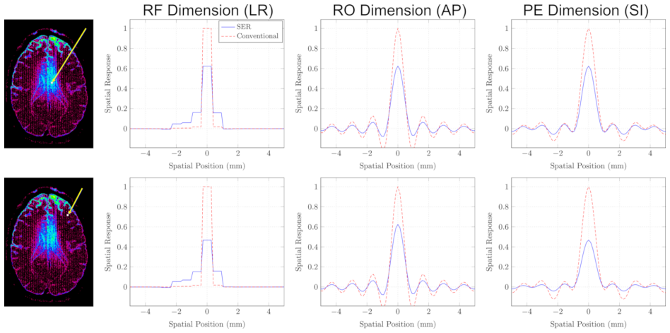
The load-bearing object is the SNR-enhancing joint reconstruction (SER) optimization, Equation [2] of the paper, which jointly estimates a real-valued high-resolution image $\mathbf{f}$ and per-slab phase maps $\mathbf{p}$ by minimizing data consistency plus a smooth-phase penalty $R(\mathbf{p})$ and an edge-preserving penalty $J(\mathbf{f})$ built from the Huber function. The phase variables account for motion-induced phase variations and partial-Fourier constraints, while $J(\mathbf{f})$ couples all 64 diffusion-weighted images so that spatial edges are shared and preserved across images, allowing strong smoothing within homogeneous regions. The optimization alternates between an iteratively-reweighted least-squares step for the image and a nonlinear conjugate-gradient step for the phase, and the same formulation yields theoretical predictions of noise-variance reduction and spatial response functions, which the paper uses to set the regularization level to achieve at least 3 times noise reduction in most brain regions.
What would settle it
Acquire the same 25-minute gSlider-SMS data and run the SER reconstruction, but compare against an independent gold standard that is not built from conventional gSlider, for example a digital phantom with known diffusion tensors and realistic noise, or an in vivo reference obtained with a different high-resolution reconstruction and much longer averaging; if SER's MD and FA errors against that independent standard are not substantially smaller than conventional gSlider's, the central claim fails.
Extended reading notes
Core claim
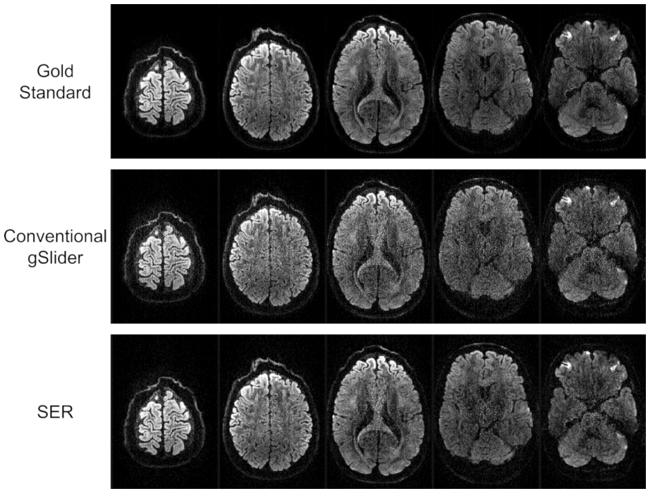
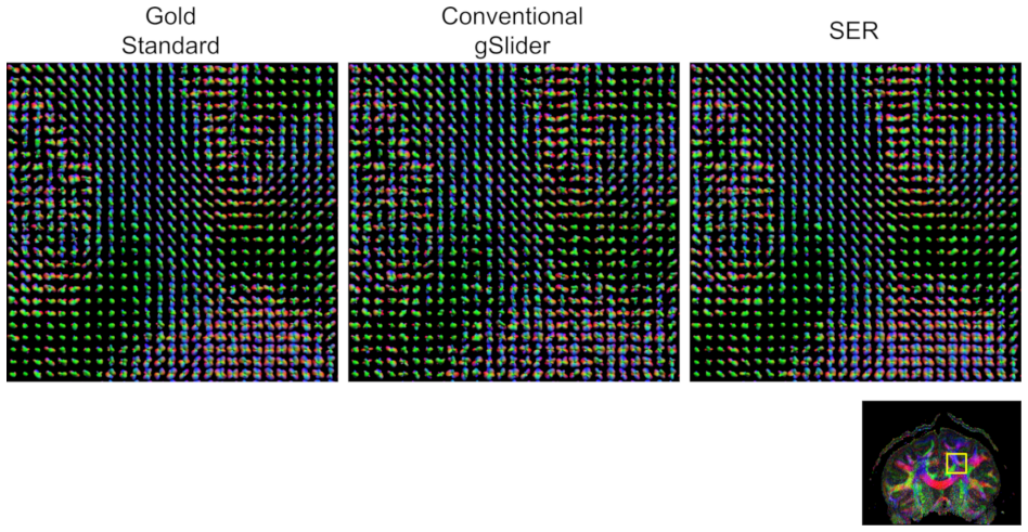
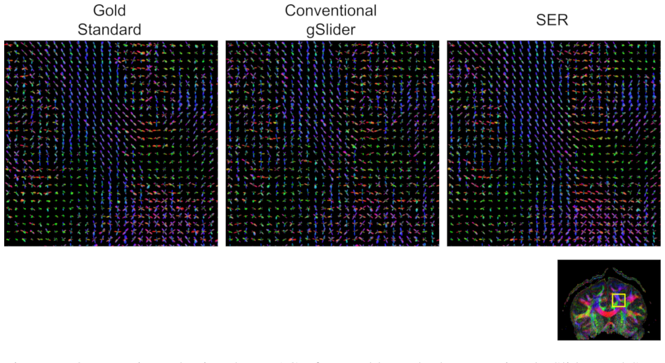
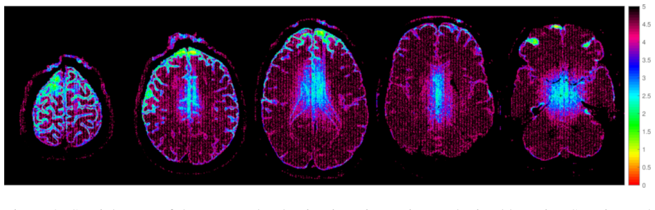
The central claim is that SNR-enhancing joint reconstruction, a regularized denoising approach that smooths within each diffusion-weighted image while preserving edges shared across all diffusion images, can be fused with gSlider-SMS RF-encoded slab acquisition, and that the fused reconstruction materially outperforms both conventional gSlider reconstruction and three low-rank matrix denoising baselines. On data from a 25-minute 3T scan (64 diffusion directions at $b=1500$ s/mm$^2$ plus 7 unweighted images), SER produced the lowest normalized root-mean-squared error among all methods for mean diffusivity ($0.056$ versus $0.152$ to $0.208$), fractional anisotropy ($0.268$ versus $0.344$ to $0.427$), and the FRT and FRACT orientation-distribution coefficients, while an oracle-tuned local PCA had only a marginal edge in raw diffusion-image NRMSE ($0.217$ versus $0.225$) and was worst on MD and FA. The same SER framework predicts 3 to 5 times noise-variance reduction with spatial-response broadening from about $724\,\mu\text{m}$ to $752$ to $778\,\mu\text{m}$ in full-volume-at-half-maximum, implying the 25-minute acquisition approximates the quality of 3-times-averaged data in smooth brain regions.
Load-bearing premise
The claim that SER substantially improves diffusion parameters rests on treating the 3-times-averaged conventional gSlider reconstruction as ground truth, and any bias that conventional gSlider has from its regularization, phase correction, or residual motion is inherited by the reference and could inflate or distort the measured improvements.
Editorial extensions
If this is right
- A 25-minute whole-brain sub-millimeter diffusion protocol is enough for quantitative MD and FA maps whose errors are roughly a third to a quarter of conventional gSlider reconstruction's errors.
- The predicted 3 to 5 times noise-variance reduction means that in smooth regions a single 25-minute scan behaves like a 75-minute scan, with only a small broadening of the effective voxel size (from about 724 to 752-778 micrometers in full-volume-at-half-maximum).
- Denoising methods that win on raw image NRMSE can still degrade downstream diffusion parameters; the oracle-tuned local PCA result shows that NRMSE-optimal denoising is not necessarily quantitatively optimal for diffusion MRI.
- Because SER's noise and resolution behavior is theoretically characterized, its regularization can be chosen prospectively to meet a target SNR improvement, which is not available for the low-rank baselines considered.
Reading between the lines
- The evaluation leans on 3-times-averaged conventional gSlider reconstruction as the gold standard; if that reconstruction carries its own regularization or phase bias, the reported SER improvements in NRMSE could partly reflect convergence to that reference rather than to the true diffusion signal.
- The paper leaves implicit that the same joint-reconstruction machinery should extend to other RF-encoded and super-resolution acquisition schemes, and to multi-shell or higher-angular-resolution diffusion data, because the shared-edge penalty $J(\mathbf{f})$ does not depend on the single-shell, 64-direction design.
- A testable extension is to combine SER with locally low-rank modeling, which the paper suggests but does not implement; it likely yields further gains, although it would sacrifice the closed-form trade-off characterizations.
- If spatially varying noise variances from the array-coil reconstruction were preserved, the center-of-brain under-denoising noted in the paper could be corrected; the paper flags this as future work.
Signed reviews
Editorial analysis
A structured set of objections, weighed in public.
Referee Report
Summary. This paper combines gSlider-SMS acquisition with SNR-enhancing joint reconstruction (SER) for fast sub-millimeter diffusion MRI. The method solves the coupled optimization in Eq. (2), which jointly estimates real-valued high-resolution image amplitudes, smooth phase maps, and edge-preserving shared structure across DWIs. On a single healthy subject scanned in approximately 25 minutes at 0.66 mm isotropic resolution, the authors compare SER against conventional gSlider reconstruction and three low-rank denoising baselines (MPPCA, LPCA, GPCA). The reported SER results have lower NRMSE than conventional gSlider for MD (0.056 vs 0.168), FA (0.268 vs 0.363), FRT (0.110 vs 0.137), and FRACT (0.639 vs 0.865), and better MD/FA/FRT/FRACT NRMSE than the low-rank baselines. The paper also provides theoretical noise-variance-reduction maps and spatial-response-function calculations showing roughly 3-5x noise variance reduction with modest resolution loss. All quantitative comparisons are made against a gold standard formed by conventional gSlider reconstruction of three averaged repetitions.
Significance. If the empirical claims hold, the proposed pipeline is significant: it demonstrates that a theoretically characterizable edge-preserving reconstruction can improve parameter-level accuracy in high-resolution diffusion MRI, outperforming oracle-tuned low-rank denoising on quantitative maps while offering predictable SNR/resolution trade-offs. The SRF/FVHM analysis and the oracle-tuned LPCA/GPCA baselines are notable strengths. However, the evidence base is a single subject with a reference that is neither noise-free nor independent of the test data, so the central quantitative claims require additional validation before they can be taken as established.
major comments (3)
- [2.3, 2.4, Eq. (9)] The gold standard used in every NRMSE computation is the average of three conventional gSlider reconstructions, but the text does not state whether the single-average data reconstructed by SER and conventional gSlider is one of the three repetitions entering that average. If it is, the reference contains one-third of the noise realization of the test image, which biases NRMSE toward methods that preserve that noise component and penalizes both a perfect noiseless reconstruction and over-smoothed reconstructions. In addition, the reference inherits any systematic bias from the Tikhonov-regularized reconstruction in Eq. (1), including its regularization parameter and phase-correction heuristics; the paper itself concedes in Section 2.4 that the gold standard is 'not entirely noise-free.' Please clarify the repetition split and, if the same repetition is used, add a held-out reference or a simulated ground-truth experiment to confirm that the reported gains reflect true signal accuracy rather than matching a biased reference.
- [Table 1] The oracle-tuned low-rank baselines LPCA and GPCA are selected to minimize DWI NRMSE, yet they produce the worst MD and FA NRMSE (0.208 and 0.427 for LPCA; 0.154 and 0.395 for GPCA), which indicates that DWI NRMSE against this reference does not track quantitative parameter accuracy. Because the paper's central claim of 'substantial improvements in estimated diffusion parameters' rests on the same type of reference, an independent evaluation with simulated ground truth or a separately acquired high-SNR reference is needed to establish that the parameter-level improvements are not artifacts of the reference definition.
- [3, Table 1] All quantitative conclusions are based on a single subject with no error bars, bootstrap estimates, or repeatability analysis. Since the abstract and conclusion make general claims about what the method 'enables' and about 'state-of-the-art performance,' the authors should either add multiple subjects or explicitly restrict the claims to illustrative single-subject results, and provide variance estimates for the reported NRMSE values.
minor comments (6)
- [Abstract] The abstract contains the typo 'SNR-ehancement' and should read 'SNR-enhancement.'
- [2.2, Eq. (8)] The gradient expression in Eq. (8) is labeled as the gradient with respect to f, but the optimization variable in Eq. (7) is p; the notation should indicate the gradient with respect to p for consistency.
- [2.2] The regularization parameters lambda_1, lambda_2, and xi are selected heuristically or by qualitative criteria; reporting the exact values used and adding a brief sensitivity analysis would improve reproducibility.
- [3] There are several typographical errors in the results and discussion sections, including 'suprising,' 'obsereved,' 'technqiues,' 'computional,' 'the the raw,' and 'In principal'; these should be corrected.
- [2.4] The NRMSE metric is computed 'within the brain' for DTI parameters and 'within the white matter' for ODFs, but the generation of these masks is not described; please specify how the brain and white-matter masks were obtained.
- [4] The authors note that SER was applied to slice-GRAPPA-reconstructed complex images rather than raw k-space data, which discards channel and spatially-varying noise information; this limitation should be stated more prominently in the conclusions because it tempers the generality of the reported noise-reduction gains.
Circularity Check
No significant circularity: SER parameters are set a priori from prior SER theory rather than fitted to the gold standard, and the empirical gains are not forced by construction; the only caveat is heavy reliance on the authors' own prior SER publications for the theoretical characterization.
full rationale
Walking the derivation chain, the SER objective (Eq. [2]) is defined directly from the gSlider encoding model A, the partial-Fourier point-spread operator G, and the smooth-phase/shared-edge regularizers; it never uses the gold-standard image fgold as an input. The regularization parameters are chosen a priori: λ2 is set so the prior SER theory predicts at least a 3× noise-variance reduction in most brain regions, and λ1 is set heuristically for phase smoothness (Sec. 2.2). The quantitative claims therefore do not reduce to a fit: Table 1 reports NRMSE of SER, conventional gSlider, MPPCA, LPCA, and GPCA against a 3×-averaged conventional gSlider reference (Secs. 2.3-2.4), and the low-rank baselines LPCA/GPCA are oracle-tuned to that same reference yet SER still outperforms GPCA on DWI NRMSE and both on parameter NRMSE, an outcome not guaranteed by construction. The concern that the gold standard is itself a conventional gSlider product (and hence imperfect or potentially biased) is a validation limitation, not a circular reduction, because SER's objective and parameters do not depend on fgold. The paper's extensive citations to the first author's prior SER work (Refs. 21, 27-32) supply the theoretical characterization and parameter-selection rules, but those are prior published results with stated assumptions, and the present empirical demonstration is computed directly from the acquired data. No self-definitional step, fitted-input-called-prediction step, author-imported uniqueness theorem, ansatz-via-citation, or renaming pattern was found. Overall, the derivation is self-contained for the claims made; the score of 1 reflects only the noticeable role of self-citations in the theoretical framing, which is not load-bearing in a circular sense.
Assumptions & free parameters
free parameters (4)
- lambda_2 (SER regularization strength) =
Chosen so that SNR improvement is at least 3x averaging in most brain regions
- lambda_1 (phase smoothness strength)
- xi (Huber parameter)
- Per-DWI median rescaling =
Median voxel intensity normalized to same magnitude across DWIs
assumptions (6)
- domain assumption The RF encoding model b = Af accurately represents gSlider acquisition after slice-GRAPPA and phase correction.
- domain assumption The diffusion image f is real-valued, enforcing partial Fourier phase constraints.
- domain assumption Image phase is smooth within each slab, as enforced by R(p) in Eq 3.
- domain assumption The joint edge-preserving penalty J(f) in Eq 4 with the Huber function is a suitable prior for diffusion MRI structure.
- domain assumption The theoretical SNR and resolution characterizations from prior SER work (Refs 21,29) remain valid when SER is combined with gSlider-SMS.
- domain assumption The 3x-averaged conventional gSlider reconstruction is an unbiased gold standard.
Cite this review
Pith. "Pith review of Fast Sub-millimeter Diffusion MRI using gSlider-SMS and SNR-Enhancing Joint Reconstruction." pith.science (2026). https://pith.science/paper/YDLWUCGY
@misc{pith2026190805698,
  author       = {Pith},
  title        = {Pith review of: Fast Sub-millimeter Diffusion MRI using gSlider-SMS and SNR-Enhancing Joint Reconstruction},
  year         = {2026},
  howpublished = {\url{https://pith.science/paper/YDLWUCGY}},
  note         = {Machine review of arXiv:1908.05698}
}
abstract
We evaluate a new approach for achieving diffusion MRI data with high spatial resolution, large volume coverage, and fast acquisition speed. A recent method called gSlider-SMS enables whole-brain sub-millimeter diffusion MRI with high signal-to-noise ratio (SNR) efficiency. However, despite the efficient acquisition, the resulting images can still suffer from low SNR due to the small size of the imaging voxels. This work proposes to mitigate the SNR problem by combining gSlider-SMS with a regularized SNR-enhancing reconstruction approach. Illustrative results show that, from gSlider-SMS data acquired over a span of only 25 minutes on a 3T scanner, the proposed method is able to produce 71 MRI images (64 diffusion encoding orientations with $b=$1500 s/mm$^2$, and 7 images without diffusion weighting) of the entire \emph{in vivo} human brain with nominal 0.66 mm spatial resolution. Using data acquired from 75 minutes of acquisition as a gold standard reference, we demonstrate that the proposed SNR-ehancement procedure leads to substantial improvements in estimated diffusion parameters compared to conventional gSlider reconstruction. Results also demonstrate that the proposed method has advantages relative to denoising methods based on low-rank matrix modeling. A theoretical analysis of the trade-off between spatial resolution and SNR suggests that the proposed approach has high efficiency. The combination of gSlider-SMS with advanced regularized reconstruction enables high-resolution quantitative diffusion MRI from a relatively fast acquisition.
Figures


Figures from the paper (6 more)


Reference graph
Works this paper leans on
- [1]
-
[2]
The quest for high spatial resolution diffusion- weighted imaging of the human brain in vivo
Holdsworth SJ, O’Halloran R, Setsompop K. The quest for high spatial resolution diffusion- weighted imaging of the human brain in vivo. NMR Biomed 2019;32:e4056. 18
work page 2019
-
[3]
Setsompop K, Gagoski BA, Polimeni JR, Witzel T, Wedeen VJ, Wald LL. Blipped-controlled aliasing in parallel imaging for simultaneous multislice echo planar imaging with reduced g-factor penalty. Magn Reson Med 2012;67:1210–1224
work page 2012
-
[4]
Pushing the limits of in vivo diffusion MRI for the Human Connectome Project
Setsompop K, Kimmlingen R, Eberlein E, Witzel T, Cohen-Adad J, McNab JA, Keil B, Tidall MD, Hoecht P, Dietz P, Cauley SF, Tountcheva V , Matschl V , Lenz VH, Heberlein K, Potthast A, Thein H, Van Horn J, Toga A, Schmitt F, Lehne D, Rosen BR, Wedeen V , Wald LL. Pushing the limits of in vivo diffusion MRI for the Human Connectome Project. NeuroImage 2013; ...
work page 2013
-
[5]
Advances in diffusion MRI acquisition and processing in the Human Con- nectome Project
Sotiropoulos SN, Jbabdi S, Xu J, Andersson JL, Moeller S, Auerbach EJ, Glasser MF, Hernan- dez M, Sapiro G, Jenkinson M, Feinberg DA, Yacoub E, Lenglet C, Van Essen DC, Ugurbil K, Behrens TEJ. Advances in diffusion MRI acquisition and processing in the Human Con- nectome Project. NeuroImage 2013;80:125–143
work page 2013
-
[6]
Jeong EK, Kim SE, Parker DL. High-resolution diffusion-weighted 3D MRI, using diffusion- weighted driven equilibrium (DW-DE) and multishot segmented 3D-SSFP without navigator echoes. Magn Reson Med 2003;50:821–829
work page 2003
-
[7]
Engstr ¨om M, Skare S. Diffusion-weighted 3D multislab echo planar imaging for high signal- to-noise ratio efficiency and isotropic image resolution. Magn Reson Med 2013;70:1507– 1514
work page 2013
-
[8]
Nguyen C, Fan Z, Sharif B, He Y , Dharmakumar R, Berman DS, Li D. In vivo three- dimensional high resolution cardiac diffusion weighted MRI: A motion compensated diffusion-prepared balanced steady-state free precession approach. Magn Reson Med 2014; 72:1257–1267
work page 2014
Show all 62 references
-
[9]
3D multi-slab diffusion-weighted readout-segmented EPI with real-time cardiac reordered k-space acquisition
Frost R, Miller KL, Tijssen RHN, Porter DA, Jezzard P. 3D multi-slab diffusion-weighted readout-segmented EPI with real-time cardiac reordered k-space acquisition. Magn Reson Med 2014;72:1565–1579. 19
2014
-
[10]
Human brain diffusion tensor imaging at submillimeter isotropic resolution on a 3 Tesla clinical MRI scanner
Chang HC, Sundman M, Petit L, Guhaniyogi S, Chu ML, Petty C, Song AW, Chen NK. Human brain diffusion tensor imaging at submillimeter isotropic resolution on a 3 Tesla clinical MRI scanner. NeuroImage 2015;118:667–675
2015
-
[11]
Slab profile encoding (PEN) for minimizing slab boundary artifact in three-dimensional diffusion-weighted multi- slab acquisition
Van AT, Aksoy M, Holdsworth SJ, Kopeinigg D, V os SB, Bammer R. Slab profile encoding (PEN) for minimizing slab boundary artifact in three-dimensional diffusion-weighted multi- slab acquisition. Magn Reson Med 2015;73:605–613
2015
-
[12]
High spatial resolution diffusion weighted imaging on clinical 3 T MRI scanners using multislab spiral acquisitions
Holtrop JL, Sutton BP. High spatial resolution diffusion weighted imaging on clinical 3 T MRI scanners using multislab spiral acquisitions. J Med Imag 2016;3:023501
2016
-
[13]
Super-resolution in medical imaging
Greenspan H. Super-resolution in medical imaging. Comput J 2009;52:43–63
2009
-
[14]
Super-resolution reconstruction to increase the spatial resolution of diffusion weighted images from orthogonal anisotropic acquisitions
Scherrer B, Gholipour A, Warfield SK. Super-resolution reconstruction to increase the spatial resolution of diffusion weighted images from orthogonal anisotropic acquisitions. Med Image Anal 2012;16:1465–1476
2012
-
[15]
Super-resolution reconstruction of diffusion parameters from diffusion-weighted images with different slice orientations
Van Steenkiste G, Jeurissen B, Veraart J, den Dekker AJ, Parizel PM, Poot DHJ, Sijbers J. Super-resolution reconstruction of diffusion parameters from diffusion-weighted images with different slice orientations. Magn Reson Med 2016;75:181–195
2016
-
[16]
A joint compressed-sensing and super-resolution approach for very high-resolution diffusion imaging
Ning L, Setsompop K, Michailovich O, Makris N, Shenton ME, Westin CF, Rathi Y . A joint compressed-sensing and super-resolution approach for very high-resolution diffusion imaging. NeuroImage 2016;125:386–400
2016
-
[17]
superresolution
Hennel F, Tian R, Engel M, Pruessmann KP. In-plane “superresolution” MRI with phaseless sub-pixel encoding. Magn Reson Med 2018;80:2384–2392
2018
-
[18]
High-resolution in vivo diffusion imaging of the human brain with generalized slice dithered enhanced resolution: simultaneous multislice (gSlider-SMS)
Setsompop K, Fan Q, Stockmann J, Bilgic B, Huang S, Cauley SF, Nummenmaa A, Wang F, Rathi Y , Witzel T, Wald LL. High-resolution in vivo diffusion imaging of the human brain with generalized slice dithered enhanced resolution: simultaneous multislice (gSlider-SMS). Magn Reson ...
2018
-
[19]
Motion-robust sub-millimeter isotropic 20 diffusion imaging through motion corrected generalized slice dithered enhanced resolution (MC-gSlider) acquisition
Wang F, Bilgic B, Dong Z, Manhard MK, Ohringer N, Zhao B, Haskell M, Cauley SF, Fan Q, Witzel T, Adalsteinsson E, Wald LL, Setsompop K. Motion-robust sub-millimeter isotropic 20 diffusion imaging through motion corrected generalized slice dithered enhanced resolution (MC-gSlid...
2018
-
[20]
High-fidelity, high-isotropic resolution diffusion imaging through gSlider acquisition with B1+ & T1 corrections and integrated δB0/Rx shim array
Liao C, Stockmann J, Tian Q, Bilgic B, Arango NS, Manhard MK, Grissom W A, Wald LL, Setsompop K. High-fidelity, high-isotropic resolution diffusion imaging through gSlider acquisition with B1+ & T1 corrections and integrated δB0/Rx shim array. Preprint 2018; arXiv:1811.05473
2018 arXiv
-
[21]
Improved dif- fusion imaging through SNR-enhancing joint reconstruction
Haldar JP, Wedeen VJ, Nezamzadeh M, Dai G, Weiner MW, Schuff N, Liang ZP. Improved dif- fusion imaging through SNR-enhancing joint reconstruction. Magn Reson Med 2013;69:277– 289
2013
-
[22]
Denoising diffusion- weighted magnitude MR images using rank and edge constraints
Lam F, Babacan SD, Haldar JP, Weiner MW, Schuff N, Liang ZP. Denoising diffusion- weighted magnitude MR images using rank and edge constraints. Magn Reson Med 2014; 71:1272–1284
2014
-
[23]
A majorize-minimize framework for Rician and non-central chi MR images
Varadarajan D, Haldar JP. A majorize-minimize framework for Rician and non-central chi MR images. IEEE Trans Med Imag 2015;34:2191–2202
2015
-
[24]
Denoising of diffusion MRI using random matrix theory
Veraart J, Novikov DS, Christiaens D, Ades-Aron B, Sijbers J, Fieremans E. Denoising of diffusion MRI using random matrix theory. NeuroImage 2016;142:394–406
2016
-
[25]
Model-based denoising in diffusion-weighted imaging using generalized spherical deconvolution
Sperl JI, Sprenger T, Tan ET, Menzel MI, Hardy CJ, Marinelli L. Model-based denoising in diffusion-weighted imaging using generalized spherical deconvolution. Magn Reson Med 2017;78:2428–2438
2017
-
[26]
Phase-correcting non-local means filtering for diffusion- weighted imaging of the spinal cord
Kafali SG, Cukur T, Saritas EU. Phase-correcting non-local means filtering for diffusion- weighted imaging of the spinal cord. Magn Reson Med 2018;80:1020–1035
2018
-
[27]
Joint reconstruction of noisy high-resolution MR image sequences
Haldar JP, Liang ZP. Joint reconstruction of noisy high-resolution MR image sequences. In: Proc. IEEE Int. Symp. Biomed. Imag.. 2008; pp. 752–755
2008
-
[28]
Signal-to-noise ratio-enhancing joint reconstruction for im- proved diffusion imaging of mouse spinal cord white matter injury
Kim JH, Song SK, Haldar JP. Signal-to-noise ratio-enhancing joint reconstruction for im- proved diffusion imaging of mouse spinal cord white matter injury. Magn Reson Med 2016; 75:852–858. 21
2016
-
[29]
Constrained Imaging: Denoising and Sparse Sampling
Haldar JP. Constrained Imaging: Denoising and Sparse Sampling. Ph.D. thesis. University of Illinois at Urbana-Champaign. Urbana, IL, USA. 2011
2011
-
[30]
Anatomically constrained reconstruction from noisy data
Haldar JP, Hernando D, Song SK, Liang ZP. Anatomically constrained reconstruction from noisy data. Magn Reson Med 2008;59:810–818
2008
-
[31]
Resolution and noise properties of linear phase-constrained partial Fourier reconstruction
Haldar JP, Sakaie K, Liang ZP. Resolution and noise properties of linear phase-constrained partial Fourier reconstruction. In: Proc. Int. Soc. Magn. Reson. Med.. 2009; p. 2862
2009
-
[32]
On MR experiment design with quadratic regularization
Haldar JP, Liang ZP. On MR experiment design with quadratic regularization. In: Proc. IEEE Int. Symp. Biomed. Imag.. 2011; pp. 1676–1679
2011
-
[33]
Whole-brain quantitative diffusion MRI at 660µm resolution in 25 minutes using gSlider-SMS and SNR-enhancing joint reconstruction
Haldar JP, Fan Q, Setsompop K. Whole-brain quantitative diffusion MRI at 660µm resolution in 25 minutes using gSlider-SMS and SNR-enhancing joint reconstruction. In: Proc. Int. Soc. Magn. Reson. Med.. 2016; p. 102
2016
-
[34]
Multiple-line-scanning spin density imaging
Maudsley AA. Multiple-line-scanning spin density imaging. J Magn Reson 1980;41:112–126
1980
-
[35]
Line-integral projection reconstruction (LPR) with slice encoding techniques: Multislice regional imaging in NMR tomography
Oh CH, Park HW, Cho ZH. Line-integral projection reconstruction (LPR) with slice encoding techniques: Multislice regional imaging in NMR tomography. IEEE Trans Med Imag 1984; MI-3:170–178
1984
-
[36]
Compressed-sensing MRI with random encoding
Haldar JP, Hernando D, Liang ZP. Compressed-sensing MRI with random encoding. IEEE Trans Med Imag 2011;30:893–903
2011
-
[37]
SMS-LORAKS: Calibrationless simultaneous multislice MRI using low- rank matrix modeling
Kim TH, Haldar JP. SMS-LORAKS: Calibrationless simultaneous multislice MRI using low- rank matrix modeling. In: Proc. IEEE Int. Symp. Biomed. Imag.. 2015; pp. 323–326
2015
-
[38]
Deconvolved spatial light interference microscopy for live cell imaging
Haldar JP, Wang Z, Popescu G, Liang ZP. Deconvolved spatial light interference microscopy for live cell imaging. IEEE Trans Biomed Eng 2011;58:2489–2497
2011
-
[39]
Separate magnitude and phase regularization via compressed sensing
Zhao F, Noll DC, Nielsen JF, Fessler JA. Separate magnitude and phase regularization via compressed sensing. IEEE Trans Med Imag 2012;31:1713–1723
2012
-
[40]
Group sparse reconstruction of vector-valued images
Trzasko J, Manduca A. Group sparse reconstruction of vector-valued images. In: Proc. Int. Soc. Magn. Reson. Med.. 2011; p. 2839. 22
2011
-
[41]
Fast multi-contrast MRI reconstruction
Huang J, Chen C, Axel L. Fast multi-contrast MRI reconstruction. Magn Reson Imag 2014; 32:1344–1352
2014
-
[42]
Global convergence properties of conjugate gradient methods for opti- mization
Gilbert JC, Nocedal J. Global convergence properties of conjugate gradient methods for opti- mization. SIAM J Optim 1992;2:21–42
1992
-
[43]
k-space and q-space: Com- bining ultra-high spatial and angular resolution in diffusion imaging using ZOOPPA at 7 T
Heidemann RM, Anwander A, Feiweier T, Kn¨osche TR, Turner R. k-space and q-space: Com- bining ultra-high spatial and angular resolution in diffusion imaging using ZOOPPA at 7 T. NeuroImage 2012;60:967–978
2012
-
[44]
Interslice leakage artifact reduction technique for simultaneous multislice acquisitions
Cauley SF, Polimeni JR, Bhat H, Wald LL, Setsompop K. Interslice leakage artifact reduction technique for simultaneous multislice acquisitions. Magn Reson Med 2014;72:93–102
2014
-
[45]
Jenkinson M, Beckmann CF, Behrens TEJ, Woolrich MW, Smith SM. FSL. NeuroImage 2012; 62:782–790
2012
-
[46]
Real diffusion-weighted MRI enabling true signal averaging and increased diffusion contrast
Eichner C, Cauley SF, Cohen-Adad J, Moeller HE, Turner R, Setsompop K, Wald LL. Real diffusion-weighted MRI enabling true signal averaging and increased diffusion contrast. Neu- roImage 2015;122:373–384
2015
-
[47]
Diffusion weighted image denoising using overcomplete local PCA
Manj ´on JV , Coup´e P, Concha L, Buades A, Collins DL, Robles M. Diffusion weighted image denoising using overcomplete local PCA. PLOS ONE 2013;8:e73021
2013
-
[48]
PCLR: Phase-constrained low-rank model for com- pressive diffusion-weighted MRI
Gao H, Li L, Zhang K, Zhou W, Hu X. PCLR: Phase-constrained low-rank model for com- pressive diffusion-weighted MRI. Magn Reson Med 2014;72:1330–1341
2014
-
[49]
Accelerated cardiac diffusion tensor imaging usint joint low-rank and sparsity constraints
Ma S, Nguyen C, Christodoulou A, Luthringer D, Kobashigawa J, Lee SE, Chang HJ, Li D. Accelerated cardiac diffusion tensor imaging usint joint low-rank and sparsity constraints. IEEE Trans Biomed Eng 2018;65:2219–2230
2018
-
[50]
Unbiased risk estimates for singular value threshold- ing and spectral estimators
Cand `es EJ, Sing-Long CA, Trzasko JD. Unbiased risk estimates for singular value threshold- ing and spectral estimators. IEEE Trans Signal Process 2013;61:4643–4657
2013
-
[51]
Optimal shrinkage of singular values
Gavish M, Donoho DL. Optimal shrinkage of singular values. IEEE Trans Inf Theory 2017; 63:2137–2152. 23
2017
-
[52]
Q-ball imaging
Tuch DS. Q-ball imaging. Magn Reson Med 2004;52:1358–1372
2004
-
[53]
Q-ball reconstruction of multimodal fiber orientations using the spherical harmonic basis
Hess CP, Mukherjee P, Han ET, Xu D, Vigneron DB. Q-ball reconstruction of multimodal fiber orientations using the spherical harmonic basis. Magn Reson Med 2006;56:104–117
2006
-
[54]
Regularized, fast, and robust analytical Q-ball imaging
Descoteaux M, Angelino E, Fitzgibbons S, Deriche R. Regularized, fast, and robust analytical Q-ball imaging. Magn Reson Med 2007;58:497–510
2007
-
[55]
Linear transforms for Fourier data on the sphere: Application to high angular resolution diffusion MRI of the brain
Haldar JP, Leahy RM. Linear transforms for Fourier data on the sphere: Application to high angular resolution diffusion MRI of the brain. NeuroImage 2013;71:233–247
2013
-
[56]
NAPALM: An algorithm for MRI reconstruction with separate magnitude and phase regularization
Liu Y , Haldar JP. NAPALM: An algorithm for MRI reconstruction with separate magnitude and phase regularization. In: Proc. Int. Soc. Magn. Reson. Med.. 2019; p. 4764
2019
-
[57]
Statistical analysis of noise in MRI: Modeling, filtering and estimation
Aja-Fernandez S, Vegas-Sanchez-Ferrero G. Statistical analysis of noise in MRI: Modeling, filtering and estimation. Springer. 2016
2016
-
[58]
Comprehensive quantification of signal-to-noise ratio and g-factor for image-based and k- space-based parallel imaging reconstructions
Robson PM, Grant AK, Madhuranthakam AJ, Lattanzi R, Sodickson DK, McKenzie CA. Comprehensive quantification of signal-to-noise ratio and g-factor for image-based and k- space-based parallel imaging reconstructions. Magn Reson Med 2008;60:895–907
2008
-
[59]
Analysis of resolution and noise properties of nonquadratically regularized image reconstruction methods for PET
Ahn S, Leahy RM. Analysis of resolution and noise properties of nonquadratically regularized image reconstruction methods for PET. IEEE Trans Med Imag 2008;27:413–424
2008
-
[60]
Regularization for uniform spatial resolution properties in penalized- likelihood image reconstruction
Stayman JW, Fessler JA. Regularization for uniform spatial resolution properties in penalized- likelihood image reconstruction. IEEE Trans Med Imag 2000;19:601–615
2000
-
[61]
Improved B 0-distortion correction in diffusion MRI using interlaced q-space sampling and constrained reconstruction
Bhushan C, Joshi AA, Leahy RM, Haldar JP. Improved B 0-distortion correction in diffusion MRI using interlaced q-space sampling and constrained reconstruction. Magn Reson Med 2014;72:1218–1232
2014
-
[62]
Fast high-resolution diffusion MRI using gSlider-SMS, interlaced subsampling, and SNR-enhancing joint reconstruction
Haldar JP, Setsompop K. Fast high-resolution diffusion MRI using gSlider-SMS, interlaced subsampling, and SNR-enhancing joint reconstruction. In: Proc. Int. Soc. Magn. Reson. Med.. 2017; p. 174. 24 Figure and Table Captions Figure 1: A graphical overview of the proposed image ...
2017
Reviewed August 14, 2026 · model on record in the stance chip above.
Discussion (0). Continue with ORCID to comment.