Low-Cost Continuous-Wave Diffusive Microtomography with Fiber-Scanned White-Light Illumination
Pith reviewed 2026-06-25 21:41 UTC · model grok-4.3
The pith
Scanned fiber white-light illumination with a smartphone microscope and machine learning produces three-dimensional reconstructions of biological samples at low cost.
A machine-rendered reading of the paper's core claim, the machinery that carries it, and where it could break.
Core claim
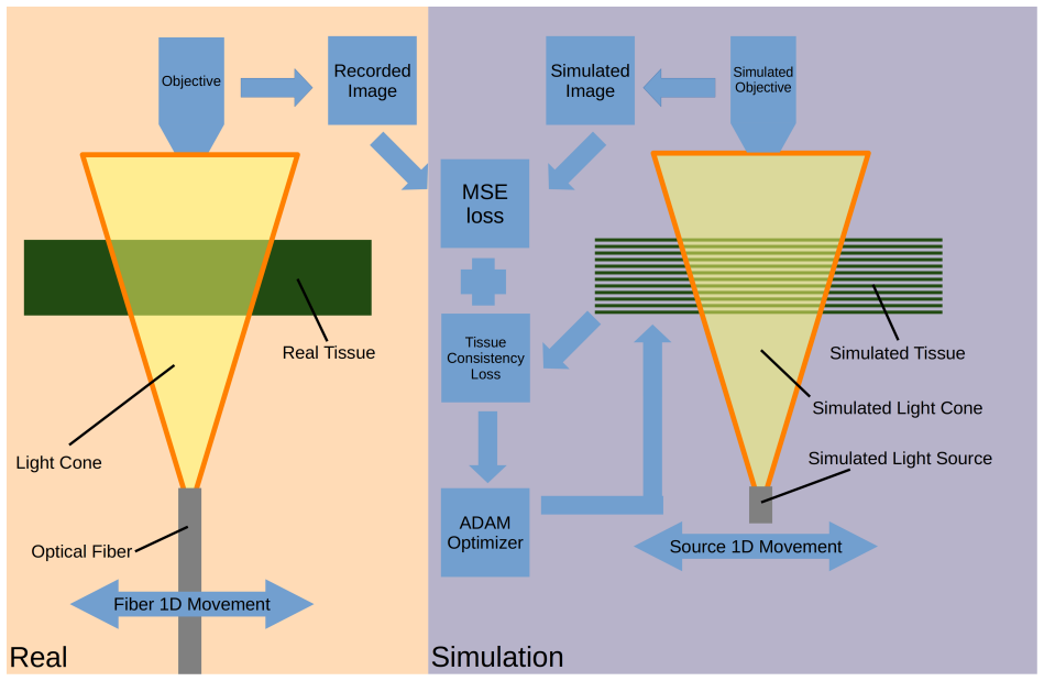
The central claim is that scanned fiber illumination from inexpensive white-light sources, paired with a machine-learning-optimized physics forward model, enables continuous-wave diffusive microtomography capable of full-color volumetric reconstructions in both cleared and scattering biological samples using only a smartphone microscope and 3D-printed hardware.
What carries the argument
The physics-based forward model optimized with machine learning, which models light transport under continuous-wave diffusive conditions to invert scanned fiber illumination data into volumetric reconstructions.
If this is right
- Scanned fiber illumination can replace more complex light sources in diffusive tomography setups.
- Machine learning optimization of the forward model improves reconstruction fidelity from low-cost data.
- The system supports imaging of both optically cleared and naturally scattering biological specimens.
- Full-color output becomes feasible without additional spectral hardware.
Where Pith is reading between the lines
- The method could support portable or educational 3D imaging in settings without access to research-grade microscopes.
- Extensions to other wavelengths or dynamic samples would require only changes to the illumination and model training.
- Combining the approach with additional smartphone sensors could yield multimodal low-cost tomography.
Load-bearing premise
The physics-based forward model, once optimized by machine learning, sufficiently captures light transport in the tested scattering and cleared samples to yield reliable volumetric outputs.
What would settle it
Quantitative comparison of the reconstructed volumes against known ground-truth structures in the same samples, such as measured feature sizes or densities obtained from higher-resolution reference imaging, showing systematic mismatch.
Figures
read the original abstract
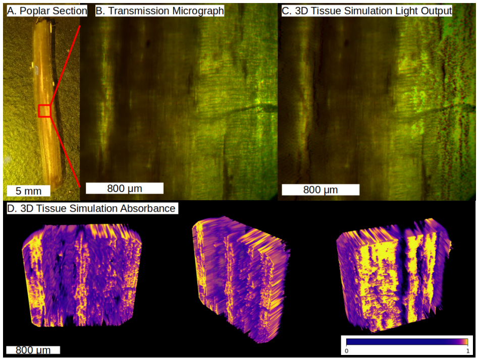
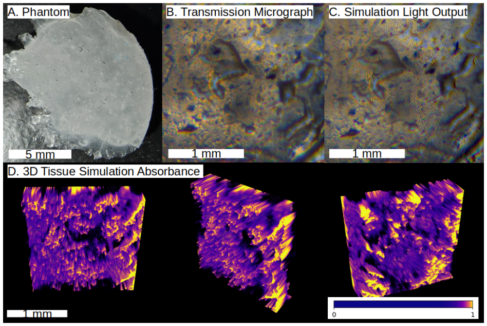
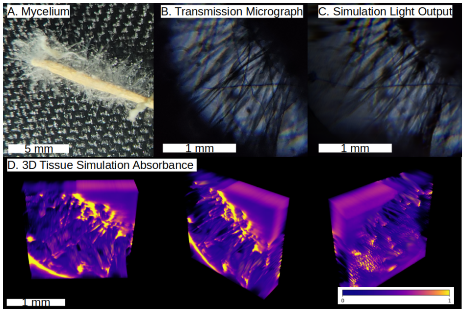
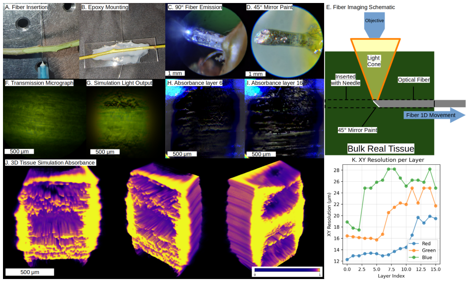
Tomographic microscopy enables three-dimensional internal imaging but often requires expensive optical or X-ray instrumentation. Here we present an ultra-low-cost continuous-wave diffusive tomography (CWDT) system for biological samples. The system uses a smartphone microscope, a white LED coupled into an optical fiber, 3D-printed micropositioners, and a physics-based forward model optimized with machine learning. We demonstrate full-color volumetric reconstructions from a tartrazine-cleared poplar section, a scattering phantom, fungal mycelium near an Arabidopsis root, and thick poplar branch imaging with an inserted side-emitting fiber. The current results are qualitative and exploratory, but they show that scanned fiber illumination and inexpensive hardware can produce useful three-dimensional reconstruction outputs for low-cost microscopy experiments.
Editorial analysis
A structured set of objections, weighed in public.
Referee Report
Summary. The manuscript presents an ultra-low-cost continuous-wave diffusive microtomography (CWDT) system for biological samples. It combines a smartphone microscope, white LED coupled to a scanned optical fiber, 3D-printed micropositioners, and a physics-based forward model whose parameters are optimized by machine learning. Qualitative full-color volumetric reconstructions are shown for a tartrazine-cleared poplar section, a scattering phantom, fungal mycelium near an Arabidopsis root, and a thick poplar branch imaged with an inserted side-emitting fiber. The work is explicitly framed as exploratory and qualitative, with the central claim that scanned fiber illumination plus inexpensive hardware can yield useful three-dimensional outputs for low-cost microscopy experiments.
Significance. If the reconstructions prove reliable, the approach could lower barriers to 3D tomographic imaging in biology by replacing costly instrumentation with commodity components and a learned forward model. The explicit use of a physics-based model optimized by ML is a constructive element that may generalize to other diffusive regimes. At present the exploratory framing limits the result to a proof-of-concept demonstration rather than a ready-to-deploy technique.
minor comments (2)
- [Abstract] Abstract: the phrase 'useful three-dimensional reconstruction outputs' is not accompanied by any operational criterion (e.g., recovery of known phantom features, consistency across illumination angles, or comparison with a reference modality). Adding a short sentence that defines what 'useful' means in the present qualitative setting would help readers evaluate the displayed volumes.
- [Methods (forward-model section)] The manuscript states that the forward model is 'optimized with machine learning' but supplies no information on the loss function, regularization, or whether the fitted parameters are held fixed when reconstructing new targets. A brief methods paragraph clarifying this point would remove ambiguity about potential circularity.
Simulated Author's Rebuttal
We thank the referee for their positive assessment of the manuscript and for recommending minor revision. The work is explicitly presented as an exploratory, qualitative demonstration, consistent with the referee's characterization. No specific major comments were provided in the report.
Circularity Check
No significant circularity
full rationale
The paper explicitly frames its outputs as qualitative and exploratory demonstrations of a low-cost CWDT system using scanned fiber illumination and a physics-based forward model optimized via ML. No derivation chain, equations, or predictions are presented in the available text that reduce by construction to fitted parameters or self-citations; the central claim remains internally consistent with the modest, hardware-focused scope without load-bearing steps that equate outputs to inputs.
Axiom & Free-Parameter Ledger
Reference graph
Works this paper leans on
-
[1]
Continuous wave-diffuse optical tomography (cw-dot) in human brain mapping: A review
Shuo Guan, Yuhang Li, Yuanyuan Gao, Yuxi Luo, Hubin Zhao, Dalin Yang, and Rihui Li. Continuous wave-diffuse optical tomography (cw-dot) in human brain mapping: A review. 15 Sensors, 25(7):2040, 2025. doi: 10.3390/s25072040
-
[2]
J. P. Culver, R. Choe, M. J. Holboke, L. Zubkov, T. Durduran, A. Slemp, V. Ntziachristos, B. Chance, and A. G. Yodh. Three-dimensional diffuse optical tomography in the parallel plane transmission geometry: Evaluation of a hybrid frequency domain/continuous wave clinical system for breast imaging.Medical Physics, 30(2):235–247, 2003. doi: 10.1118/1.1534109
-
[3]
Alfonso Galderisi, Sabrina Brigadoi, Simone Cutini, Sara Basso Moro, Elisabetta Lolli, Feder- ica Meconi, Silvia Benavides-Varela, Eugenio Baraldi, Piero Amodio, Claudio Cobelli, Daniele Trevisanuto, and Roberto Dell’Acqua. Long-term continuous monitoring of the preterm brain with diffuse optical tomography and electroencephalography: A technical note on ...
-
[5]
J. J. Davenport, M. Hickey, J. P. Phillips, and P. A. Kyriacou. Method for producing angled optical fiber tips in the laboratory.Optical Engineering, 55(2):026120, 2016. doi: 10.1117/1. oe.55.2.026120
work page doi:10.1117/1 2016
-
[6]
Triaxis micropositioner.https://makerworld.com/en/models/ 1256154-triaxis-micropositioner, 2025
Alexander Ingold. Triaxis micropositioner.https://makerworld.com/en/models/ 1256154-triaxis-micropositioner, 2025. MakerWorld 3D-print model page
2025
-
[7]
Triaxis micropositioner micromanipulator 3-axis stage.https://www
Alexander Ingold. Triaxis micropositioner micromanipulator 3-axis stage.https://www. thingiverse.com/thing:6976184, 2025. Thingiverse 3D-print model page
2025
-
[8]
Triaxis micropositioner cad.https://cad.onshape
Alexander Ingold. Triaxis micropositioner cad.https://cad.onshape. com/documents/c31396d958296393a0cbb499/w/10e122b0be94b2b2a2f3787d/e/ 975a9698867d3d77a59824b4, 2025. Editable Onshape CAD document
2025
-
[9]
Penelope Lindsay, Kyle W. Swentowsky, and David Jackson. Cultivating potential: Harnessing plant stem cells for agricultural crop improvement.Molecular Plant, 17(1):50–74, 2024. doi: 10.1016/j.molp.2023.12.014
-
[10]
Juan Du, Yichen Wang, Wenfan Chen, Mingling Xu, Ruhong Zhou, Huixia Shou, and Jun Chen. High-resolution anatomical and spatial transcriptome analyses reveal two types of meristematic cell pools within the secondary vascular tissue of poplar stem.Molecular Plant, 16(5):809–828, 2023. doi: 10.1016/j.molp.2023.03.005
-
[11]
Alexander Ingold, Gayatri Mishra, Reed Sorenson, Andrew Groover, Leslie Sieburth, and Rajesh Menon. Live cell imaging of cellular dynamics in poplar wood using computational cannula microscopy.Applied Optics, 63(28):G47–G53, 2024. doi: 10.1364/AO.523456
-
[12]
Zihao Ou, Yi-Shiou Duh, Nicholas J. Rommelfanger, Carl H. C. Keck, Shan Jiang, Kenneth Brinson, Su Zhao, Elizabeth L. Schmidt, Xiang Wu, Fan Yang, Betty Cai, Han Cui, Wei Qi, Shifu Wu, Adarsh Tantry, Richard Roth, Jun Ding, Xiaoke Chen, Julia A. Kaltschmidt, Mark L. Brongersma, and Guosong Hong. Achieving optical transparency in live animals with absorbin...
-
[13]
Miniscope v4.https://open-ephys.org/miniscope-v4, 2024
Open Ephys. Miniscope v4.https://open-ephys.org/miniscope-v4, 2024
2024
-
[14]
Miniscope v4 assembly wiki.https://github.com/AharoniLab/ Miniscope-v4/wiki/Assembly, 2024
Aharoni Lab. Miniscope v4 assembly wiki.https://github.com/AharoniLab/ Miniscope-v4/wiki/Assembly, 2024
2024
-
[15]
Autofluorescence in plants.Molecules, 25(10):2393, 2020
Lloyd Donaldson. Autofluorescence in plants.Molecules, 25(10):2393, 2020. doi: 10.3390/ molecules25102393
2020
-
[16]
Miniscope-daq-qt-software.https://github.com/Aharoni-Lab/ Miniscope-DAQ-QT-Software, 2024
Aharoni Lab. Miniscope-daq-qt-software.https://github.com/Aharoni-Lab/ Miniscope-DAQ-QT-Software, 2024. GitHub repository
2024
-
[17]
Forster, D
B. Forster, D. Van De Ville, J. Berent, D. Sage, and M. Unser. Complex wavelets for extended depth-of-field: A new method for the fusion of multichannel microscopy images.Microscopy Research and Technique, 65(1–2):33–42, 2004
2004
-
[18]
3d gaussian splatting for real-time radiance field rendering.ACM Transactions on Graphics, 42(4):139,
Bernhard Kerbl, Georgios Kopanas, Thomas Leimkuehler, and George Drettakis. 3d gaussian splatting for real-time radiance field rendering.ACM Transactions on Graphics, 42(4):139,
-
[19]
doi: 10.1145/3592433
-
[20]
torch-splatting: A pure pytorch implementation of 3d gaussian splatting.https: //github.com/hbb1/torch-splatting, 2025
hbb1. torch-splatting: A pure pytorch implementation of 3d gaussian splatting.https: //github.com/hbb1/torch-splatting, 2025. GitHub repository, MIT license. 17 Figure S2: Raw chicken breast, sectioned with 0–3 mm thickness. A. Sample stained with tartrazine for 15 minutes; grid lines are 0.125 inches. B. Sample stained with tartrazine for 18 hours; grid ...
2025
discussion (0)
Sign in with ORCID, Apple, or X to comment. Anyone can read and Pith papers without signing in.