REVIEW 4 major objections 3 minor 40 references
Correlative Study of Enhanced Excitonic Emission in ZnO Coated with Al Nanoparticles using Electron and Laser Excitation
T0 review · 4 major / 3 minor · reviewed 2026-08-14 · deepseek-v4-flash
Pith's one-line read A 2-nm aluminum coating enhances ZnO ultraviolet emission up to 12 times, and the gain depends on how the crystal is oriented relative to the laser polarization.
desk verdict Competent correlative study with a useful polarization-alignment rule, but the LSP attribution is softer than the headline enhancement numbers suggest. read the letter →
The pith
A machine-rendered reading of the paper's core claim, the machinery that carries it, and where it could break.
The reading
What carries the argument
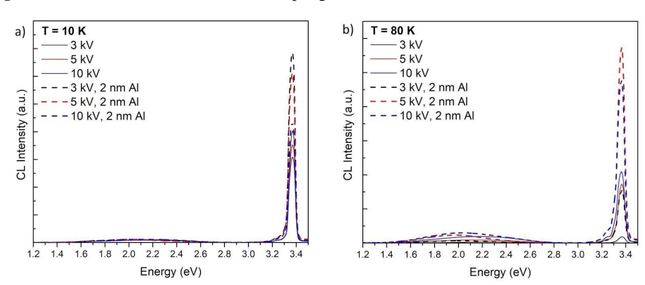
The central object is the aluminum nanoparticle localized surface plasmon (LSP), a collective electron oscillation on the 2 nm sputtered Al layer that can couple to the excitonic emission of ZnO. The argument is carried by comparing PL and CL excitation at different depths, by temperature-dependent spectra that separate FX from DBX contributions, and by time-resolved PL that measures the lifetime reduction. The polarization dependence is the deciding piece: FX-A and FX-B are polarized perpendicular to the c-axis, while FX-C is parallel to it, so rotating the ZnO crystal relative to the laser polarization selects which exciton couples most. This mechanism explains why PL enhancement (up to 12 times) exceeds CL enhancement (about 4 times): laser excitation generates more excitons within the first few nanometers where LSP coupling is strong.
What would settle it
Measure the same samples with a technique that separates emission channels, for instance depth-profiling the Al concentration and mapping the 3.343 eV interface band and the 3.358 eV Al-donor line against the FX-C-LO features. If the enhanced UV signal persists when the LSP resonance is detuned by changing particle size, or if it scales with Al doping rather than with the modeled plasmon response, the LSP attribution would be falsified.
Extended reading notes
Core claim
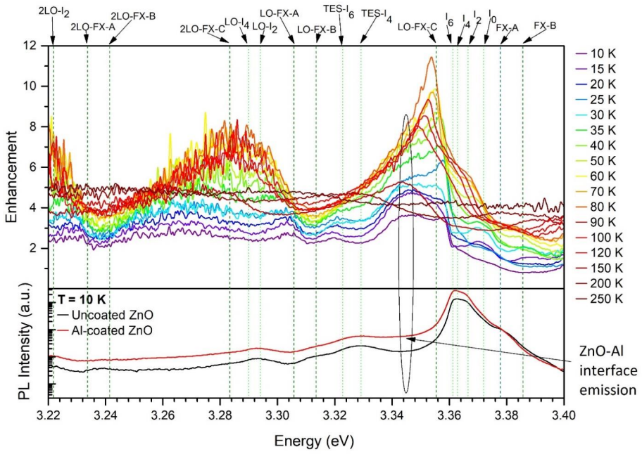
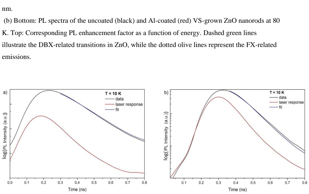
The central claim is that aluminum nanoparticle LSPs form a faster, non-radiative relaxation channel that selectively couples to free excitons (FX) in ZnO, enhancing their spontaneous emission rate. On an a-plane ZnO crystal the UV emission is enhanced up to 12 times at 80 K under laser excitation, and the enhancement tracks the FX-C emission. The LSP coupling is polarization-selective: when the FX transition is polarized in the same plane as the electric field of the incident laser, its enhancement is larger. The paper also reports a lifetime reduction from (161 ± 4) ps to at most 53 ps, which yields a lower-bound Purcell factor of 3.0.
Load-bearing premise
The argument assumes the 2 nm aluminum layer is actually a population of metallic nanoparticles whose localized surface plasmon resonances drive the enhancement, rather than the Al-donor in-diffusion and interface defects that the spectra also show.
Editorial extensions
If this is right
- On a-plane ZnO, aligning the sample so FX-C lies parallel to the laser's electric vector gives the strongest UV enhancement; in c-oriented nanorods the same rule enhances FX-A and FX-B instead.
- The enhancement is strongest near the ZnO/Al interface, so excitons generated within the first few nanometers matter most; shallow excitation therefore yields larger gains.
- At 80 K, where donor-bound excitons are thermally dissociated, free-exciton coupling to Al LSPs peaks at roughly 12 times; the same mechanism fades as temperature rises further and phonon replicas dominate.
- The observed Purcell factor of at least 3.0 from the lifetime drop independently supports an increased spontaneous emission rate, not simply a change in absorption or collection.
Reading between the lines
- If the polarization rule is general, ZnO-based UV light-emitting devices could be engineered by choosing the crystal orientation so that the desired exciton transition aligns with the pump polarization, without altering the metal coating.
- A testable extension would be to vary Al nanoparticle size and shape while tracking the enhancement peak; the LSP resonance should shift accordingly, and the 12-fold maximum should follow it.
- The paper's own spectra show Al-donor in-diffusion and interface defects contributing at 3.358 eV and 3.343 eV; disentangling those channels from the LSP signal is the next step before quantitative design rules can be fixed.
Signed reviews
Editorial analysis
A structured set of objections, weighed in public.
Referee Report
Summary. This paper reports a correlative photoluminescence (PL), cathodoluminescence (CL), and time-resolved PL study of a-plane ZnO single crystals and c-axis ZnO nanorods coated with a 2 nm Al nanoparticle layer. The authors observe up to 12-fold enhancement of the integrated UV (3.00-3.45 eV) emission at 80 K under 325 nm laser excitation, larger near-surface enhancement under low-voltage CL, a temperature dependence that peaks when donor-bound excitons are thermally dissociated, a polarization dependence consistent with FX-C vs FX-A/B selection depending on crystal orientation, and a lifetime reduction from (161 ± 4) ps to ≤ 53 ps yielding a Purcell factor lower bound of 3.0. The central claim is that Al LSPs couple more strongly to free excitons than to donor-bound excitons and that the enhancement is polarization-selective with respect to the laser electric vector.
Significance. If the attribution holds, this work provides a useful demonstration of polarization- and orientation-selective UV emission enhancement in ZnO via Al nanoparticle LSPs, with a self-consistent set of correlative measurements: depth-resolved CL with CASINO modeling, temperature-dependent enhancement tracking the FX-C intensity, and an opposite polarization pattern in c-axis nanorods that serves as an internal control. The paper does not rely on fitting free parameters to produce the enhancement factors; the SI oscillator model is used only to characterize the Al film. However, the quantitative claims would be materially strengthened by spectral decomposition of overlapping emission lines, error bars, and control experiments, as detailed in the major comments.
major comments (4)
- [Section 2, Figures 3-5] The integrated UV enhancement window (3.00-3.45 eV) mentioned in the text for the factors in Figures 2-5 includes the I6 donor-bound exciton line at 3.358 eV (attributed to Al in-diffusion) and an unassigned emission at 3.343 eV (attributed to interface defects), both of which are enhanced in the Al-coated spectra. Because these features overlap the FX-C LO replicas (e.g., 3.353 eV), the reported up-to-12-times enhancement of the FX emission is not cleanly separated from Al-related radiative channels. The authors should spectrally decompose the energy-resolved enhancement spectra (e.g., by fitting FX-C-LO replicas, I6, and the defect band) to quantify the FX-specific enhancement. As written, the central attribution of the enhancement to LSP-exciton coupling is not quantitatively established.
- [Section 2, Figure 7, Eq. (1)] The interpretation of the TR-PL lifetime reduction from (161 ± 4) ps to ≤ 53 ps as evidence for increased spontaneous emission rate via LSP-exciton coupling is ambiguous. A shorter lifetime can equally result from an added non-radiative recombination channel associated with the Al layer or interface defects, which the paper itself identifies. The lower bound FP ≥ 3.0 is therefore not a direct confirmation of a Purcell enhancement of the radiative rate; it only bounds the total decay rate increase. The authors should either provide a spectrally resolved decay measurement of the FX line, measure the internal quantum efficiency, or temper the wording in the conclusion ('clearly confirms an increased spontaneous emission rate').
- [Section 2, Figures 4 and 6] The polarization-selective enhancement claim for the a-plane single crystal requires that the in-plane laser electric field be aligned with the c-axis (the FX-C polarization direction). The text states that the laser E-vector is parallel to the sample surface, but it does not specify the azimuthal orientation of the crystal or the laser polarization relative to the c-axis. Without this control, the larger FX-C enhancement relative to FX-A/FX-B could depend on an uncontrolled projection. The nanorod data in Figure 6 provide a useful control, but the planar crystal data would be much stronger if the sample orientation were stated and, ideally, rotated to verify the dependence.
- [Section 2, Figures 2-6] The enhancement factors are reported without error bars, replicate statistics, or information on the number of measurement locations. Given the spatial inhomogeneity of the sputtered Al NP film and the known sensitivity of CL/PL to surface conditions, the quantitative claims (e.g., 12x maximum, 8.3x at 3 kV CL) need to be supported by standard deviations from repeated measurements on at least several spots. In addition, a control sample coated with an inert, non-plasmonic layer (e.g., SiO2 or Al2O3) would help distinguish LSP coupling from non-radiative and chemical effects of the Al coating.
minor comments (3)
- [Supporting Information, Figure SI 2] The Supporting Information contains a typographical error: 'the sample Al hot contains' should read 'the sample Al film contains'.
- [Figure captions (Figures 3-6)] The color/line-style conventions for the enhancement spectra are described in the captions of Figures 3 and 4, but later figures (e.g., Figure 5) repeat similar overlays without a shared legend; a single figure-level legend or a table in the main text would improve readability.
- [Section 1, Introduction] The phrasing 'the polarization of the FX in ZnO are different' is grammatically incorrect; consider 'the polarization directions of the FX in ZnO are different' for clarity.
Circularity Check
No circularity: measured enhancement ratios and lifetimes are compared against uncoated controls; the LSP attribution is an inference, not a fitted prediction.
full rationale
The paper's central claims are derived from direct experimental comparisons. UV enhancement factors are computed as integrated emission ratios between the Al-coated and uncoated halves of the same sample, and the Purcell factor lower bound is computed from measured lifetimes (161 ps versus at most 53 ps). No parameter is fitted to a subset of data and then renamed as a prediction; the oscillator model in the Supporting Information is used only to characterize the Al nanoparticle film, not to produce the enhancement factors. The paper openly identifies confounding contributions, including the I6 Al-donor line and the 3.343 eV interface defect emission, and acknowledges the lifetime measurement is limited by the laser response. Citations to prior work by overlapping authors are used for standard ZnO exciton peak assignments and polarization properties; they do not carry the central LSP-coupling claim. Therefore the derivation chain is self-contained with respect to its experimental inputs, and no circular step is present.
Assumptions & free parameters
free parameters (1)
- Nanoparticle depolarization factor L =
0.20 to 0.33 (spheroidal to spherical)
assumptions (5)
- domain assumption ZnO exciton polarization selection rules: FX-A and FX-B are polarized perpendicular to the c-axis, and FX-C is polarized parallel to the c-axis.
- domain assumption Laser excitation at normal incidence produces an electric vector parallel to the sample surface.
- domain assumption Electron beam excitation produces LSPs polarized normal to the surface via the electron and its image charge dipole.
- domain assumption The Beer-Lambert absorption profile for 325 nm light in ZnO with roughly 8% absorbed in the first 5 nm, and the CASINO-simulated CL energy loss profile.
- domain assumption The 2 nm Al film forms metallic nanoparticles with UV LSP resonances in an Al2O3 matrix.
Cite this review
Pith. "Pith review of Correlative Study of Enhanced Excitonic Emission in ZnO Coated with Al Nanoparticles using Electron and Laser Excitation." pith.science (2026). https://pith.science/paper/Z3VXXNDL
@misc{pith2026190805856,
  author       = {Pith},
  title        = {Pith review of: Correlative Study of Enhanced Excitonic Emission in ZnO Coated with Al Nanoparticles using Electron and Laser Excitation},
  year         = {2026},
  howpublished = {\url{https://pith.science/paper/Z3VXXNDL}},
  note         = {Machine review of arXiv:1908.05856}
}
read the original abstract
Metal nanoparticle (NP) surface coatings are known to significantly enhance the ultra-violet luminescence intensity of ZnO. Although there is general agreement that resonantly excited Localized Surface Plasmons (LSPs) in metal NPs can directly couple to excitons in the semiconductor increasing their spontaneous emission rate, the exact mechanisms involved in this phenomenon are currently not fully understood. In this work, LSP-exciton coupling in a ZnO single crystal and ZnO nanorods coated with a 2 nm Al layer has been investigated using correlative photoluminescence and depth-resolved cathodoluminescence and time-resolved photoluminescence spectroscopy. Temperature-resolved cathodoluminescence and photoluminescence measurements from 10 K to 250 K show enhancement factors up to 12 times of the free exciton (FX) emission at 80 K. The FX couple more efficiently to the LSPs in Al compared to the localized donor-bound excitons. Furthermore, a strong polarization dependence of the LSPs with respect to the FX was observed with higher enhanced FX transitions polarized in the same direction as the electric field of the incident excitation. These results indicate that selective enhancement of the ultra-violet excitonic PL in ZnO can be achieved by careful alignment of the crystallographic axes of the ZnO relative to the electric vector of the excitation source.
Figures


Figures from the paper (4 more)


Reference graph
Works this paper leans on
-
[1]
J. Lu, J. Li, C. Xu, Y. Li, J. Dai, Y. Wang, Y. Lin, S. Wang, ACS Appl. Mater. Interfaces 2014, 6, 18301
work page 2014
-
[2]
J. Lu, Z. Shi, Y. Wang, Y. Lin, Q. Zhu, Z. Tian, J. Dai, S. Wang, C. Xu, Sci. Rep. 2016, 6, 25645
work page 2016
-
[3]
J. Lu, C. Xu, J. Dai, J. Li, Y. Wang, Y. Lin, P. Li, Nanoscale 2015, 7, 3396
work page 2015
- [4]
-
[5]
N. Gogurla, A. K. Sinha, S. Santra, S. Manna, S. K. Ray, Sci. Rep. 2014, 4, 6483
work page 2014
-
[6]
C. W. Cheng, E. J. Sie, B. Liu, C. H. A. Huan, T. C. Sum, H. D. Sun, H. J. Fan, Appl. Phys. Lett. 2010, 96, 071107
work page 2010
-
[7]
J. Yin, C. Yue, Y. Zang, C.-H. Chiu, J. Li, H.-C. Kuo, Z. Wu, J. Li, Y. Fang, C. Chen, Nanoscale 2013, 5, 4436
work page 2013
-
[8]
G. Bertoni, F. Fabbri, M. Villani, L. Lazzarini, S. Turner, G. Van Tendeloo, D. Calestani, S. Gradečak, A. Zappettini, G. Salviati, Sci. Rep. 2016, 6, 19168
work page 2016
Show all 40 references
-
[9]
Hillman, J
C. Hillman, J. Arnold, S. Binfield, J. Seppi, C. Park, SMTA Inter., Oct. 2007
2007
-
[10]
M. Erol, Y. Han, S. K. Stanley, C. M. Stafford, H. Du, S. Sukhishvili, J. Am. Chem. Soc. 2009, 131, 7480
2009
-
[11]
Cabrera, N
N. Cabrera, N. F. Mott, Rep. Prog. Phys. 1949, 12, 163
1949
-
[12]
M. W. Knight, L. Liu, Y. Wang, L. Brown, S. Mukherjee, N. S. King, H. O. Everitt, P. Nordlander, N. J. Halas, Nano Lett. 2012, 12, 6000
2012
-
[13]
Gérard, S
D. Gérard, S. K. Gray, J. Phys. Appl. Phys. 2015, 48, 184001
2015
-
[14]
Langhammer, M
C. Langhammer, M. Schwind, B. Kasemo, I. Zorić, Nano Lett. 2008, 8, 1461
2008
-
[15]
Taguchi, Y
A. Taguchi, Y. Saito, K. Watanabe, S. Yijian, S. Kawata, Appl. Phys. Lett. 2012, 101, 081110
2012
-
[16]
D. G. Thomas, J. Phys. Chem. Solids 1960, 15, 86
1960
-
[17]
Miller, Doctoral Thesis, University of Canterbury, 2008
P. Miller, Doctoral Thesis, University of Canterbury, 2008. 14
2008
-
[18]
Özgür, Ya
Ü. Özgür, Ya. I. Alivov, C. Liu, A. Teke, M. A. Reshchikov, S. Doğan, V. Avrutin, S.-J. Cho, H. Morkoç, J. Appl. Phys. 2005, 98, 041301
2005
-
[19]
M. R. Wagner, G. Callsen, J. S. Reparaz, J.-H. Schulze, R. Kirste, M. Cobet, I. A. Ostapenko, S. Rodt, C. Nenstiel, M. Kaiser, A. Hoffmann, A. V. Rodina, M. R. Phillips, S. Lautenschläger, S. Eisermann, B. K. Meyer, Phys. Rev. B 2011, 84
2011
-
[20]
M. R. Wagner, Doctoral Thesis, Technische Universität Berlin, Dec. 2010
2010
-
[21]
W. Y. Liang, A. D. Yoffe, Phys. Rev. Lett. 1968, 20, 59
1968
-
[22]
A. A. Toropov, O. V. Nekrutkina, T. V. Shubina, T. Gruber, C. Kirchner, A. Waag, K. F. Karlsson, B. Monemar, Phys. Status Solidi A 2005, 202, 392
2005
-
[23]
A. Mang, K. Reimann, St. Rübenacke, Solid State Commun. 1995, 94, 251
1995
-
[24]
J. M. Calleja, M. Cardona, Phys. Rev. B 1977, 16, 3753
1977
-
[25]
X. Ding, Y. Fang, M. Zhao, W. Wang, J. Sha, Y. Wang, Opt. Mater. Express 2017, 7, 1898
2017
-
[26]
K. Wu, Y. Lu, H. He, J. Huang, B. Zhao, Z. Ye, J. Appl. Phys. 2011, 110, 023510
2011
-
[27]
M. Liu, R. Chen, G. Adamo, K. F. MacDonald, E. J. Sie, T. C. Sum, N. I. Zheludev, H. Sun, H. J. Fan, Nanophotonics 2013, 2
2013
-
[28]
C. W. Lai, J. An, H. C. Ong, Appl. Phys. Lett. 2005, 86, 251105
2005
-
[29]
J. Lu, Q. Zhu, Z. Zhu, Y. Liu, M. Wei, Z. Shi, C. Xu, J Mater Chem C 2016, 4, 7718
2016
-
[30]
Dixit, I
T. Dixit, I. A. Palani, V. Singh, J Phys Chem C 2017, 121, 3540
2017
-
[31]
B. K. Meyer, H. Alves, D. M. Hofmann, W. Kriegseis, D. Forster, F. Bertram, J. Christen, A. Hoffmann, M. Straßburg, M. Dworzak, U. Haboeck, A. V. Rodina, Phys. Status Solidi B 2004, 241, 231
2004
-
[32]
N. C. Giles, C. Xu, M. J. Callahan, B. Wang, J. S. Neal, L. A. Boatner, Appl. Phys. Lett. 2006, 89, 251906
2006
-
[33]
L. Wang, N. C. Giles, J. Appl. Phys. 2003, 94, 973
2003
-
[34]
Coenen, E
T. Coenen, E. J. R. Vesseur, A. Polman, Appl. Phys. Lett. 2011, 99, 143103. 15
2011
-
[35]
E. J. R. Vesseur, J. Aizpurua, T. Coenen, A. Reyes-Coronado, P. E. Batson, A. Polman, MRS Bull. 2012, 37, 752
2012
-
[36]
Coenen, E
T. Coenen, E. J. R. Vesseur, A. Polman, A. F. Koenderink, Nano Lett. 2011, 11, 3779
2011
-
[37]
Y. Zang, X. He, J. Li, J. Yin, K. Li, C. Yue, Z. Wu, S. Wu, J. Kang, Nanoscale 2013, 5, 574
2013
-
[38]
Q. J. Ren, S. Filippov, S. L. Chen, M. Devika, N. Koteeswara Reddy, C. W. Tu, W. M. Chen, I. A. Buyanova, Nanotechnology 2012, 23, 425201
2012
-
[39]
Ton-That, M
C. Ton-That, M. Foley, M. R. Phillips, Nanotechnology 2008, 19, 415606
2008
-
[40]
Drouin, A
D. Drouin, A. R. Couture, D. Joly, X. Tastet, V. Aimez, R. Gauvin, Scanning 2007, 29, 92. Figure 1. Illustration of the LSP-exciton coupling mechanism. Figure 2. Depth-resolved CL of uncoated (solid lines) and Al-coated ZnO (dashed lines) at a temperature of (a) 10 K and (b) 8...
2007
Reviewed August 14, 2026 · model on record in the stance chip above.
Discussion (0). Continue with ORCID to comment.