REVIEW 3 major objections 5 minor 64 references
Feasibility of spectral-element modeling of wave propagation through the anatomy of marine mammals
T0 review · 3 major / 5 minor · reviewed 2026-08-06 · deepseek-v4-flash
Pith's one-line read This paper shows that the spectral-element method can simulate 3D ultrasonic wave propagation through a bottlenose dolphin head, making high-frequency bioacoustic modeling scalable.
desk verdict Genuine first for 3D spectral-element modeling of a marine mammal head, with a practical meshing workflow, but the numerical validation is too thin to support the quantitative claims. read the letter →
The pith
A machine-rendered reading of the paper's core claim, the machinery that carries it, and where it could break.
The reading
What carries the argument
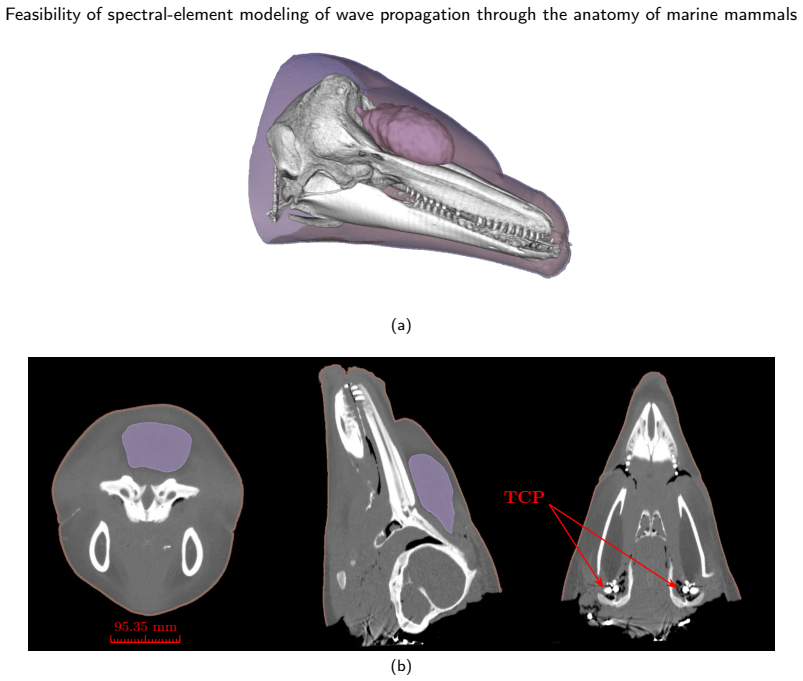
The spectral-element method itself: a high-order finite-element formulation using Gauss-Legendre-Lobatto points, whose global mass matrix is diagonal by construction, enabling explicit, matrix-inversion-free time stepping. The authors use SPECFEM3D, which couples acoustic (fluid) and elastic (solid) wave equations at interfaces. The mesh is generated with the SCULPT tool from a conformal, boolean-fragmented geometry, using adaptive hexahedral cells down to 0.1 mm at complex features. The machinery also includes the CT-to-STL-to-CUBIT pipeline with per-tissue homogeneous material parameters derived from Hounsfield units.
What would settle it
If a 120 kHz simulation that adds air-filled nasal passages, teeth, and tissue gradients produces wavefields at the tympano-periotic complex that differ from the simplified model by more than the model's numerical noise level, then the feasibility claim for real echolocation conditions would not hold.
Extended reading notes
Core claim
On the paper's own terms, the central discovery is that SEM, implemented through SPECFEM3D, can successfully simulate 3D time-domain wave propagation through a real bottlenose dolphin head built from CT data. The authors demonstrate this with a 16-million-element hexahedral mesh with adaptive refinement at jaws and acoustic fats, a plane-wave simulation recorded near the tympano-periotic complex, and a point-source simulation of an outgoing click-like signal through the melon. They validate the numerical solver with a reciprocity test whose mismatch is 40 dB below signal level, and they show the mesh quality is acceptable (average scaled Jacobian 0.93). This is presented as the first use of SEM for marine mammal bioacoustics, and as a feasible alternative to 3D FEM models.
Load-bearing premise
The central claim rests on the assumption that a CT-derived, manually segmented head with homogeneous per-tissue acoustic properties and no internal air spaces is realistic enough that simulated wave propagation represents real dolphin anatomy.
Editorial extensions
If this is right
- If SEM proves feasible, 3D time-domain simulations of dolphin biosonar at realistic click frequencies (around 120 kHz) become tractable on HPC clusters, where the authors estimate runs could complete in minutes with GPU acceleration.
- The same pipeline can be applied to other marine mammal species and to other anatomical regions, enabling comparative studies of sound reception and emission.
- The approach provides a numerical testbed for hypotheses about the roles of the melon, jaws, acoustic fats, and skull in beam formation and hearing, complementing existing 2D and FEM studies.
- With future inclusion of air spaces, attenuation, and tissue gradients, the model can be used to study anthropogenic noise impacts on marine mammals.
Reading between the lines
- The paper's feasibility claim is conditional on the simplified anatomy; a natural extension would be to quantify how much the omitted air sacs and teeth change TPC wavefields at 120 kHz, which would test whether the simplified model suffices for echolocation studies.
- The reciprocity validation suggests numerical error is low, but the lack of physical validation means the next decisive experiments are comparisons against measured sound fields in dolphin heads (e.g., in vitro hydrophone measurements).
- The same SEM pipeline could be transferred to medical ultrasound applications such as transcranial focused ultrasound, where similar hexahedral meshing challenges arise, though that is beyond the paper's scope.
Editorial analysis
A structured set of objections, weighed in public.
Referee Report
Summary. This paper reports the first 3D spectral-element (SEM) simulation of time-domain ultrasonic wave propagation in a CT-derived bottlenose dolphin head. The authors build a hexahedral mesh from segmented CT data using 3D Slicer, Blender, MeshLab, FreeCAD, and Coreform CUBIT/SCULPT, assign homogeneous acoustic properties to four tissue classes plus water, and run two SPECFEM3D simulations: a 40 kHz plane wave incident on the head and a point source near the phonic lips. Signals are recorded near the left and right tympano-periotic complexes, and a reciprocity test is used as numerical validation. The paper concludes that SEM is feasible and scalable for marine mammal bioacoustics, with detailed meshing commands provided for reproducibility.
Significance. If the numerical results are trustworthy, this is a useful and timely contribution: it is the first application of SEM to marine mammal head acoustics, it uses an open-source and reproducible software stack, and it demonstrates the complete pipeline from CT segmentation to high-performance time-domain wavefield simulation. The authors are appropriately transparent about anatomical simplifications, and the forward model uses no fitted parameters. The main reservation is that the only quantitative validation, a reciprocity check, is a symmetry test rather than a convergence test; the significance of the 'feasibility' claim therefore depends on whether the authors can demonstrate that the discrete solution is accurate, for example via h/p-refinement or comparison with an independent solver.
major comments (3)
- [§3.6, §4, §5] The reciprocity test in §3.6 is a symmetry property of the discretized operator and does not certify that the computed wavefields are accurate solutions of the continuous equations; a severely under-resolved mesh can satisfy reciprocity to machine precision while producing wrong amplitudes and phases. This matters because the central claim of the paper ('successfully simulate 3D wave propagation') rests on the numerical trustworthiness of the TPC signals in §4. The minimum scaled Jacobian of 0.0028 reported in Table 1 is far below the threshold of 0.2 cited from the CUBIT documentation, and the run uses N_GLL=3 with no h- or p-refinement study. I recommend adding a convergence test—for example a mesh-coarsening/refinement sequence or a comparison against an analytical solution in a simplified geometry (homogeneous sphere/cylinder) and, if feasible, against k-Wave or COMSOL on the same anatomical model—and reporting how the TPC signals change under refinement.
- [§3.4, §5] The simulations are run at f0=40 kHz with three GLL points per element, while the motivating dolphin-click applications are at roughly 120 kHz. The statement in §5 that the 2.5 mm mesh supports up to 200 kHz using three points per wavelength is an extrapolation, not a demonstrated result; at high impedance contrasts and complex interfaces, three points per wavelength is likely to be insufficient for phase-accurate transmission and reflection. To support the claimed scalability to full-bandwidth biosonar frequencies, the authors should either run at least one higher-frequency case (with a corresponding resolution analysis) or explicitly limit the feasibility claim to the demonstrated frequency range.
- [Abstract, §5] The abstract and Section 5 state that SEM can successfully simulate wave propagation through a bottlenose dolphin head, but the model omits internal air spaces, teeth, attenuation, and tissue-property gradients, as acknowledged in §5. Air spaces in particular are known to be acoustically important (e.g., near the phonic lips and nasal passages), and their omission may change the wavefield substantially. Since these omissions are intentional and documented, the manuscript should qualify its central claim to 'a simplified CT-derived head model' rather than the anatomy of a dolphin head, or it should justify why these features are not needed for the feasibility conclusion.
minor comments (5)
- [§3.6, Fig. 6] The text says the difference is 'four orders of magnitude (i.e. 40 dB)'; if the plotted quantity is particle velocity (an amplitude), a factor of 10^4 corresponds to 80 dB, not 40 dB. Please correct and specify whether the comparison is made on amplitude or power.
- [§3.2] The description of the 1-mm shift to avoid coplanar faces is a bit unclear: after subtraction, how is the remaining 1-mm section removed and does this alter the head-water interface? Please clarify.
- [Keywords] The keyword 'SPECEFM3D' contains a typo and should be 'SPECFEM3D'.
- [§3.4, Fig. 5] Please specify how the 'plane wave' is generated: the text says multiple point sources fired simultaneously, but the source-time function and amplitude tapering near the boundaries should be given as a formula or a reference; currently the reader cannot reproduce the source.
- [Table 3] The SCULPT command table is useful, but a few entries are not self-explanatory (e.g., thicken_material, adapt_material); a short explanation or pointer to CUBIT documentation would help.
Circularity Check
No significant circularity: forward SEM simulation with no fitted parameters; self-citations are not load-bearing.
full rationale
The paper's central claim is that SPECFEM3D, an externally validated spectral-element code, can produce time-domain 3D wavefields through a CT-derived hexahedral mesh of a dolphin head. The material properties in Table 2 are assigned from Hounsfield-unit tissue classification and literature values, not inverted or tuned to match any simulated output; the paper reports no parameter fitting. The only self-citation that enters the results is the qualitative statement that the recorded TPC signals are consistent with the qualitative findings of Hejazi Nooghabi et al. [5], made in Section 4.1; this comparison is illustrative and not load-bearing for the feasibility claim, and the forward simulation would stand independently without it. The reciprocity test in Section 3.6 checks a symmetry that the discretized operator should satisfy; it is a weak accuracy check, but it is not a step that defines the output in terms of the input. The acknowledged omissions of air spaces, teeth, attenuation, and tissue gradients are modeling limitations, not circularity. No quoted equation or parameter is equivalent by construction to the claimed demonstration, so no circular step is identified.
Assumptions & free parameters
free parameters (1)
- Per-tissue homogeneous acoustic properties (density, vp, vs) =
soft tissue: 1013/1536/215; acoustic fat: 928/1390/186; melon: 884/1316/184; bone: 2035/3400/1817; water: 1028/1480/0…
assumptions (5)
- domain assumption SPECFEM3D correctly solves the coupled acoustic-elastic wave equation for this hexahedral mesh.
- domain assumption Homogeneous, isotropic material properties per tissue type are adequate for the feasibility claim.
- domain assumption Omitted features (teeth, air sacs, ducts, attenuation) do not change the feasibility conclusion for SEM.
- standard math The reciprocity theorem provides a valid numerical error estimate.
- domain assumption CT Hounsfield unit thresholds correctly classify tissue types.
Cite this review
Pith. "Pith review of Feasibility of spectral-element modeling of wave propagation through the anatomy of marine mammals." pith.science (2026). https://pith.science/paper/ZMGNPTTI
@misc{pith2026250622944,
  author       = {Pith},
  title        = {Pith review of: Feasibility of spectral-element modeling of wave propagation through the anatomy of marine mammals},
  year         = {2026},
  howpublished = {\url{https://pith.science/paper/ZMGNPTTI}},
  note         = {Machine review of arXiv:2506.22944}
}
read the original abstract
This study introduces the first 3D spectral-element method (SEM) simulation of ultrasonic wave propagation in a bottlenose dolphin (Tursiops truncatus) head. Unlike traditional finite-element methods (FEM), which struggle with high-frequency simulations due to costly linear-system inversions and slower convergence, SEM offers exponential convergence and efficient parallel computation. Using Computed Tomography (CT) scan data, we developed a detailed hexahedral mesh capturing complex anatomical features, such as acoustic fats and jaws. Our simulations of plane and spherical waves confirm SEM's effectiveness for ultrasonic time-domain modeling. This approach opens new avenues for marine biology, contributing to research in echolocation, the impacts of anthropogenic marine noise pollution and the biophysics of hearing and click generation in marine mammals. By overcoming FEM's limitations, SEM provides a powerful scalable tool to test hypotheses about dolphin bioacoustics, with significant implications for conservation and understanding marine mammal auditory systems under increasing environmental challenges.
Figures


Figures from the paper (6 more)


Reference graph
Works this paper leans on
-
[1]
Q. Grimal, B. A. Gama, S. Naili, A. Watzky, J. W. Gillespie, Finite element study of high-speed blunt impact on thorax: Linear elastic considerations, International Journal of Impact Engineering 30 (2004) 665–683. doi:10.1016/j.ijimpeng.2003.08.002
-
[2]
E.van’tWout,P.Gélat,T.Betcke,S.Arridge, Afastboundaryelementmethodforthescatteringanalysisofhigh-intensityfocusedultrasound, The Journal of the Acoustical Society of America 138 (2015) 2726–2737. doi:10.1121/1.4932166. C. García A.et al.: Preprint Page 13 of 15 Feasibility of spectral-element modeling of wave propagation through the anatomy of marine mammals
- [3]
-
[4]
E. Bachmann, J. Tromp, Source encoding for viscoacoustic ultrasound computed tomographya), The Journal of the Acoustical Society of America 147 (2020) 3221–3235. doi:10.1121/10.0001191
-
[5]
A.HejaziNooghabi,Q.Grimal,A.Herrel,M.Reinwald,L.Boschi, Contributionofbone-reverberatedwavestosoundlocalizationofdolphins: A numerical model, Acta Acust. 5 (2021) 3. doi:10.1051/aacus/2020030
-
[6]
C. Wei, C. Erbe, A. B. Smith, W.-C. Yang, Validated 3D finite-element model of the Risso’s dolphin (Grampus griseus) head anatomy demonstrates gular sound reception and channelling through the mandibular fats, Bioinspir. Biomim. 19 (2024) 056025. doi:10.1088/ 1748-3190/ad7344
work page 2024
-
[7]
G. Castellazzi, P. Krysl, L. Rojas, T. W. Cranford, Assessment of the effect of natural and anthropogenic aquatic noise on vaquita (Phocoena sinus) through a numerical simulation, Advances in experimental medicine and biology 730 (2012). doi:10.1007/978-1-4419-7311-5_68
-
[8]
P. E. Nachtigall, Biosonar and Sound Localization in Dolphins, in: Oxford Research Encyclopedia of Neuroscience, 2016. doi:10.1093/ acrefore/9780190264086.013.103
arXiv 2016
Show all 64 references
-
[9]
Kazemi, M
A. Kazemi, M. Kesba, P. Provini, Realistic three-dimensional avian vocal tract model demonstrates how shape affects sound filtering (Passer domesticus), Journal of The Royal Society Interface 20 (2023) 20220728. doi:10.1098/rsif.2022.0728
2023
-
[10]
W. Feng, Y. Zhang, C. Wei, A biosonar model of finless porpoise (Neophocaena phocaenoides) for material composition discrimination of cylinders, The Journal of the Acoustical Society of America 146 (2019) 1362–1370. doi:10.1121/1.5122981
2019 doi
-
[11]
COMSOL Multiphysics, COMSOL Multiphysics (version 6.0) [computer program],, www.comsol.com (Last viewed Jun 25, 2025), 2024
2025
-
[12]
Alnaes, J
M. Alnaes, J. Blechta, J. Hake, A. Johansson, B. Kehlet, A. Logg, C. Richardson, J. Ring, M. E. Rognes, G. N. Wells, The FEniCS Project Version 1.5, Archive of Numerical Software 3 (2015). doi:10.11588/ans.2015.100.20553
2015 doi
-
[13]
Hecht, New development in freefem++, Journal of Numerical Mathematics 20 (2012) 1–14
F. Hecht, New development in freefem++, Journal of Numerical Mathematics 20 (2012) 1–14. doi:10.1515/jnum-2012-0013
2012 doi
-
[14]
Arndt, W
D. Arndt, W. Bangerth, D. Davydov, T. Heister, L. Heltai, M. Kronbichler, M. Maier, J.-P. Pelteret, B. Turcksin, D. Wells, The deal.II finite element library: Design, features, and insights, Computers & Mathematics with Applications 81 (2021) 407–422. doi:10.1016/j.camwa. 2020...
2021 arXiv
-
[15]
S. R. Haqshenas, P. Gélat, E. van ’t Wout, T. Betcke, N. Saffari, A fast full-wave solver for calculating ultrasound propagation in the body, Ultrasonics 110 (2021) 106240. doi:10.1016/j.ultras.2020.106240
2021
-
[16]
doi:10.21105/joss.02879
T.Betcke,M.W.Scroggs, Bempp-cl:AfastPythonbasedjust-in-timecompilingboundaryelementlibrary., JournalofOpenSourceSoftware 6 (2021) 2879. doi:10.21105/joss.02879
2021 doi
-
[17]
M. B. Porter, H. P. Bucker, Gaussian beam tracing for computing ocean acoustic fields, The Journal of the Acoustical Society of America 82 (1987) 1349–1359. doi:10.1121/1.395269
1987 doi
-
[18]
B. E. Treeby, J. Jaros, D. Rohrbach, B. T. Cox, Modelling elastic wave propagation using the k-Wave MATLAB Toolbox, in: 2014 IEEE International Ultrasonics Symposium, 2014, pp. 146–149. doi:10.1109/ULTSYM.2014.0037
2014
-
[19]
doi:10.1016/0021-9991(84)90128-1
A.T.Patera, Aspectralelementmethodforfluiddynamics:Laminarflowinachannelexpansion, JournalofComputationalPhysics54(1984) 468–488. doi:10.1016/0021-9991(84)90128-1
1984 doi
-
[20]
Komatitsch, J
D. Komatitsch, J. Tromp, Introduction to the spectral element method for three-dimensional seismic wave propagation, Geophysical Journal International 139 (1999) 806–822. doi:10.1046/j.1365-246x.1999.00967.x
1999
-
[21]
Komatitsch, C
D. Komatitsch, C. Barnes, J. Tromp, Wave propagation near a fluid-solid interface: A spectral-element approach, GEOPHYSICS 65 (2000) 623–631. doi:10.1190/1.1444758
2000 doi
-
[22]
4035–4039
M.Afanasiev,C.Boehm,M.vanDriel,L.Krischer,A.Fichtner, Flexiblehigh-performancemultiphysicswaveformmodelingonunstructured spectral-element meshes, in: SEG Technical Program Expanded Abstracts 2018, SEG Technical Program Expanded Abstracts, Society of Exploration Geophysicists, ...
2018 doi
-
[23]
doi:10.1046/j.1365-246X.2002.01653.x
D.Komatitsch,J.Tromp, Spectral-elementsimulationsofglobalseismicwavepropagation—I.Validation, GeophysicalJournalInternational 149 (2002) 390–412. doi:10.1046/j.1365-246X.2002.01653.x
2002 arXiv
-
[24]
Komatitsch, J
D. Komatitsch, J. Tromp, Spectral-element simulations of global seismic wave propagation—II. Three-dimensional models, oceans, rotation and self-gravitation, Geophysical Journal International 150 (2002) 303–318. doi:10.1046/j.1365-246X.2002.01716.x
2002 arXiv
-
[25]
Marty, C
P. Marty, C. Boehm, C. Paverd, M. Rominger, A. Fichtner, Full-waveform ultrasound modeling of soft tissue-bone interactions using conforming hexahedral meshes, in: Medical Imaging 2022: Physics of Medical Imaging, volume 12031, SPIE, 2022, pp. 877–891. doi:10.1117/12.2611548
2022 doi
-
[26]
Marty, C
P. Marty, C. Boehm, A. Fichtner, Elastic Full-Waveform Inversion for Transcranial Ultrasound Computed Tomography using Optimal Transport, in: 2022 IEEE International Ultrasonics Symposium (IUS), 2022, pp. 1–4. doi:10.1109/IUS54386.2022.9957394
2022
-
[27]
Marty, C
P. Marty, C. Boehm, M. van Driel, A. Fichtner, Transcranial ultrasound modeling using the spectral-element method, The Journal of the Acoustical Society of America 156 (2024) 3674–3693. doi:10.1121/10.0034474
2024 doi
-
[28]
Ferroni, Discontinuous Galerkin Spectral Element Methods for the Elastodynamics Equation on Hybrid Hexahedral-Tetrahedral Grids, Ph.D
A. Ferroni, Discontinuous Galerkin Spectral Element Methods for the Elastodynamics Equation on Hybrid Hexahedral-Tetrahedral Grids, Ph.D. thesis, Politecnico Milano, 2017
2017
-
[29]
Blacker, Meeting the Challenge for Automated Conformal Hexahedral Meshing, 2000
T. Blacker, Meeting the Challenge for Automated Conformal Hexahedral Meshing, 2000
2000
-
[30]
C. Wei, W. Au, D. Ketten, Y. Zhang, Finite element simulation of broadband biosonar signal propagation in the near- and far-field of an echolocating Atlantic bottlenose dolphin ( Tursiops truncatus ), The Journal of the Acoustical Society of America 143 (2018) 2611–2620. doi:1...
2018 doi
-
[31]
C. Wei, W. Au, D. Ketten, Modeling of the near to far acoustic fields of an echolocating bottlenose dolphin and harbor porpoise, The Journal of the Acoustical Society of America 147 (2020) 1790–1801. doi:10.1121/10.0000918
2020 doi
-
[32]
Zhang, Z
Y. Zhang, Z. Song, X. Wang, W. Cao, W. Au, Directional Acoustic Wave Manipulation by a Porpoise via Multiphase Forehead Structure, Physical Review Applied 8 (2017). doi:10.1103/PhysRevApplied.8.064002. C. García A.et al.: Preprint Page 14 of 15 Feasibility of spectral-element ...
2017 doi
-
[33]
doi:10.3390/ani12243472
C.Wei,L.G.Gill,C.Erbe,A.B.Smith,W.-C.Yang, TheDistinctiveForeheadCleftoftheRisso’sDolphin(Grampusgriseus)HardlyAffects Biosonar Beam Formation, Animals 12 (2022) 3472. doi:10.3390/ani12243472
2022 doi
-
[34]
doi:10.1121/1.4941780
C.Wei,W.W.L.Au,Z.Song,Y.Zhang, Theroleofvariousstructuresintheheadontheformationofthebiosonarbeamofthebaiji(Lipotes vexillifer), The Journal of the Acoustical Society of America 139 (2016) 875–880. doi:10.1121/1.4941780
2016 doi
-
[35]
Z.Song,Y.Zhang,C.Wei,X.Wang, Inducingrostruminterfacialwavesbyfluid-solidcouplinginaChineseriverdolphin(Lipotesvexillifer), Phys. Rev. E 93 (2016) 012411. doi:10.1103/PhysRevE.93.012411
2016 doi
-
[36]
doi:10.1142/S2591728518500093
C.Wei,Z.Song,W.W.L.Au,Y.Zhang,D.Wang, ANumericalEvidenceofBiosonarBeamFormationofaNeonateYangtzeFinlessPorpoise (Neophocaena asiaeorientalis), Journal of Theoretical and Computational Acoustics (2018). doi:10.1142/S2591728518500093
2018 doi
-
[37]
E. Dong, Y. Zhang, Z. Song, T. Zhang, C. Cai, N. X. Fang, Physical modeling and validation of porpoises’ directional emission via hybrid metamaterials, National Science Review 6 (2019) 921–928. doi:10.1093/nsr/nwz085
2019 doi
-
[38]
doi:10.1785/BSSA0880020368
D.Komatitsch,J.-P.Vilotte,Thespectralelementmethod:Anefficienttooltosimulatetheseismicresponseof2Dand3Dgeologicalstructures, Bulletin of the Seismological Society of America 88 (1998) 368–392. doi:10.1785/BSSA0880020368
1998 doi
-
[39]
Igel, Computational Seismology: A Practical Introduction, Oxford University Press, 2016
H. Igel, Computational Seismology: A Practical Introduction, Oxford University Press, 2016. doi:10.1093/acprof:oso/9780198717409. 001.0001
2016
-
[40]
Fedorov, R
A. Fedorov, R. Beichel, J. Kalpathy-Cramer, J. Finet, J.-C. Fillion-Robin, S. Pujol, C. Bauer, D. Jennings, F. Fennessy, M. Sonka, J. Buatti, S. Aylward, J. V. Miller, S. Pieper, R. Kikinis, 3D Slicer as an Image Computing Platform for the Quantitative Imaging Network, Magneti...
2012 doi
-
[41]
R. A. Brooks, A quantitative theory of the Hounsfield unit and its application to dual energy scanning, J Comput Assist Tomogr 1 (1977) 487–493. doi:10.1097/00004728-197710000-00016
1977 doi
-
[42]
Blender Foundation, Blender (version 4.4.0) [computer program],, https://www.blender.org/ (Last viewed Jun 25, 2025), 2022
2025
-
[43]
Cignoni, M
P. Cignoni, M. Callieri, M. Corsini, M. Dellepiane, F. Ganovelli, G. Ranzuglia, MeshLab: An Open-Source Mesh Processing Tool, in: Sixth Eurographics Italian Chapter Conference, 2008, pp. 129–136
2008
-
[44]
Muntoni, P
A. Muntoni, P. Cignoni, Pymeshlab 2023.12.post3 : A Python interface to MeshLab, 2021
2023
-
[45]
Rieger, W
J. Rieger, W. Mayer, Y. van Havre, FreeCad, 2001-2024
2001
-
[46]
Coreform LLC, Coreform Cubit (version 2024.8) [computer program],, http://coreform.com (Last viewed Jun 25, 2025), 2024
2024
-
[47]
S. J. Owen, M. L. Staten, M. C. Sorensen, Parallel hexahedral meshing from volume fractions, Eng. with Comput. 30 (2014) 301–313. doi:10.1007/s00366-012-0292-8
2014 doi
-
[48]
P. M. Knupp, Algebraic Mesh Quality Metrics, SIAM J. Sci. Comput. 23 (2001) 193–218. doi:10.1137/S1064827500371499
2001 doi
-
[49]
S. J. Owen, Parallel Smoothing for Grid-Based Methods., Technical Report SAND2012-6355C, Sandia National Laboratories (SNL), Albuquerque, NM, and Livermore, CA (United States), 2012
2012
-
[50]
J. Gao, Z. Xiao, S. Shen, C. Xu, J. Cai, G. Xu, Improved hexahedral mesh generation from quadrilateral surface meshes, Computers & Structures 307 (2025) 107620. doi:10.1016/j.compstruc.2024.107620
2025
-
[51]
T. J. Baker, Mesh generation: Art or science?, Progress in Aerospace Sciences 41 (2005) 29–63. doi:10.1016/j.paerosci.2005.02.002
2005 doi
-
[52]
Ahrens, B
J. Ahrens, B. Geveci, C. Law, ParaView: An End-User Tool for Large-Data Visualization, in: Visualization Handbook, Elsevier, 2005, pp. 717–731. doi:10.1016/B978-012387582-2/50038-1
2005 doi
-
[53]
Komatitsch, Fluid–solid coupling on a cluster of GPU graphics cards for seismic wave propagation, Comptes Rendus
D. Komatitsch, Fluid–solid coupling on a cluster of GPU graphics cards for seismic wave propagation, Comptes Rendus. Mécanique 339 (2011) 125–135. doi:10.1016/j.crme.2010.11.007
2011 doi
-
[54]
Cozzi, S
B. Cozzi, S. Huggenberger, H. Oelschläger, Chapter 5 - Head and Senses, in: B. Cozzi, S. Huggenberger, H. Oelschläger (Eds.), Anatomy of Dolphins, Academic Press, San Diego, 2017, pp. 133–196. doi:10.1016/B978-0-12-407229-9.00005-1
2017 doi
-
[55]
T. W. Cranford, W. R. Elsberry, W. G. Van Bonn, J. A. Jeffress, M. S. Chaplin, D. J. Blackwood, D. A. Carder, T. Kamolnick, M. A. Todd, S. H. Ridgway, Observation and analysis of sonar signal generation in the bottlenose dolphin (Tursiops truncatus): Evidence for two sonar sou...
2011 doi
-
[56]
C. Wei, Z. Wang, Z. Song, K. Wang, D. Wang, W. W. L. Au, Y. Zhang, Acoustic Property Reconstruction of a Neonate Yangtze Finless Porpoise’s (Neophocaena asiaeorientalis) Head Based on CT Imaging, PLOS ONE 10 (2015) e0121442. doi:10.1371/journal.pone. 0121442
2015 doi
-
[57]
K. Aki, P. G. Richards, Quantitative Seismology. Theory and Methods., Univ Science Books, Freeman, San Francisco, 1980
1980
-
[58]
W. W. L. Au, The Sonar of Dolphins, Springer, New York, NY, 1993. doi:10.1007/978-1-4612-4356-4
1993 doi
-
[59]
Dobbins, Dolphin sonar—modelling a new receiver concept, Bioinspir
P. Dobbins, Dolphin sonar—modelling a new receiver concept, Bioinspir. Biomim. 2 (2007) 19. doi:10.1088/1748-3182/2/1/003
2007 doi
-
[60]
Bériot, A
H. Bériot, A. Modave, An automatic perfectly matched layer for acoustic finite element simulations in convex domains of general shape, International Journal for Numerical Methods in Engineering 122 (2021) 1239–1261. doi:10.1002/nme.6560
2021 doi
-
[61]
K. S. Norris, G. W. Harvey, Sound transmission in the porpoise head, The Journal of the Acoustical Society of America 56 (1974) 659–664. doi:10.1121/1.1903305
1974 doi
-
[62]
V.A.Ryabov,TheRoleofAsymmetryoftheLeftandRightExternalEarofBottlenoseDolphin(Tursiopstruncatus)intheSpatialLocalization of Sound, Acoust. Phys. 69 (2023) 119–131. doi:10.1134/S1063771022060112
2023 doi
-
[63]
Ryabov, Role of the mental foramens in dolphin hearing, Natural Science 2 (2010) 646–653
V. Ryabov, Role of the mental foramens in dolphin hearing, Natural Science 2 (2010) 646–653. doi:10.4236/ns.2010.26081
2010
-
[64]
Ryabov, Mechanisms of Sound Reception and Conduction in the Dolphin V
V. Ryabov, Mechanisms of Sound Reception and Conduction in the Dolphin V. A. Ryabov, Biophysics 59 (2014) 475–483. doi:10.1134/ S0006350914030208. C. García A.et al.: Preprint Page 15 of 15
2014
Reviewed August 6, 2026 · model on record in the stance chip above.
Discussion (0). Continue with ORCID to comment.