DUET: Dual-Paradigm Adaptive Expert Triage with Single-cell Inductive Prior for Spatial Transcriptomics Prediction
Pith reviewed 2026-05-15 05:21 UTC · model grok-4.3
The pith
DUET predicts spatial gene expression from histology images by combining regression and retrieval under single-cell constraints.
A machine-rendered reading of the paper's core claim, the machinery that carries it, and where it could break.
Core claim
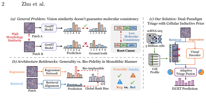
DUET implements a parallel regression-retrieval paradigm that adaptively reconciles the outputs of its complementary pathways, incorporates large-scale single-cell references to impose molecular states as biological constraints, and employs a lightweight adapter to dynamically assign branch preference across spatial contexts, achieving state-of-the-art performance with consistent gains from each component on three public datasets.
What carries the argument
Dual-paradigm adaptive expert triage that runs parametric regression and memory-based retrieval in parallel, then uses a lightweight adapter to weight their outputs under single-cell inductive priors.
If this is right
- Prediction accuracy improves consistently when both the regression and retrieval branches are active rather than used alone.
- Biological fidelity increases because single-cell priors penalize visually plausible but molecularly inconsistent outputs.
- The same architecture delivers gains across different gene panel sizes and tissue types on the tested public datasets.
- A lightweight adapter suffices to route decisions without retraining the full model for new spatial contexts.
Where Pith is reading between the lines
- The approach may generalize to other histology-based prediction tasks such as protein localization or cell-type deconvolution if suitable reference panels exist.
- It could lower the cost barrier for high-resolution molecular mapping in clinical cohorts where only H&E slides are routinely collected.
- Performance may degrade in rare cell states or disease contexts where the single-cell reference distribution diverges sharply from the imaged tissue.
- An ablation that replaces the adapter with a fixed average of the two branches would test whether dynamic triage is essential or whether the dual streams alone suffice.
Load-bearing premise
Large-scale single-cell references can reliably impose molecular states as biological constraints to mitigate aleatoric vision ambiguity in histology images.
What would settle it
Performance on a held-out tissue type drops to or below prior single-paradigm baselines when the single-cell reference panel is removed or mismatched in cellular composition.
Figures


read the original abstract
Inferring spatially resolved gene expression from histology images offers a cost-effective complement to spatial transcriptomics (ST). However, existing methods reduce this task to a simple morphology-to-expression mapping, where visual similarity does not guarantee molecular consistency. Meanwhile, single-cell data has amassed rich resources far surpassing the scale of ST data, yet it remains underexplored in vision-omics modeling. Furthermore, current approaches commit to a monolithic paradigm with bottlenecks, unable to balance expressive flexibility with biological fidelity. To bridge these gaps, we propose DUET, a novel dual-paradigm framework that synergizes parametric prediction and memory-based retrieval under cellular inductive priors. DUET implements a parallel regression-retrieval paradigm, adaptively reconciling the outputs of its complementary pathways. To mitigate aleatoric vision ambiguity, we incorporate large-scale single-cell references to impose molecular states as biological constraints for faithful learning. Building upon structural refinement, we further design a lightweight adapter to dynamically assign branch preference across spatial contexts to achieve optimal performance. Extensive experiments on three public datasets across varied gene scales demonstrate that DUET achieves SOTA performance, with consistent gains contributed by each proposed component. Code is available at https://github.com/Junchao-Zhu/DUET
Editorial analysis
A structured set of objections, weighed in public.
Referee Report
Summary. The manuscript proposes DUET, a dual-paradigm framework for inferring spatially resolved gene expression from histology images. It combines a parametric regression pathway with a memory-based retrieval pathway, reconciled adaptively via a lightweight adapter, while incorporating large-scale single-cell data as inductive priors to impose molecular-state constraints and mitigate visual ambiguity. Experiments across three public datasets at varying gene scales are reported to achieve state-of-the-art performance, with each component (dual paradigm, single-cell prior, adaptive adapter) contributing consistent additive gains. Code is released.
Significance. If the empirical results hold under rigorous validation, the work could meaningfully advance spatial transcriptomics prediction by demonstrating how abundant single-cell resources can serve as biological constraints within a hybrid regression-retrieval architecture. The adaptive triage mechanism offers a concrete way to balance flexibility and fidelity, potentially improving upon monolithic morphology-to-expression models and enabling more reliable cost-effective ST alternatives.
minor comments (3)
- Abstract: the SOTA claim and statements of 'consistent gains contributed by each proposed component' are presented without any quantitative metrics, baseline names, or effect sizes; while the full experimental section presumably supplies these, the abstract should at least indicate the magnitude of improvement (e.g., average PCC or RMSE delta) to allow immediate assessment of the central claim.
- §3 (Method) and §4 (Experiments): the description of how single-cell references are converted into 'molecular states as biological constraints' should include a precise formulation (e.g., loss term, embedding alignment, or retrieval key) and an ablation that isolates this prior from the dual-paradigm structure; without it, the claimed mitigation of aleatoric ambiguity remains difficult to verify independently.
- Table/Figure captions and §4.2: ensure all reported metrics (PCC, RMSE, etc.) are accompanied by standard deviations across multiple runs or cross-validation folds, and that the three datasets are characterized by gene count, spot count, and tissue type so readers can judge generalizability.
Simulated Author's Rebuttal
We thank the referee for the positive summary of our work, the recognition of its potential significance in advancing spatial transcriptomics prediction via hybrid regression-retrieval with single-cell priors, and the recommendation for minor revision. We are pleased that the core contributions—adaptive dual-paradigm triage and inductive priors—are viewed favorably.
Circularity Check
No significant circularity detected
full rationale
The DUET framework is an empirical architecture combining parametric regression, memory-based retrieval, and a lightweight adaptive adapter, with single-cell references used as an external inductive prior. No equations, derivations, or load-bearing steps in the provided abstract or described components reduce any claimed prediction or performance gain to a fitted parameter or self-citation by construction. The SOTA claims rest on standard validation across public datasets rather than internal redefinition of inputs as outputs. The single-cell prior is invoked as independent biological constraint data, not derived from the model's own predictions.
Axiom & Free-Parameter Ledger
axioms (1)
- domain assumption Single-cell data provides valid molecular state constraints for histology-based prediction
Reference graph
Works this paper leans on
-
[1]
Spatial transcriptomics coming of age.Nature Reviews Genetics, 20(6):317–317, 2019
Darren J Burgess. Spatial transcriptomics coming of age.Nature Reviews Genetics, 20(6):317–317, 2019
work page 2019
-
[2]
Michaela Asp, Stefania Giacomello, Ludvig Larsson, Chenglin Wu, Daniel Fürth, Xiaoyan Qian, Eva Wärdell, Joaquin Custodio, Johan Reimegård, Fredrik Salmén, et al. A spatiotemporal organ-wide gene expression and cell atlas of the developing human heart.Cell, 179(7):1647–1660, 2019
work page 2019
-
[3]
Michaela Asp, Joseph Bergenstråhle, and Joakim Lundeberg. Spatially re- solved transcriptomes—next generation tools for tissue exploration.Bioessays, 42(10):1900221, 2020
work page 2020
-
[4]
Kyongho Choe, Unil Pak, Yu Pang, Wanjun Hao, and Xiuqin Yang. Advances and challenges in spatial transcriptomics for developmental biology.Biomolecules, 13(1):156, 2023
work page 2023
-
[5]
Yang Jin, Yuanli Zuo, Gang Li, Wenrong Liu, Yitong Pan, Ting Fan, Xin Fu, Xiao- jun Yao, and Yong Peng. Advances in spatial transcriptomics and its applications in cancer research.Molecular Cancer, 23(1):129, 2024
work page 2024
-
[6]
Clusterseg: A crowd cluster pinpointed nucleus segmentation framework with cross-modality datasets
Jing Ke, Yizhou Lu, Yiqing Shen, Junchao Zhu, Yijin Zhou, Jinghan Huang, Ji- eteng Yao, Xiaoyao Liang, Yi Guo, Zhonghua Wei, et al. Clusterseg: A crowd cluster pinpointed nucleus segmentation framework with cross-modality datasets. Medical Image Analysis, 85:102758, 2023
work page 2023
-
[7]
Hiren Madhu, João Felipe Rocha, Tinglin Huang, Siddharth Viswanath, Smita Krishnaswamy, and Rex Ying. Heist: A graph foundation model for spatial tran- scriptomics and proteomics data.ArXiv, pages arXiv–2506, 2025
work page 2025
-
[8]
Chongyu Qu, Ritchie Zhao, Ye Yu, Bin Liu, Tianyuan Yao, Junchao Zhu, Ben- nett A Landman, Yucheng Tang, and Yuankai Huo. Post-training quantization for 3d medical image segmentation: A practical study on real inference engines.arXiv preprint arXiv:2501.17343, 2025
-
[9]
Junchao Zhu, Yiqing Shen, Haolin Zhang, and Jing Ke. An anti-biased tbsrtc- category aware nuclei segmentation framework with a multi-label thyroid cytol- ogy benchmark. InInternational Conference on Medical Image Computing and Computer-Assisted Intervention, pages 580–590. Springer, 2023. 10 Zhu et al
work page 2023
-
[10]
TinglinHuang,TianyuLiu,MehrtashBabadi,RexYing,andWengongJin. Stpath: a generative foundation model for integrating spatial transcriptomics and whole- slide images.NPJ Digital Medicine, 8(1):659, 2025
work page 2025
-
[11]
Bryan He, Ludvig Bergenstråhle, Linnea Stenbeck, Abubakar Abid, Alma Ander- sson, Åke Borg, Jonas Maaskola, Joakim Lundeberg, and James Zou. Integrating spatial gene expression and breast tumour morphology via deep learning.Nature biomedical engineering, 4(8):827–834, 2020
work page 2020
-
[12]
Yan Yang, Md Zakir Hossain, Eric A Stone, and Shafin Rahman. Exemplar guided deep neural network for spatial transcriptomics analysis of gene expression pre- diction. InProceedings of the IEEE/CVF Winter Conference on Applications of Computer Vision, pages 5039–5048, 2023
work page 2023
-
[13]
Ronald Xie, Kuan Pang, Sai Chung, Catia Perciani, Sonya MacParland, Bo Wang, and Gary Bader. Spatially resolved gene expression prediction from histology im- ages via bi-modal contrastive learning.Advances in Neural Information Processing Systems, 36, 2024
work page 2024
-
[14]
Asign: an anatomy- aware spatial imputation graphic network for 3d spatial transcriptomics
Junchao Zhu, Ruining Deng, Tianyuan Yao, Juming Xiong, Chongyu Qu, Junlin Guo, Siqi Lu, Mengmeng Yin, Yu Wang, Shilin Zhao, et al. Asign: an anatomy- aware spatial imputation graphic network for 3d spatial transcriptomics. InPro- ceedings of the Computer Vision and Pattern Recognition Conference, pages 30829– 30838, 2025
work page 2025
-
[15]
Yitao Yang, Yang Cui, Xin Zeng, Yubo Zhang, Martin Loza, Sung-Joon Park, and Kenta Nakai. Staig: Spatial transcriptomics analysis via image-aided graph contrastive learning for domain exploration and alignment-free integration.Nature Communications, 16(1):1067, 2025
work page 2025
-
[16]
Magnet: Multi- levelattentiongraphnetworkforpredictinghigh-resolutionspatialtranscriptomics
Junchao Zhu, Ruining Deng, Tianyuan Yao, Juming Xiong, Chongyu Qu, Junlin Guo, Siqi Lu, Yucheng Tang, Daguang Xu, Mengmeng Yin, et al. Magnet: Multi- levelattentiongraphnetworkforpredictinghigh-resolutionspatialtranscriptomics. arXiv preprint arXiv:2502.21011, 2025
-
[17]
The human cell atlas.elife, 6:e27041, 2017
Aviv Regev, Sarah A Teichmann, Eric S Lander, Ido Amit, Christophe Benoist, Ewan Birney, Bernd Bodenmiller, Peter Campbell, Piero Carninci, Menna Clat- worthy, et al. The human cell atlas.elife, 6:e27041, 2017
work page 2017
-
[18]
Computer vision methods for spatial transcriptomics: A survey.bioRxiv, pages 2025–10, 2025
Junchao Zhu, Ruining Deng, Junlin Guo, Tianyuan Yao, Siqi Lu, Chongyu Qu, Juming Xiong, Yanfan Zhu, Zhengyi Lu, Yuechen Yang, et al. Computer vision methods for spatial transcriptomics: A survey.bioRxiv, pages 2025–10, 2025
work page 2025
-
[19]
Wenwen Min, Zhiceng Shi, Jun Zhang, Jun Wan, and Changmiao Wang. Multi- modal contrastive learning for spatial gene expression prediction using histology images.Briefings in Bioinformatics, 25(6):bbae551, 2024
work page 2024
-
[20]
Minxing Pang, Kenong Su, and Mingyao Li. Leveraging information in spatial transcriptomics to predict super-resolution gene expression from histology images in tumors.BioRxiv, pages 2021–11, 2021
work page 2021
-
[21]
Junchao Zhu, Ruining Deng, Junlin Guo, Tianyuan Yao, Juming Xiong, Chongyu Qu, Mengmeng Yin, Yu Wang, Shilin Zhao, Haichun Yang, et al. Img2st-net: efficient high-resolution spatial omics prediction from whole-slide histology im- ages via fully convolutional image-to-image learning.Journal of Medical Imaging, 12(6):061410–061410, 2025
work page 2025
-
[22]
Minghao Han, Dingkang Yang, Jiabei Cheng, Xukun Zhang, Zizhi Chen, Haopeng Kuang, and Lihua Zhang. Towards unified molecule-enhanced pathology image representation learning via integrating spatial transcriptomics.Pattern Recogni- tion, page 112458, 2025. DUET 11
work page 2025
-
[23]
Weiqing Chen, Pengzhi Zhang, Tu N Tran, Yiwei Xiao, Shengyu Li, Vrutant V Shah, Hao Cheng, Kristopher W Brannan, Keith Youker, Li Lai, et al. A visual– omics foundation model to bridge histopathology with spatial transcriptomics.Na- ture Methods, pages 1–15, 2025
work page 2025
-
[24]
Chongyu Qu, Allen J Luna, Thomas Z Li, Junchao Zhu, Junlin Guo, Juming Xiong, Kim L Sandler, Bennett A Landman, and Yuankai Huo. Cohort-aware agents for individualized lung cancer risk prediction using a retrieval-augmented model selection framework. InProceedings of SPIE–the International Society for Optical Engineering, volume 13926, page 139262M, 2026
work page 2026
-
[25]
Junchao Zhu, Ruining Deng, Junlin Guo, Tianyuan Yao, Chongyu Qu, Juming Xiong, Siqi Lu, Zhengyi Lu, Yanfan Zhu, Marilyn Lionts, et al. Scr2-st: Combine single cell with spatial transcriptomics for efficient active sampling via reinforce- ment learning.arXiv preprint arXiv:2512.13635, 2025
-
[26]
Cell-type deconvolution methods for spatial transcriptomics
Lucie C Gaspard-Boulinc, Luca Gortana, Thomas Walter, Emmanuel Barillot, and Florence MG Cavalli. Cell-type deconvolution methods for spatial transcriptomics. Nature Reviews Genetics, 26(12):828–846, 2025
work page 2025
-
[27]
Vitalii Kleshchevnikov, Artem Shmatko, Emma Dann, Alexander Aivazidis, Hamish W King, Tong Li, Rasa Elmentaite, Artem Lomakin, Veronika Kedlian, Adam Gayoso, et al. Cell2location maps fine-grained cell types in spatial tran- scriptomics.Nature biotechnology, 40(5):661–671, 2022
work page 2022
-
[28]
Fabian Hörst, Moritz Rempe, Lukas Heine, Constantin Seibold, Julius Keyl, Giulia Baldini, Selma Ugurel, Jens Siveke, Barbara Grünwald, Jan Egger, et al. Cellvit: Vision transformers for precise cell segmentation and classification.Medical image analysis, 94:103143, 2024
work page 2024
-
[29]
Densely connected convolutional networks
Gao Huang, Zhuang Liu, Laurens Van Der Maaten, and Kilian Q Weinberger. Densely connected convolutional networks. InProceedings of the IEEE conference on computer vision and pattern recognition, pages 4700–4708, 2017
work page 2017
-
[30]
A visual-language foundation model for computational pathology.Nature medicine, 30(3):863–874, 2024
Ming Y Lu, Bowen Chen, Drew FK Williamson, Richard J Chen, Ivy Liang, Tong Ding, Guillaume Jaume, Igor Odintsov, Long Phi Le, Georg Gerber, et al. A visual-language foundation model for computational pathology.Nature medicine, 30(3):863–874, 2024
work page 2024
-
[31]
AlmaAndersson,LudvigLarsson,LinneaStenbeck,FredrikSalmén,AnnaEhinger, Sunny Z Wu, Ghamdan Al-Eryani, Daniel Roden, Alex Swarbrick, Åke Borg, et al. Spatial deconvolution of her2-positive breast cancer delineates tumor-associated cell type interactions.Nature communications, 12(1):6012, 2021
work page 2021
-
[32]
Blue B Lake, Rajasree Menon, Seth Winfree, Qiwen Hu, Ricardo Melo Ferreira, Kian Kalhor, Daria Barwinska, Edgar A Otto, Michael Ferkowicz, Dinh Diep, et al. An atlas of healthy and injured cell states and niches in the human kidney.Nature, 619(7970):585–594, 2023
work page 2023
-
[33]
Blue B Lake, Ricardo Melo Ferreira, Jens Hansen, Rajasree Menon, Jeannine Basta, Heather Thiessen Philbrook, Stephanie Reinert, Robin Fallegger, Asmita K Lagwankar, Xi Chen, et al. Cellular and spatial drivers of unresolved injury and functional decline in the human kidney.BioRxiv, pages 2025–09, 2025
work page 2025
-
[34]
A highly resolved integrated transcriptomic atlas of human breast cancers
Andrew Chen, Lina Kroehling, Christina S Ennis, Gerald V Denis, and Stefano Monti. A highly resolved integrated transcriptomic atlas of human breast cancers. bioRxiv, pages 2025–03, 2025
work page 2025
-
[35]
A single- cell atlas enables mapping of homeostatic cellular shifts in the adult human breast
Austin D Reed, Sara Pensa, Adi Steif, Jack Stenning, Daniel J Kunz, Linsey J Porter, Kui Hua, Peng He, Alecia-Jane Twigger, Abigail JQ Siu, et al. A single- cell atlas enables mapping of homeostatic cellular shifts in the adult human breast. Nature genetics, 56(4):652–662, 2024. 12 Zhu et al
work page 2024
-
[36]
Johanna Klughammer, Daniel L Abravanel, Åsa Segerstolpe, Timothy R Blosser, Yury Goltsev, Yi Cui, Daniel R Goodwin, Anubhav Sinha, Orr Ashenberg, Michal Slyper, et al. A multi-modal single-cell and spatial expression map of metastatic breast cancer biopsies across clinicopathological features.Nature Medicine, 30(11):3236–3249, 2024
work page 2024
-
[37]
Yuansong Zeng, Zhuoyi Wei, Weijiang Yu, Rui Yin, Yuchen Yuan, Bingling Li, Zhonghui Tang, Yutong Lu, and Yuedong Yang. Spatial transcriptomics prediction from histology jointly through transformer and graph neural networks.Briefings in Bioinformatics, 23(5):bbac297, 2022
work page 2022
-
[38]
Sichen Zhu, Yuchen Zhu, Molei Tao, and Peng Qiu. Diffusion generative model- ing for spatially resolved gene expression inference from histology images.arXiv preprint arXiv:2501.15598, 2025
-
[39]
Neng Wang, Qi Wang, Hailin Tang, Fengxue Zhang, Yifeng Zheng, Shengqi Wang, Jin Zhang, Zhiyu Wang, and Xiaoming Xie. Direct inhibition of actn4 by ellagic acid limits breast cancer metastasis via regulation ofβ-catenin stabilization in cancer stem cells.Journal of Experimental & Clinical Cancer Research, 36(1):172, 2017
work page 2017
-
[40]
Role of actn4 in tumorigenesis, metastasis, and emt.Cells, 8(11):1427, 2019
Dmitri Tentler, Ekaterina Lomert, Ksenia Novitskaya, and Nikolai A Barlev. Role of actn4 in tumorigenesis, metastasis, and emt.Cells, 8(11):1427, 2019
work page 2019
discussion (0)
Sign in with ORCID, Apple, or X to comment. Anyone can read and Pith papers without signing in.