Tracking atomic-scale interdiffusion in immiscible bimetallic nanoparticles via four-dimensional electron tomography
Pith reviewed 2026-06-27 09:11 UTC · model grok-4.3
The pith
Immiscible PdIr nanoparticles intermix at the atomic level starting at 200°C through distinct surface and diffusion steps.
A machine-rendered reading of the paper's core claim, the machinery that carries it, and where it could break.
Core claim
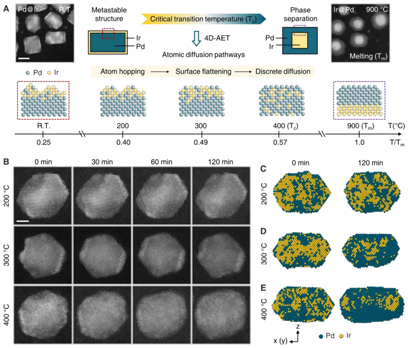
Ex situ four-dimensional atomic resolution electron tomography combined with in situ scanning transmission electron microscopy reveals the atomic scale miscible transition driven by interdiffusion in immiscible PdIr nanoparticles at temperatures far below the melting point. The pathway involves surface reconstruction atom hopping at 200°C and surface flattening at 300°C, followed by a critical transition at 400°C where Ir interfacial diffusion and discrete Ir intermediates drive miscible intermixing. Upon reaching the nanoscale melting point 900°C, collective inward Ir diffusion yields the thermodynamically stable IrPd configuration.
What carries the argument
Four-dimensional atomic resolution electron tomography combined with in situ scanning transmission electron microscopy that tracks the temperature-dependent sequence of surface atom hopping, flattening, and Ir interfacial diffusion through discrete intermediates.
Load-bearing premise
The observed structural changes result from thermally driven interdiffusion rather than electron-beam artifacts, sample preparation damage, or uncontrolled temperature gradients.
What would settle it
If PdIr nanoparticles heated to the same temperatures without any electron beam exposure show no atomic intermixing when subsequently imaged, the claim that the changes are caused by interdiffusion would be falsified.
Figures
read the original abstract
The interdiffusion of immiscible elements is generally considered both thermodynamically unfavorable and kinetically hindered. At the nanoscale, however, the mixing behavior of multielements materials often diverges from bulk equilibrium, yet a quantitative, atomically resolved description of this transformation has remained challenging. Using ex situ four dimensional atomic resolution electron tomography combined with in situ scanning transmission electron microscopy, here we reveal the atomic scale miscible transition driven by interdiffusion in immiscible PdIr nanoparticles at temperatures far below the melting point. The pathway involves surface reconstruction atom hopping at 200oC and surface flattening at 300oC, followed by a critical transition at 400oC where Ir interfacial diffusion and discrete Ir intermediates drive miscible intermixing. Upon reaching the nanoscale melting point 900oC, collective inward Ir diffusion yields the thermodynamically stable IrPd configuration. Our findings provide quantitative atomic scale insights into how metastable nanostructures evolve through distinct intermediates, offering a design framework for advanced multielement materials.
Editorial analysis
A structured set of objections, weighed in public.
Referee Report
Summary. The manuscript reports the use of ex situ four-dimensional atomic-resolution electron tomography combined with in situ scanning transmission electron microscopy to track atomic-scale interdiffusion in immiscible PdIr nanoparticles. It describes a temperature-dependent pathway involving surface reconstruction and atom hopping at 200°C, surface flattening at 300°C, a critical transition at 400°C driven by Ir interfacial diffusion and discrete intermediates leading to miscible intermixing, and collective inward Ir diffusion at 900°C to the thermodynamically stable configuration.
Significance. If the structural changes are confirmed to arise from thermally driven interdiffusion, the work would deliver quantitative atomic-scale observations of how metastable nanostructures evolve through distinct intermediates in immiscible bimetallic systems below the bulk melting point. The combination of in situ STEM heating with ex situ 4D tomography is a methodological strength that enables direct visualization of atom trajectories and could inform design principles for multielement nanomaterials.
major comments (2)
- [Abstract] Abstract: the central claim that distinct transitions at 200°C, 300°C, and especially the critical 400°C transition are 'driven by interdiffusion' is load-bearing, yet the described experimental workflow provides no beam-off controls, dose-series comparisons, or beam-heating estimates to exclude electron-beam-induced artifacts (local heating, knock-on displacement, or radiolysis) during in situ STEM or ex situ tomography.
- [Experimental workflow] Experimental methods (as summarized): without quantitative reporting of electron dose, temperature calibration under beam illumination, or control experiments separating thermal from beam effects, attribution of the observed Ir diffusion and intermixing specifically to thermal activation at 400°C cannot be rigorously distinguished from beam-assisted processes.
minor comments (2)
- [Abstract] Abstract: temperatures are inconsistently formatted as '200oC' rather than the conventional '200 °C'.
- [Abstract] Abstract: 'four dimensional' should be hyphenated as 'four-dimensional' for standard technical usage.
Simulated Author's Rebuttal
We thank the referee for the constructive feedback on potential electron-beam artifacts. We address each major comment below and will revise the manuscript to strengthen the distinction between thermal and beam-induced effects where possible.
read point-by-point responses
-
Referee: [Abstract] Abstract: the central claim that distinct transitions at 200°C, 300°C, and especially the critical 400°C transition are 'driven by interdiffusion' is load-bearing, yet the described experimental workflow provides no beam-off controls, dose-series comparisons, or beam-heating estimates to exclude electron-beam-induced artifacts (local heating, knock-on displacement, or radiolysis) during in situ STEM or ex situ tomography.
Authors: We agree that explicit controls are important for rigorous attribution. The ex situ 4D tomography workflow heats particles without the electron beam present during the temperature ramp, with atomic-resolution imaging performed afterward at ambient temperature; the in situ STEM component images changes only after the target temperature is reached. In the revised manuscript we will add quantitative electron-dose values for both modalities, literature-based estimates of beam-induced local heating in nanoparticles, and a discussion of why the observed temperature-specific stepwise transitions (surface hopping at 200 °C, flattening at 300 °C, interfacial diffusion at 400 °C) are inconsistent with constant beam-driven processes. Dedicated beam-off heating controls and full dose-series comparisons were not performed in the original study; we will note this limitation explicitly while arguing that the multi-particle reproducibility and thermodynamic consistency support the thermal interpretation. revision: yes
-
Referee: [Experimental workflow] Experimental methods (as summarized): without quantitative reporting of electron dose, temperature calibration under beam illumination, or control experiments separating thermal from beam effects, attribution of the observed Ir diffusion and intermixing specifically to thermal activation at 400°C cannot be rigorously distinguished from beam-assisted processes.
Authors: We accept that the current methods section lacks the requested quantitative details. The revised manuscript will report the electron doses used during in situ STEM heating and ex situ tomography, clarify the heating-stage temperature calibration procedure, and include an estimate of beam-heating contributions using established models for metallic nanoparticles. While separate beam-off control experiments were not included in the original workflow (the ex situ design intentionally minimizes beam exposure during heating), the distinct, temperature-thresholded structural pathways observed across multiple particles provide internal evidence favoring thermal activation. We will expand the methods and discussion sections accordingly to address this point directly. revision: yes
Circularity Check
No circularity: purely observational experimental report with no derivations or fitted predictions
full rationale
The paper reports experimental observations of atomic-scale structural changes in PdIr nanoparticles using in-situ STEM and ex-situ 4D tomography. The abstract and described pathway detail temperature-dependent transitions (surface reconstruction at 200°C, flattening at 300°C, intermixing at 400°C) without any equations, parameters, predictions, or first-principles derivations. No self-citations, ansatzes, or uniqueness theorems are invoked in a load-bearing mathematical sense. The central claim is an attribution of observed dynamics to thermal interdiffusion, which is an interpretive step but does not reduce to a self-definitional or fitted-input circularity by construction. This matches the default expectation for non-circular experimental papers.
Axiom & Free-Parameter Ledger
Reference graph
Works this paper leans on
-
[1]
H. Li, R. Zeng, Z. Shi, H. Wang, D. Leshchev, E. Stavitski, M. M. Tellez -Cruz, W. Xu, M.-J. Kim, A. Molina Villarino, Q. Li, D. A. Muller, H. D. Abruñ a, Rational design of high - performance low-loading oxygen reduction catalysts for alkaline fuel cells. Nat. Mater. 25, 447-455 (2026). doi: 10.1038/s41563-025-02422-4
-
[2]
M. Peng, Y. Ge, R. Gao, J. Yang, A. Li, Z. Xie, Q. Yu, J. Zhang, H. Asakura, H. Zhang, Z. Liu, Q. Zhang, J. Deng, J. Zhou, W. Zhou, G. J. Hutchings, D. Ma, Thermal catalytic reforming for hydrogen production with zero CO 2 emission. Science 387, 769 –775 (2025). doi: 10.1126/science.adt0682
-
[3]
W. Zuo, F. Ren, P. Barai, D. Hou, S. Zhou, G. Wang, T. Li, X. Jia, Y. Qin, Z. Yang, W. Xu, Y. Liu, H. Yan, Y. S. Chu, Y. Yang, V. Srinivasan, X. Xiao, K. Amine, G.-L. Xu, Gas-mediated defect engineering in earth -abundant Mn-rich layered oxides for non -aqueous sodium-based batteries. Nat. Nanotechnol. 20, 1667–1677 (2025). doi: 10.1038/s41565-025-01998-x
-
[4]
M. Cai, Y. Dong, M. Xie, W. Dong, C. Dong, P. Dai, H. Zhang, X. Wang, X. Sun, S. Zhang, M. Yoon, H. Xu, Y. Ge, J. Li, F. Huang, Stalling oxygen evolution in high -voltage cathodes by lanthurization. Nat. Energy 8, 159–168 (2023). doi: 10.1038/s41560-022-01179-3
-
[5]
B. Jiang, Y. Yu, J. Cui, X. Liu, L. Xie, J. Liao, Q. Zhang, Y. Huang, S. Ning, B. Jia, B. Zhu, S. Bai, L. Chen, S. J. Pennycook, J. He, High -entropy-stabilized chalcogenides with high thermoelectric performance. Science 371, 830–834 (2021). doi: 10.1126/science.abe1292
-
[6]
B. Jiang, W. Wang, S. Liu, Y. Wang, C. Wang, Y. Chen, L. Xie, M. Huang, J. He, High figure- of-merit and power generation in high-entropy GeTe-based thermoelectrics. Science 377, 208– 213 (2022). doi: 10.1126/science.abq5815
-
[7]
R. A. van Santen, Complementary structure sensitive and insensitive catalytic relationships. Acc. Chem. Res. 42, 57–66 (2009). doi: 10.1021/ar800022m
-
[8]
A. S. Barnard, Size -dependent phase transitions and phase reversal at the nanoscale (Oxford University Press, 2017)
2017
-
[9]
Z. Fan, M. Bosman, X. Huang, D. Huang, Y. Yu, K. P. Ong, Y. A. Akimov, L. Wu, B. Li, J. Wu, Y. Huang, Q. Liu, C. Eng Png, C. Lip Gan, P. Yang, H. Zhang, Stabilization of 4H hexagonal phase in gold nanoribbons. Nat. Commun. 6, 7684 (2015). doi: 10.1038/ncomms8684
-
[10]
Q. Zhang, K. Kusada, D. Wu, T. Yamamoto, T. Toriyama, S. Matsumura, S. Kawaguchi, Y. Kubota, H. Kitagawa, Selective control of fcc and hcp crystal structures in Au –Ru solid - solution alloy nanoparticles. Nat. Commun. 9, 510 (2018). doi: 10.1038/s41467-018-02933-6
-
[12]
P.-C. Chen, C. Chen, Y. Yang, A. L. Maulana, J. Jin, J. Feijoo, P. Yang, Chemical and structural evolution of AgCu catalysts in electrochemical CO 2 reduction. J. Am. Chem. Soc. 145, 10116–10125 (2023). doi: 10.1021/jacs.3c00467
-
[13]
T. He, W. Wang, F. Shi, X. Yang, X. Li, J. Wu, Y. Yin, M. Jin, Mastering the surface strain of platinum catalysts for efficient electrocatalysis. Nature 598, 76 –81 (2021). doi: 10.1038/s41586-021-03870-z. 12
-
[14]
X. Li, Z. Liu, A. Gao, Q. Zhang, H. Zhong, F. Meng, T. Lin, S. Wang, D. Su, K. Jin, C. Ge, L. Gu, Ferroelastically protected reversible orthorhombic to monoclinic -like phase transition in ZrO2 nanocrystals. Nat. Mater. 23, 1077–1084 (2024). doi: 10.1038/s41563-024-01853-9
-
[15]
P. Schweizer, A. Sharma, L. Pethö , E. Huszar, L. M. Vogl, J. Michler, X. Maeder, Atomic scale volume and grain boundary diffusion elucidated by in situ STEM. Nat. Commun. 14, 1– 6 (2023). doi: 10.1038/s41467-023-43103-7
-
[16]
X. Han, M. Niu, Y. Luo, R. Li, J. Dan, Y. Hong, X. Wu, A. V. Trukhanov, W. Ji, Y. Wang, J. Zhou, J. Qiao, J. Zhang, X. Zhao, Atomically engineering metal vacancies in monolayer transition metal dichalcogenides. Nat. Synth. 3, 586 –594 (2024). doi: 10.1038/s44160 -024- 00501-z
-
[17]
M. Chi, C. Wang, Y. Lei, G. Wang, D. Li, K. L. More, A. Lupini, L. F. Allard, N. M. Markovic, V. R. Stamenkovic, Surface faceting and elemental diffusion behaviour at atomic scale for alloy nanoparticles during in situ annealing. Nat. Commun. 6, 8925 (2015). doi: 10.1038/ncomms9925
-
[18]
S. Dai, Y. You, S. Zhang, W. Cai, M. Xu, L. Xie, R. Wu, G. W. Graham, X. Pan, In situ atomic- scale observation of oxygen-driven core-shell formation in Pt3Co nanoparticles. Nat. Commun. 8, 204 (2017). doi: 10.1038/s41467-017-00161-y
-
[19]
X. Zhang, S. Han, B. Zhu, G. Zhang, X. Li, Y. Gao, Z. Wu, B. Yang, Y. Liu, W. Baaziz, O. Ersen, M. Gu, J. T. Miller, W. Liu, Reversible loss of core–shell structure for Ni–Au bimetallic nanoparticles during CO2 hydrogenation. Nat. Catal. 3, 411–417 (2020). doi: 10.1038/s41929- 020-0440-2
-
[20]
R. Xu, C.-C. Chen, L. Wu, M. C. Scott, W. Theis, C. Ophus, M. Bartels, Y. Yang, H. Ramezani- Dakhel, M. R. Sawaya, H. Heinz, L. D. Marks, P. Ercius, J. Miao, Three -dimensional coordinates of individual atoms in materials revealed by electron tomography. Nat. Mater. 14, 1099–1103 (2015). doi: 10.1038/nmat4426. 21.J. Miao, P. Ercius, S. J. L. Billinge, Ato...
-
[21]
Y. Zhang, Z. Li, X. Tong, Z. Xie, S. Huang, Y. -E. Zhang, H. -B. Ke, W. -H. Wang, J. Zhou, Three-dimensional atomic insights into the metal-oxide interface in Zr-ZrO2 nanoparticles. Nat. Commun. 15, 7624 (2024). doi: 10.1038/s41467-024-52026-w
-
[22]
Z. Sun, Y. Zhang, Z. Li, Z. Xie, Y. Dai, X. Du, C. Ophus, J. Zhou, Strain release by 3D atomic misfit in fivefold twinned icosahedral nanoparticles with amorphization and dislocations. Nat. Commun. 16, 1595 (2025). doi: 10.1038/s41467-025-56842-6
-
[23]
Y. Zhang, L. Cao, Z. Sun, J. Zhou, “Physics-aware neural networks enable robust and full atomic structure determination via low -dose atomic electron tomography ”. (2026; https://arxiv.org/pdf/2603.19942)
arXiv 2026
-
[24]
Y. Yang, C. -C. Chen, M. C. Scott, C. Ophus, R. Xu, A. Pryor, L. Wu, F. Sun, W. Theis, J. Zhou, M. Eisenbach, P. R. C. Kent, R. F. Sabirianov, H. Zeng, P. Ercius, J. Miao, Deciphering chemical order/disorder and material properties at the single -atom level. Nature 542, 75–79 (2017). doi: 10.1038/nature21042
-
[25]
Y. Yang, J. Zhou, F. Zhu, Y. Yuan, D. J. Chang, D. S. Kim, M. Pham, A. Rana, X. Tian, Y. Yao, S. J. Osher, A. K. Schmid, L. Hu, P. Ercius, J. Miao, Determining the three -dimensional atomic structure of an amorphous solid. Nature 592, 60–64 (2021). doi: 10.1038/s41586-021- 03354-0
-
[26]
S. Moniri, Y. Yang, J. Ding, Y. Yuan, J. Zhou, L. Yang, F. Zhu, Y. Liao, Y. Yao, L. Hu, P. Ercius, J. Miao, Three-dimensional atomic structure and local chemical order of medium- and high-entropy nanoalloys. Nature 624, 564–569 (2023). doi: 10.1038/s41586-023-06785-z. 13
-
[27]
Y. Yang, J. Zhou, Z. Zhao, G. Sun, S. Moniri, C. Ophus, Y. Yang, Z. Wei, Y. Yuan, C. Zhu, Y. Liu, Q. Sun, Q. Jia, H. Heinz, J. Ciston, P. Ercius, P. Sautet, Y. Huang, J. Miao, Atomic - scale identification of active sites of oxygen reduction nanocatalysts. Nat. Catal. 7, 796–806 (2024). doi: 10.1038/s41929-024-01175-8
-
[28]
Y. Dai, Z. Xie, Y. Zhang, X. Du, Z. Li, J. Xie, Z. Sun, J. Zhou, Mapping surface and subsurface atomic structures of Au@Pd core-shell nanoparticles in three dimensions. ACS Nano 19, 9006– 9016 (2025). doi: 10.1021/acsnano.4c17462
-
[29]
Y. Liao, H. Sha, C. M. O’Leary, H. Zhong, Y. Yang, J. Miao, Accurate determination of the 3D atomic structure of amorphous materials. Nature 649, 1123 –1129 (2026). doi: 10.1038/s41586-025-09857-4
-
[30]
J. Zhou, Y. Yang, Y. Yang, D. S. Kim, A. Yuan, X. Tian, C. Ophus, F. Sun, A. K. Schmid, M. Nathanson, H. Heinz, Q. An, H. Zeng, P. Ercius, J. Miao, Observing crystal nucleation in four dimensions using atomic electron tomography. Nature 570, 500 –503 (2019). doi: 10.1038/s41586-019-1317-x
-
[31]
Tracking four-dimensional atomic evolutions of single nanocatalysts throughout the life cycles
J. Xie, Z. Xie, D. Jiang, S. Li, Y. Dai, Y. Zhang, M. Li, J. Zhou, “Tracking four-dimensional atomic evolutions of single nanocatalysts throughout the life cycles ”. (2025; https://arxiv.org/pdf/2509.17438)
arXiv 2025
-
[32]
S. N. Tripathl, S. R. Bharadwoj, M. S. Chandrasekharalah, The Ir -Pd (iridium -palladium) system. J. Phase Equilib. 12, 603–605 (1991). doi: 10.1007/BF02645078
-
[33]
H. Guo, L. Li, Y. Chen, W. Zhang, C. Shang, X. Cao, M. Li, Q. Zhang, H. Tan, Y. Nie, L. Gu, S. Guo, Precise strain tuning boosts electrocatalytic hydrogen generation. Adv. Mater. 35, 2302285 (2023). doi: 10.1002/adma.202302285
-
[34]
H. L. Skriver, N. M. Rosengaard, Surface energy and work function of elemental metals. Phys. Rev. B 46, 7157–7168 (1992). doi: 10.1103/physrevb.46.7157
-
[35]
J. Xie, Z. Xie, Z. Li, Y. Dai, Y. Zhang, S. Li, D. Jiang, Y. Bu, C. Liu, X. Chang, J. Wang, H. Jiang, M. Li, J. Zhou, Revealing the interface -driven atomic local chemical heterogeneity in bimetallic catalysts in three dimensions. J. Am. Chem. Soc. 147, 41573–41585 (2025). doi: 10.1021/jacs.5c12285
-
[36]
S. Hu, W.-X. Li, Sabatier principle of metal-support interaction for design of ultrastable metal nanocatalysts. Science 374, 1360–1365 (2021). doi: 10.1126/science.abi9828
-
[37]
S. Zhang, C. Chen, M. Cargnello, P. Fornasiero, R. J. Gorte, G. W. Graham, X. Pan, Dynamic structural evolution of supported palladium –ceria core –shell catalysts revealed by in situ electron microscopy. Nat. Commun. 6, 7778 (2015). doi: 10.1038/ncomms8778
-
[38]
Z. Li, Z. Xie, Y. Zhang, X. Mu, J. Xie, H. -J. Yin, Y.-W. Zhang, C. Ophus, J. Zhou, Probing the atomically diffuse interfaces in Pd@Pt core -shell nanoparticles in three dimensions. Nat. Commun. 14, 2934 (2023). doi: 10.1038/s41467-023-38536-z
-
[39]
I. V. Chepkasov, V. S. Baidyshev, A. V. Iosimovska, I. S. Zamulin, A. G. Kvashnin, Adsorption properties of crystalline and amorphous PdIr nanoparticles. A systematic first - principles study. J. Catal. 447, 116102 (2025). doi: 10.1016/j.jcat.2025.116102
-
[40]
A. Fick, Ueber diffusion. Ann. Phys. 170, 59–86 (1855). doi: 10.1002/andp.18551700105
-
[41]
M. Polak, L. Rubinovich, The thermal stability of asymmetric separated configurations inside alloy nanoparticles: atomic -scale modeling of Pd -Ir nanophase diagrams. ACS Nano 16, 20186–20196 (2022). doi: 10.1021/acsnano.2c05419
-
[42]
M. Jin, H. Liu, H. Zhang, Z. Xie, J. Liu, Y. Xia, Synthesis of Pd nanocrystals enclosed by {100} facets and with sizes <10 nm for application in CO oxidation. Nano Res. 4, 83–91 (2011). doi: 10.1007/s12274-010-0051-3. 14
-
[43]
M. Pham, Y. Yuan, A. Rana, S. Osher, J. Miao, Accurate real space iterative reconstruction (RESIRE) algorithm for tomography. Sci. Rep. 13, 5624 (2023). doi: 10.1038/s41598 -023- 31124-7
-
[44]
K. Dabov, A. Foi, V. Katkovnik, K. Egiazarian, Image denoising by sparse 3 -D transform- domain collaborative filtering. IEEE Trans. on Image Process. 16, 2080–2095 (2007). doi: 10.1109/TIP.2007.901238
-
[45]
N. Otsu, A threshold selection method from gray -level histograms. IEEE Trans. Syst. Man Cybern. 9, 62–66 (1979). doi: 10.1109/TSMC.1979.4310076
-
[46]
S. S. Rogers, T. A. Waigh, X. Zhao, J. R. Lu, Precise particle tracking against a complicated background: polynomial fitting with Gaussian weight. Phys. Biol. 4, 220 –227 (2007). doi: 10.1088/1478-3975/4/3/008
-
[47]
M. C. Scott, C.-C. Chen, M. Mecklenburg, C. Zhu, R. Xu, P. Ercius, U. Dahmen, B. C. Regan, J. Miao, Electron tomography at 2.4 -å ngströ m resolution. Nature 483, 444–447 (2012). doi: 10.1038/nature10934
-
[48]
C.-C. Chen, C. Zhu, E. R. White, C.-Y. Chiu, M. C. Scott, B. C. Regan, L. D. Marks, Y. Huang, J. Miao, Three -dimensional imaging of dislocations in a nanoparticle at atomic resolution. Nature 496, 74–77 (2013). doi: 10.1038/nature12009
-
[49]
Least squares quantization in PCM,
S. Lloyd, Least squares quantization in PCM. IEEE Trans. Inf. Theory 28, 129–137 (1982). doi: 10.1109/TIT.1982.1056489
-
[50]
A. Pryor, C. Ophus, J. Miao, A streaming multi -GPU implementation of image simulation algorithms for scanning transmission electron microscopy. Adv. Struct. Chem. Imaging 3, 15 (2017). doi: 10.1186/s40679-017-0048-z
-
[51]
M. P. d. Carmo, Differential geometry of curves & surfaces (Dover Publications, 2018)
2018
-
[52]
Stukowski, Computational analysis methods in atomistic modeling of crystals
A. Stukowski, Computational analysis methods in atomistic modeling of crystals. JOM 66, 399–407 (2014). doi: 10.1007/s11837-013-0827-5
-
[53]
Q.-J. Li, H. Sheng, E. Ma, Strengthening in multi-principal element alloys with local-chemical- order roughened dislocation pathways. Nat. Commun. 10, 3563 (2019). doi: 10.1038/s41467 - 019-11464-7. Acknowledgments: The authors thank the Analysis Center at Tsinghua University for the use of aberration-corrected electron microscopes. This work is also supp...
-
[54]
1017269) were purchased from Leyan, Shanghai, China
Chemicals Sodium tetrachloropalladate(II) (Na2PdCl4, ≥99.99%) was purchased from Sigma -Aldrich; iridium (III) acetylacetonate (Ir(acac)3) was purchased from Bide Pharmatech, Shanghai, China; ethanol, cyclohexane and acetone were purchased from TGchem, Beijing, China; potassium bromide (KBr, 99.7%) was purchased from MREDA, Beijing, China; potassium chlor...
-
[55]
Sample preparation Pd@Ir NPs were synthesized by a modified two-step method described in detail elsewhere (34, 43)
-
[56]
Na2PdCl4 (30 mg) was dissolved in 9 mL deionized water, sonicated for 10 min, and then combined with the preheated solution
Synthesis of Pd NPs PVP (315 mg), KBr (15 mg), KCl (610.5 mg) and L-ascorbic acid (180 mg) were dissolved in 24 mL of deionized water , and the solution was heated to 80° C. Na2PdCl4 (30 mg) was dissolved in 9 mL deionized water, sonicated for 10 min, and then combined with the preheated solution. The mixture was stirred at 80° C for 4 h, and allowed to c...
-
[57]
The resulting solution was then cooled to room temperature
Synthesis of Pd@Ir NPs The Pd seeds dispersion obtained above (4.7 mL) was first mixed with 6 mL of benzyl alcohol and stirred at 200° C in an open vessel for 5 min to complete phase transfer of the seeds from ethanol to benzyl alcohol. The resulting solution was then cooled to room temperature. An Ir precursor solution was prepared by dissolving 30.6 mg ...
-
[58]
In situ heating was performed using a Chip-Nova volcano series in situ ultra-heating single-tilt holder
Material characterizations Annular dark -field scanning transmission electron microscopy (ADF -STEM), energy- dispersive X-ray spectroscopy (EDS) and in situ STEM during heating experiments were carried out on an aberration -corrected FEI Themis Z microscope at 300 kV. In situ heating was performed using a Chip-Nova volcano series in situ ultra-heating si...
-
[59]
Four-dimensional atomic-resolution tomography
-
[60]
To further enhance the conductivity of the membrane, a 4 -nm-thick carbon layer was deposited onto the TEM window using a sputter coater (pulse mode , Leica EM ACE600)
Specimen preparation 18 A single-slot silicon TEM window with a SiN x membrane (purchased from NORCADA , Canada) was used for the specimen preparation. To further enhance the conductivity of the membrane, a 4 -nm-thick carbon layer was deposited onto the TEM window using a sputter coater (pulse mode , Leica EM ACE600). The grid was then heated at 350 ℃ fo...
-
[61]
To obtain NPs in different diffusion states, the three NPs were annealed under an argon flow at 200, 300, and 400° C, respectively
4D Tomography experiment Three Pd@Ir NPs were randomly selected for the 4D tomography experiment. To obtain NPs in different diffusion states, the three NPs were annealed under an argon flow at 200, 300, and 400° C, respectively. For each particle, four tomographic tilt series were collected at cumulative annealing times of 0, 30, 60, and 120 min (figs. S...
-
[62]
Operation parameters used in acquisitions of tomographic tilt series All tomographic tilt series were acquired in ADF-STEM mode on a n aberration-corrected FEI Themis Z microscope . The operation parameters were: accelerating voltage 300 kV, convergence semi-angle 25 mrad, HAADF detector inner semi -angle 40.6 mrad and HAADF detector outer semi-angle 200 ...
-
[63]
3D atomic-resolution reconstruction
-
[64]
Image pre-processing For e ach tilt series, the images w ere pre-processed prior to reconstruction following an established procedure (27, 39). First, at each tilt angle, the three acquired frames were registered by normalized cross-correlation and then averag ed to improve the signal-to-noise ratio; during this step, linear drifts were estimated and corr...
-
[65]
3D atomic-resolution reconstruction The Real Space Iterative Reconstruction (RESIRE) algorithm (44) was employed for the reconstruction, with the specific parameters listed in Supplementary Table s 1-3. To correct the residual misalignment and angular inaccuracies caused by stage and holder instability, spatial realignment and angular refinement were perf...
-
[66]
Determination of 3D atomic coordinates and chemical species Using the final 3D volume obtained above, a series of steps were performed to determine the atomic coordinates and identify the chemical species
-
[67]
missing wedge
Identification of local intensity maxima A polynomial fitting method (26, 47) was applied within a voxel region of 7× 7× 7 box to locate peak locations. From the preliminary list, local maxima in the reconstructed density were selected as candidate atomic positions, with a minimum separation of 2.2 Å enforced between adjacent candidate s. Three-dimensiona...
-
[68]
missing wedge
Element classification Initial element classification was conducted using the K-means clustering algorithm (50). For each atomic coordinate, the integrated intensity was calculated by summing the intensity signal within a 7× 7× 7 box centered on the atom . These integrated intensit ies were then grouped into two classes: Pd (lower intensity) and Ir (highe...
-
[69]
Structural analysis of final atomic models
-
[70]
CNs were determined by setting the first minima of the PDFs as the cutoff distance
Calculation of CN, curvature, specific surface area, and atomic distance to surface Pair distribution functions (PDF) were computed from the final atomic models. CNs were determined by setting the first minima of the PDFs as the cutoff distance. The atoms with CN<12 were classified as surface atoms. The local curvature at each surface atom was calculated ...
-
[71]
Next, we compared the two atomic models obtained from the same NP at different annealing times
Identification of common positions and chemically consistent atoms First, an ideal FCC lattice was fitted to each dataset by a least-squares fitting approach (20), matching all atomic positions well. Next, we compared the two atomic models obtained from the same NP at different annealing times. Atomic pairs were identified by requiring that the two atoms ...
-
[72]
In this work, we used the first shell (m=1) to calculate 𝛼𝑃𝑑−𝑃𝑑 and 𝛼𝐼𝑟−𝑃𝑑
Quantification of chemical short-range-order parameter (CSROP) To evaluate short -range order in each NP, a pairwise multicomponent short -range-order parameter was applied (54), which is denoted as: 𝛼𝑖𝑗 𝑚 = (𝑝𝑖𝑗 𝑚 − 𝐶𝑗) / (𝛿𝑖𝑗 − 𝐶𝑗) Here, m indicates the m-th nearest-neighbor shell around a central atom i, 𝑝𝑖𝑗 𝑚 represents the averaged probability of det...
-
[73]
onion peeling
Calculation of interfacial diffusion coefficients of Ir atoms The radial concentration profiles of Ir at different depths were quantified based on the “onion peeling” method described in the previous section (f ig. S32). To eliminate interference from particle reshaping during surface reconstruction, we selected the change s in Ir concentration s with the...
1915
discussion (0)
Sign in with ORCID, Apple, or X to comment. Anyone can read and Pith papers without signing in.