Atom Probe Tomography as an Emerging Tool for Understanding Defect-driven Mechanisms in HfO₂-based Ferroelectrics
Pith reviewed 2026-06-27 03:38 UTC · model grok-4.3
The pith
Atom probe tomography can map individual dopants, oxygen-vacancy clusters, and interfacial segregation in HfO2-based ferroelectrics at atomic scale.
A machine-rendered reading of the paper's core claim, the machinery that carries it, and where it could break.
Core claim
By resolving individual dopants, vacancy clustering, and interfacial segregation in three dimensions, atom probe tomography can supply the quantitative defect maps needed to establish direct defect-property relations that govern polar-phase stabilization, wake-up, fatigue, and imprint in HfO2-based ferroelectrics.
What carries the argument
Atom probe tomography (APT) for three-dimensional, atomic-scale chemical mapping of all constituent species inside ferroelectric device stacks.
If this is right
- Quantitative maps of oxygen-vacancy clusters would allow direct testing of proposed mechanisms for the wake-up effect.
- Interfacial segregation profiles would constrain models of imprint and retention loss.
- Dopant distributions at the atomic level would guide doping strategies that stabilize the desired polar phase.
- Three-dimensional defect statistics would replace averaged or two-dimensional proxies in reliability simulations.
Where Pith is reading between the lines
- If APT succeeds on hafnia stacks it would likely be applied to other CMOS-compatible ferroelectrics such as doped zirconia or aluminum scandium nitride.
- Routine APT on device stacks could shift defect engineering from post-fabrication analysis to in-line process control.
- Combined APT and electrical measurements on the same nanoscale volume would test whether local defect density predicts local switching behavior.
Load-bearing premise
That sample-preparation, reconstruction-fidelity, and data-interpretation difficulties can be solved well enough to yield reliable atomic-scale maps inside actual device stacks.
What would settle it
A set of APT reconstructions on the same hafnia stack that show inconsistent dopant or vacancy positions when cross-checked against transmission-electron-microscopy or secondary-ion-mass-spectrometry depth profiles.
Figures
read the original abstract
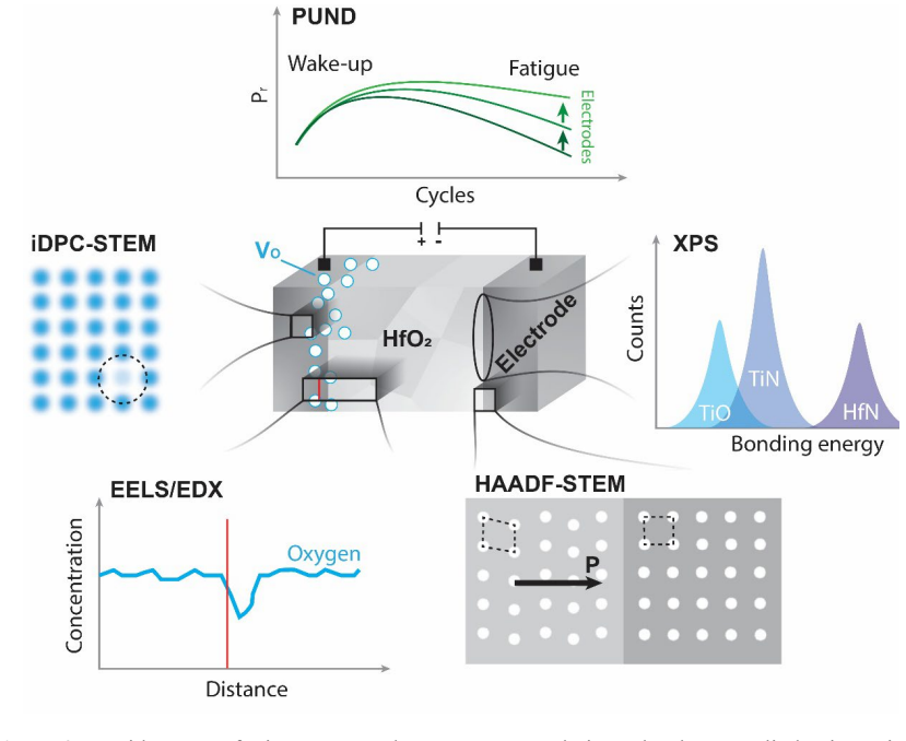
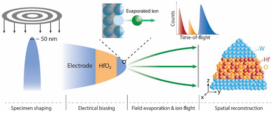
HfO$_{2}$-based ferroelectrics are essential for the next generation of CMOS-compatible memory and logic devices, yet their performance is governed by a complex interplay between oxygen vacancies, dopants, and structural defects that remains an active area of investigation. These defects shape the function-critical dynamic phenomena, such as polar phase stabilization, wake-up, fatigue, and imprint. In this Perspective, we review the limitations of established high-resolution structural characterization techniques and propose atom probe tomography (APT) as a powerful tool for the three-dimensional (3D), atomic-scale mapping of all constituent species in hafnia-based ferroelectric systems. By resolving individual dopants, vacancy clustering, and interfacial segregation, APT can facilitate a quantitative understanding of defect-property relations in hafnia-based ferroelectrics. We discuss current experimental challenges for APT application to ferroelectric oxides, demonstrate a proof-of-concept of atomic-scale reconstruction in a hafnia-based device stack, and highlight the potential of APT to guide the development of ferroelectric structures with enhanced reliability and performance.
Editorial analysis
A structured set of objections, weighed in public.
Referee Report
Summary. The manuscript is a Perspective proposing atom probe tomography (APT) as a tool for 3D atomic-scale mapping of oxygen vacancies, dopants, and interfacial segregation in HfO2-based ferroelectrics to enable quantitative defect-property relations. It reviews limitations of existing techniques, discusses experimental challenges for APT on oxides, presents a proof-of-concept atomic-scale reconstruction in a device stack, and outlines potential benefits for device reliability.
Significance. If the proposal is realized with validated quantitative mapping, APT could supply 3D compositional data on defect clustering and segregation that complements existing methods and accelerates optimization of wake-up, fatigue, and imprint in hafnia ferroelectrics. The perspective usefully identifies an application domain where APT's strengths in multi-species detection could address open questions.
major comments (1)
- [Proof-of-concept section / abstract] The proof-of-concept reconstruction (mentioned in the abstract and corresponding discussion) demonstrates atomic-scale imaging but supplies no metrics on composition accuracy, oxygen vs. hafnium detection efficiency, preferential evaporation control, or cluster identification fidelity. This is load-bearing for the central claim that APT can deliver quantitative defect-property relations, as reconstruction alone does not establish that vacancy or dopant distributions are reliably mapped in device stacks.
minor comments (1)
- [Challenges discussion] The discussion of APT challenges for ferroelectric oxides would benefit from explicit comparison to published APT results on other oxide systems (e.g., perovskites) to clarify what is HfO2-specific.
Simulated Author's Rebuttal
We thank the referee for their thoughtful review and constructive comment on our Perspective manuscript. We address the major comment point by point below.
read point-by-point responses
-
Referee: [Proof-of-concept section / abstract] The proof-of-concept reconstruction (mentioned in the abstract and corresponding discussion) demonstrates atomic-scale imaging but supplies no metrics on composition accuracy, oxygen vs. hafnium detection efficiency, preferential evaporation control, or cluster identification fidelity. This is load-bearing for the central claim that APT can deliver quantitative defect-property relations, as reconstruction alone does not establish that vacancy or dopant distributions are reliably mapped in device stacks.
Authors: We agree that the proof-of-concept section demonstrates atomic-scale reconstruction in a device stack but does not include quantitative metrics on composition accuracy, detection efficiencies, or cluster fidelity. As this is a Perspective proposing APT as an emerging tool rather than a methods validation study, the reconstruction serves to illustrate feasibility of 3D atomic-scale imaging in hafnia stacks. We acknowledge that this limits the strength of claims regarding immediate quantitative defect mapping. We will revise the manuscript to (i) explicitly state the qualitative nature of the current proof-of-concept, (ii) discuss known APT challenges for oxides (preferential evaporation, oxygen detection efficiency) with references to the literature, and (iii) outline the additional calibration and validation steps required to achieve reliable vacancy/dopant quantification. This will better align the text with the Perspective scope while addressing the concern. revision: partial
Circularity Check
No circularity: perspective review with no derivations or self-referential predictions
full rationale
The manuscript is a perspective article that reviews existing characterization limitations and proposes APT for defect mapping in HfO2 ferroelectrics, supported by a brief proof-of-concept mention. It contains no equations, fitted parameters, predictions, or derivation chains of any kind. No self-citations function as load-bearing uniqueness theorems or ansatzes. The central claim is a forward-looking suggestion rather than a result derived from prior inputs within the paper. This matches the default expectation of no significant circularity for non-derivational review/proposal texts.
Axiom & Free-Parameter Ledger
Reference graph
Works this paper leans on
-
[1]
Kittl, J. A. et al. High-k dielectrics for future generation memory devices (Invited Paper). Microelectron. Eng. 86, 1789–1795 (2009)
2009
-
[2]
S., Müller, J., Bräuhaus, D., Schröder, U
Böscke, T. S., Müller, J., Bräuhaus, D., Schröder, U. & Böttger, U. Ferroelectricity in hafnium oxide thin films. Appl. Phys. Lett. 99, 102903 (2011)
2011
-
[3]
Schroeder, U., Hwang, C. S. & Funakubo, H. Ferroelectricity in Doped Hafnium Oxide. (Elsevier, 2025). doi:10.1016/C2023-0-51447-8
-
[4]
Müller, J. et al. Ferroelectricity in simple binary ZrO 2 and HfO2. Nano Lett. 12, 4318–4323 (2012)
2012
-
[5]
H., Mikolajick, T
Schroeder, U., Park, M. H., Mikolajick, T. & Hwang, C. S. The fundamentals and applications of ferroelectric HfO2. Nat. Rev. Mater. 7, 653–669 (2022)
2022
-
[6]
& Acuautla, M
Noheda, B., Nukala, P. & Acuautla, M. Lessons from hafnium dioxide-based ferroelectrics. Nat. Mater. 22, 562–569 (2023)
2023
-
[7]
Ma, L. Y. & Liu, S. Structural Polymorphism Kinetics Promoted by Charged Oxygen Vacancies in HfO2. Phys. Rev. Lett. 130, 096801 (2023)
2023
-
[8]
Zhang, L. et al. Mapping electric fields and observation of ferroelectric domain switching in hafnia-zirconia devices by electron holography. Nat. Commun. 16, 11233 (2025)
2025
-
[9]
Yang, W. et al. Effect and mechanism of point charge defects on ferroelectric domain switching properties of HfO2-based ferroelectric thin film. Comput. Mater. Sci. 213, 111607 (2022)
2022
-
[10]
Park, M. H. et al. Ferroelectricity and Antiferroelectricity of Doped Thin HfO2- Based Films. Adv. Mater. 27, 1811–1831 (2015)
2015
-
[11]
Park, J. Y. et al. Revival of Ferroelectric Memories Based on Emerging Fluorite- Structured Ferroelectrics. Adv. Mater. 35, 2204904 (2023)
2023
-
[12]
Mikolajick, T. et al. Next generation ferroelectric materials for semiconductor process integration and their applications. J. Appl. Phys. 129, 100901 (2021)
2021
-
[13]
H., Karpov, I., Olsson, R
Kim, K. H., Karpov, I., Olsson, R. H. & Jariwala, D. Wurtzite and fluorite ferroelectric materials for electronic memory. Nat. Nanotechnol. 18, 422–441 (2023)
2023
-
[14]
Defect chemistry in metal oxides
Kofstad, P. Defect chemistry in metal oxides. Phase Transitions 58, 75–93 (1996)
1996
-
[15]
Smyth, D. M. Defects and Order in Perovskite-Related Oxides. Annu. Rev. Mater. Sci. 15, 329–357 (1985)
1985
-
[16]
Tuller, H. L. & Bishop, S. R. Point defects in oxides: Tailoring materials through defect engineering. Annu. Rev. Mater. Res. 41, 369–398 (2011). 14
2011
-
[17]
Queisser, H. J. & Haller, E. E. Defects in semiconductors: Some fatal, some vital. Science. 281, 945–950 (1998)
1998
-
[18]
V., Chen, Y
Gunkel, F., Christensen, D. V., Chen, Y. Z. & Pryds, N. Oxygen vacancies: The (in)visible friend of oxide electronics. Appl. Phys. Lett. 116, 120505 (2020)
2020
-
[19]
D., Rossetti, G
Batra, R., Huan, T. D., Rossetti, G. A. & Ramprasad, R. Dopants Promoting Ferroelectricity in Hafnia: Insights from a comprehensive Chemical Space Exploration. Chem. Mater. 29, 9102–9109 (2017)
2017
-
[20]
Materano, M. et al. Interplay between oxygen defects and dopants: Effect on structure and performance of HfO 2-based ferroelectrics. Inorg. Chem. Front. 8, 2650–2672 (2021)
2021
-
[21]
& Liu, S
Zhu, T., Ma, L., Deng, S. & Liu, S. Progress in computational understanding of ferroelectric mechanisms in HfO
-
[22]
npj Comput. Mater. 10, 188 (2024)
2024
-
[23]
Zhou, D. et al. Wake-up effects in Si-doped hafnium oxide ferroelectric thin films. Appl. Phys. Lett. 103, 192904 (2013)
2013
-
[24]
A., Glaum, J., Hoffmann, M
Genenko, Y. A., Glaum, J., Hoffmann, M. J. & Albe, K. Mechanisms of aging and fatigue in ferroelectrics. Mater. Sci. Eng. B 192, 52–82 (2015)
2015
-
[25]
Pešić, M. et al. Physical Mechanisms behind the Field-Cycling Behavior of HfO 2- Based Ferroelectric Capacitors. Adv. Funct. Mater. 26, 4601–4612 (2016)
2016
-
[26]
Lu, H. et al. Electrically induced cancellation and inversion of piezoelectricity in ferroelectric Hf0.5Zr0.5O2. Nat. Commun. 15, 860 (2024)
2024
-
[27]
& Yoshimura, T
Takada, K., Takarae, S., Shimamoto, K., Fujimura, N. & Yoshimura, T. Time- Dependent Imprint in Hf0.5Zr0.5O2 Ferroelectric Thin Films. Adv. Electron. Mater. 7, 2100151 (2021)
2021
-
[28]
Fengler, F. P. G., Hoffmann, M., Slesazeck, S., Mikolajick, T. & Schroeder, U. On the relationship between field cycling and imprint in ferroelectric Hf 0.5Zr0.5O2. J. Appl. Phys. 123, 204101 (2018)
2018
-
[29]
Wei, T. et al. Three-Dimensional Reconstruction of Conductive Filaments in HfOx. Adv. Mater. 35, 2209925 (2023)
2023
-
[30]
Nukala, P. et al. Reversible oxygen migration and phase transitions in hafnia-based ferroelectric devices. Science. 372, 630–635 (2021)
2021
-
[31]
Szyjka, T. et al. Enhanced ferroelectric polarization in TiN/HfO2/TiN capacitors by interface design. ACS Appl. Electron. Mater. 2, 3152–3159 (2020)
2020
-
[32]
Alcala, R. et al. The Electrode-Ferroelectric Interface as the Primary Constraint on Endurance and Retention in HZO-Based Ferroelectric Capacitors. Adv. Funct. Mater. 33, 2303261 (2023)
2023
-
[33]
Tasneem, N. et al. Remote Oxygen Scavenging of the Interfacial Oxide Layer in 15 Ferroelectric Hafnium-Zirconium Oxide-Based Metal-Oxide-Semiconductor Structures. ACS Appl. Mater. Interfaces 14, 43897–43906 (2022)
2022
-
[34]
Hamouda, W. et al. Physical chemistry of the TiN/Hf0.5Zr0.5O2 interface. J. Appl. Phys. 127, 064105 (2020)
2020
-
[35]
& Böttger, U
Starschich, S., Menzel, S. & Böttger, U. Evidence for oxygen vacancies movement during wake-up in ferroelectric hafnium oxide. Appl. Phys. Lett. 108, 032903 (2016)
2016
-
[36]
Lee, J. et al. Role of oxygen vacancies in ferroelectric or resistive switching hafnium oxide. Nano Converg. 10, 1–39 (2023)
2023
-
[37]
Li, C. et al. Dynamic observation of oxygen vacancies in hafnia layer by in situ transmission electron microscopy. Nano Res. 8, 3571–3579 (2015)
2015
-
[38]
R., Chernikova, A
Khakimov, R. R., Chernikova, A. G., Koroleva, A. A. & Markeev, A. M. On the Reliability of HZO-Based Ferroelectric Capacitors: The Cases of Ru and TiN Electrodes. Nanomaterials 12, 3059 (2022)
2022
-
[39]
W., Fina, I., Sánchez, F., Bakaul, S
Adkins, J. W., Fina, I., Sánchez, F., Bakaul, S. R. & Abiade, J. T. Thermal evolution of ferroelectric behavior in epitaxial Hf 0.5Zr0.5O2. Appl. Phys. Lett. 117, 142902 (2020)
2020
-
[40]
Calka, P. et al. Chemical and structural properties of conducting nanofilaments in TiN/HfO2-based resistive switching structures. Nanotechnology 24, 085706 (2013)
2013
-
[41]
Gault, B. et al. Atom probe tomography. Nat. Rev. Methods Prim. 1, 1–30 (2021)
2021
-
[42]
& Pareigea, P
Blavette, D., Duguaya, S. & Pareigea, P. Atom probe tomography: From physical metallurgy towards microelectronics. Int. J. Mater. Res. 102, 1074–1081 (2011)
2011
-
[43]
Barnes, J. P. et al. Atom probe tomography for advanced nanoelectronic devices: Current status and perspectives. Scr. Mater. 148, 91–97 (2018)
2018
-
[44]
& Pérez-Huerta, A
Cappelli, C., Smart, S., Stowell, H. & Pérez-Huerta, A. Exploring Biases in Atom Probe Tomography Compositional Analysis of Minerals. Geostand. Geoanalytical Res. 45, 457–476 (2021)
2021
-
[45]
Reddy, S. M. et al. Atom Probe Tomography: Development and Application to the Geosciences. Geostand. Geoanalytical Res. 44, 5–50 (2020)
2020
-
[46]
& Ringer, S
Gault, B., Moody, M., Cairney, J. & Ringer, S. Atom probe tomography. (Springer, New York, NY, 2012)
2012
-
[47]
J., Prosa, T
Larson, D. J., Prosa, T. J., Perea, D. E., Inoue, K. & Mangelinck, D. Atom probe tomography of nanoscale electronic materials. MRS Bull. 41, 30–34 (2016)
2016
-
[48]
Hunnestad, K. A. et al. Atomic-scale 3D imaging of individual dopant atoms in an oxide semiconductor. Nat. Commun. 13, 4783 (2022). 16
2022
-
[49]
Xu, X. et al. Variability and origins of grain boundary electric potential detected by electron holography and atom-probe tomography. Nat. Mater. 19, 887–893 (2020)
2020
-
[50]
Hunnestad, K. A. et al. Quantitative Mapping of Chemical Defects at Charged Grain Boundaries in a Ferroelectric Oxide. Adv. Mater. 35, 2302543 (2023)
2023
-
[51]
Zhao, H. et al. Hydrogen trapping and embrittlement in high-strength Al alloys. Nature 602, 437–441 (2022)
2022
-
[52]
Chen, Y.-S. et al. Atom Probe Tomography for the Observation of Hydrogen in Materials : A Review Atom Probe Tomography for the Observation of Hydrogen in Materials : A Review. Microsc. Microanal. 29, 1–15 (2022)
2022
-
[53]
Kirchhofer, R. et al. Quantifying Compositional Homogeneity in Pb(Zr, Ti)O3 Using Atom Probe Tomography. J. Am. Ceram. Soc. 97, 2677–2697 (2014)
2014
-
[54]
A., Meier, Q
Gradauskaite, E., Hunnestad, K. A., Meier, Q. N., Meier, D. & Trassin, M. Ferroelectric Domain Engineering Using Structural Defect Ordering. Chem. Mater. 34, 6468–6475 (2022)
2022
-
[55]
Hunnestad, K. A. et al. 3D oxygen vacancy distribution and defect-property relations in an oxide heterostructure. Nat. Commun. 15, 5400 (2024)
2024
-
[56]
Panciera, F. et al. Three-dimensional distribution of Al in high-k metal gate: Impact on transistor voltage threshold. Appl. Phys. Lett. 100, 201909 (2012)
2012
-
[57]
Lotharukpong, C. et al. Specimen preparation methods for elemental characterisation of grain boundaries and isolated dislocations in multicrystalline silicon using atom probe tomography. Mater. Charact. 131, 472–479 (2017)
2017
-
[58]
Thompson, K. et al. In situ site-specific specimen preparation for atom probe tomography. Ultramicroscopy 107, 131–139 (2007)
2007
-
[59]
Kim, S. H. et al. Atom probe analysis of BaTiO 3 enabled by metallic shielding. Scr. Mater. 229, 115370 (2023)
2023
discussion (0)
Sign in with ORCID, Apple, or X to comment. Anyone can read and Pith papers without signing in.