GUMP-Net: An interpretable model-data-driven intelligent algorithm for multi-class pelvic segmentation
Pith reviewed 2026-06-26 21:41 UTC · model grok-4.3
The pith
GUMP-Net fuses an improved geodesic active contour model with three deep network modules to segment pelvic bones accurately even with small training sets.
A machine-rendered reading of the paper's core claim, the machinery that carries it, and where it could break.
Core claim
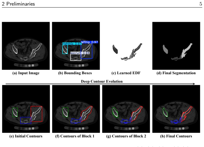
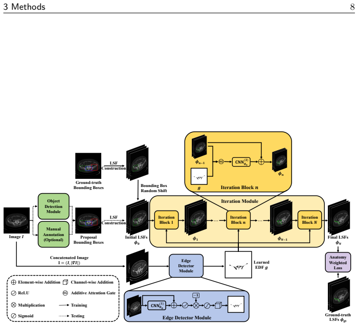
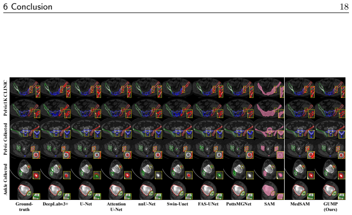
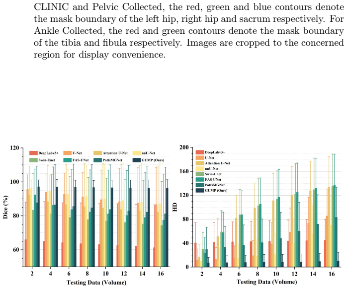
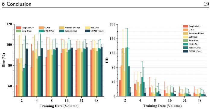
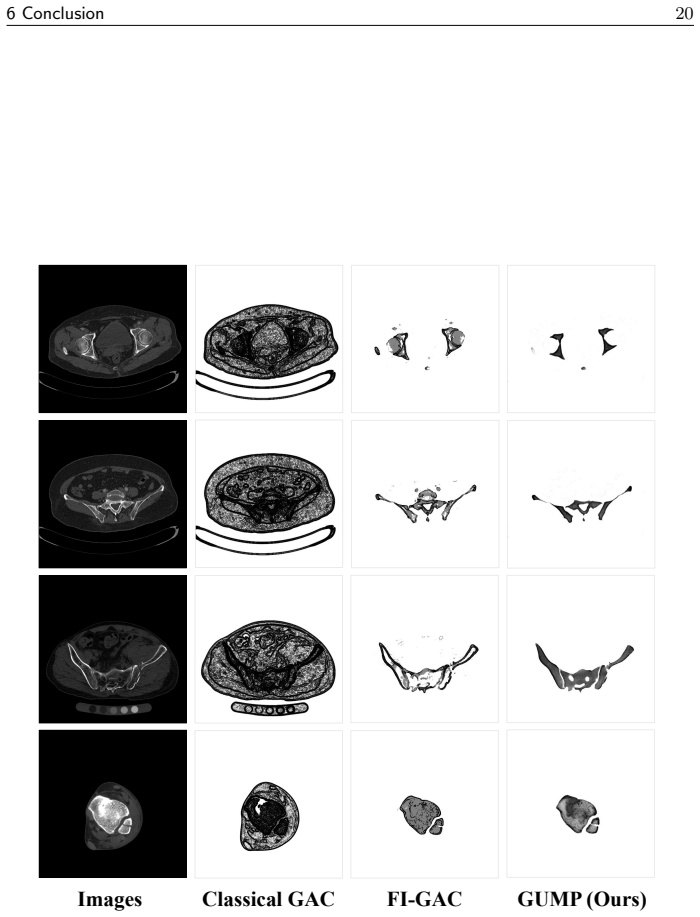
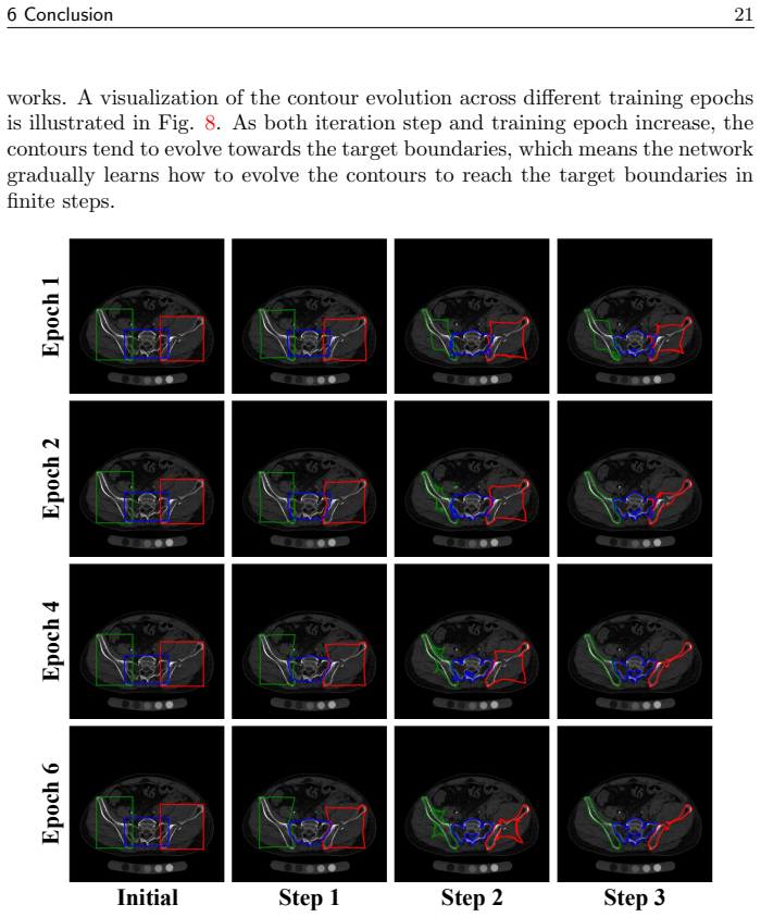
By integrating an improved geodesic active contour model with an object detection module for level set initialization, an edge detector module for anatomy-aware functions, and an iteration module for deep level set evolution, GUMP-Net achieves more accurate, robust, and consistent multi-class pelvic segmentation than state-of-the-art methods, particularly when training data is limited. Extensive experiments on pelvic datasets demonstrate the rationality and effectiveness of the proposed algorithm, with further experiments on ankle data indicating broader applications to other anatomies.
What carries the argument
The object detection module for level set initialization, edge detector module for anatomy-aware edge function, and iteration module for deep level set evolution, all integrated with the improved geodesic active contour model.
Load-bearing premise
The assumption that the three designed network modules can be effectively integrated with an improved geodesic active contour model to deliver the claimed performance gains without instability or loss of accuracy.
What would settle it
If experiments on an independent pelvic imaging dataset with small training samples show that GUMP-Net does not achieve higher accuracy metrics such as Dice scores than existing methods across bone classes, the central performance claim would be falsified.
Figures
read the original abstract
Pelvic segmentation is one of the most important and fundamental research problems in precise and intelligent diagnosis and treatment, as well as surgical planning and navigation for pelvic fractures. By combining an improved geodesic active contour model with deep neural networks, we propose GUMP-Net, an interpretable model-data-driven intelligent algorithm for multi-class pelvic segmentation, in which three network modules are designed to constitute the overall segmentation framework together: the object detection module for automatic level set initialization, the edge detector module for learning an anatomy-aware edge detector function and the iteration module for deep level set evolution. Leveraging the advantages of level set representation and deep learning, GUMP-Net shows more accurate, robust and consistent segmentation performance, especially in small training data situation, compared to the state-of-the-art methods. Extensive experiments on pelvic datasets demonstrate the rationality and effectiveness of the proposed algorithm. Further experiments extended to ankle dataset indicate broader applications to other anatomies. The proposed algorithm not only provides an efficient segmentation method for complex fracture reduction, but also gives an interpretable geometric perspective for understanding deep learning segmentation.
Editorial analysis
A structured set of objections, weighed in public.
Referee Report
Summary. The manuscript proposes GUMP-Net, an interpretable hybrid algorithm for multi-class pelvic segmentation that integrates an improved geodesic active contour model with three deep neural network modules: object detection for level set initialization, edge detector for anatomy-aware function, and iteration module for deep level set evolution. It claims superior accuracy, robustness, and consistency over state-of-the-art methods, particularly in small training data regimes, supported by experiments on pelvic and ankle datasets, and highlights the geometric interpretability of the approach.
Significance. If the performance claims are validated, this work contributes a model-data-driven framework that combines the strengths of traditional level set methods with deep learning for improved segmentation in medical imaging, especially under data scarcity. The interpretability aspect and extension to other anatomies like ankle could advance understanding and application of hybrid methods in computer vision for healthcare.
minor comments (2)
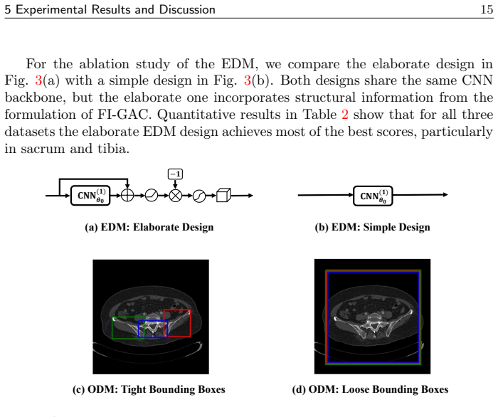
- [Experiments] Table reporting the small-data regime results should explicitly state the number of training samples used in each ablation to allow direct comparison with the SOTA baselines.
- [Method] The loss function formulation in the iteration module section would benefit from an explicit equation numbering and a short derivation sketch showing how the edge-detector output enters the level-set speed term.
Simulated Author's Rebuttal
We thank the referee for the positive assessment of GUMP-Net and the recommendation for minor revision. The referee summary accurately captures the hybrid model-data-driven framework, its interpretability, and the experimental results on pelvic and ankle datasets.
Circularity Check
No significant circularity identified
full rationale
The paper presents GUMP-Net as a novel integration of an improved geodesic active contour model with three deep network modules (object detection for level-set initialization, edge detector for anatomy-aware function, and iteration module for evolution). No equations, parameter-fitting procedures, or derivation steps are described that reduce any claimed prediction or result to the inputs by construction. Performance claims rest on experimental metrics across pelvic and ankle datasets rather than internal redefinitions or self-citation chains. The method is offered as an architectural combination, not a tautological renaming or self-referential fit.
Axiom & Free-Parameter Ledger
Reference graph
Works this paper leans on
-
[1]
H. Cao, Y. Wang, J. Chen, D. Jiang, X. Zhang, Q. Tian, and M. Wang. Swin-Unet: Unet-like Pure Transformer for Medical Image Segmentation. InECCV, pages 205–218, 2021
2021
-
[2]
Caselles, R
V. Caselles, R. Kimmel, and G. Sapiro. Geodesic Active Contours. InProc. IEEE/CVF Int. Conf. Comput. Vis. (ICCV), pages 694–699, 1995
1995
-
[3]
T. F. Chan and L. A. Vese. Active Contours without Edges.IEEE Trans. Image Process., 10(2):266–277, 2001
2001
-
[4]
C. Chen, J. Leng, and G. Xu. A General Framework of Piecewise- polynomial Mumford–Shah Model for Image Segmentation.Int. J. Comput. Math., 94:1981–1997, 2017
1981
-
[5]
L.-C. Chen, Y. Zhu, G. Papandreou, F. Schroff, and H. Adam. Encoder- Decoder with Atrous Separable Convolution for Semantic Image Segmen- tation. InECCV, pages 833–851, 2018. 6 Conclusion23
2018
-
[6]
H. Gu, R. Colglazier, H. Dong, J. Zhang, Y. Chen, Z. Yildiz, et al. Seg- mentAnyBone: A universal model that segments any bone at any location on MRI.Med. Image Anal., 101:103469, 2025
2025
-
[7]
L. Gui, J. Ma, and X. Yang.Variational Models and Their Combinations with Deep Learning in Medical Image Segmentation: A Survey, pages 1001–
-
[8]
Springer International Publishing, Cham, 2023
2023
-
[9]
Han and Y
B. Han and Y. Wu. Active contour model for inhomogenous image segmen- tation based on Jeffreys divergence.Pattern Recogn., 107:107520, 2020
2020
-
[10]
R. Han, A. Uneri, R. C. Vijayan, P. Wu, P. Vagdargi, N. Sheth, S. Vogt, G. Kleinszig, G. M. Osgood, and J. H. Siewerdsen. Fracture reduction planning and guidance in orthopaedic trauma surgery via multi-body image registration.Med. Image Anal., 68:101917, 2021
2021
-
[11]
X. Han, J. Wang, S. Ying, J. Shi, and D. Shen. ML-DSVM+: A meta- learning based deep SVM+ for computer-aided diagnosis.Pattern Recogn., 134:109076, 2023
2023
-
[12]
Hatamizadeh, A
A. Hatamizadeh, A. Hoogi, D. Sengupta, W. Lu, B. Wilcox, D. Rubin, and D. Terzopoulos. Deep active lesion segmentation. InMachine Learn- ing in Medical Imaging, pages 98–105, Cham, 2019. Springer International Publishing
2019
-
[13]
Hatamizadeh, D
A. Hatamizadeh, D. Sengupta, and D. Terzopoulos. End-to-End Trainable Deep Active Contour Models for Automated Image Segmentation: Delin- eating Buildings in Aerial Imagery. InECCV, pages 730–746, 2020
2020
-
[14]
Hoang Ngan Le, K
T. Hoang Ngan Le, K. Luu, C. N. Duong, K. G. Quach, T. D. Truong, K. Sadler, and M. Savvides.Active Contour Model in Deep Learning Era: A Revise and Review, pages 231–260. Springer International Publishing, Cham, 2020
2020
-
[15]
Isensee, P
F. Isensee, P. F. Jaeger, S. A. A. Kohl, J. Petersen, and K. H. Maier-Hein. nnU-Net: A Self-Configuring Method for Deep Learning-Based Biomedical Image Segmentation.Nat. Methods, 18(2):203–211, 2021
2021
-
[16]
Jia and Y
Y. Jia and Y. Jiang. Active Contour Model with Shape Constraints for Bone Fracture Detection. InInt. Conf. Comput. Graphics, Imaging and Visual., pages 90–95, 2006
2006
-
[17]
Z. Jin, H. Wang, M. Ng, and L. Min. Regularized CNN with Geodesic Active Contour and Edge Predictor for Image Segmentation.SIAM J. Imaging Sci., 17(4):2392–2417, 2024
2024
-
[18]
Kim and J
B. Kim and J. C. Ye. Mumford–Shah Loss Functional for Image Segmen- tation With Deep Learning.IEEE Trans. Image Process., 29:1856–1866, 2020. 6 Conclusion24
2020
-
[19]
Kirillov, E
A. Kirillov, E. Mintun, N. Ravi, H. Mao, C. Rolland, L. Gustafson, T. Xiao, S. Whitehead, A. C. Berg, W.-Y. Lo, P. Doll´ ar, and R. B. Girshick. Segment Anything.Proc. IEEE/CVF Int. Conf. Comput. Vis. (ICCV), pages 3992– 4003, 2023
2023
-
[20]
Li, C.-Y
C. Li, C.-Y. Kao, J. C. Gore, and Z. Ding. Minimization of Region-Scalable Fitting Energy for Image Segmentation.IEEE Trans. Image Process., 17(10):1940–1949, 2008
1940
-
[21]
X. Li, H. Chen, X. Qi, Q. Dou, C.-W. Fu, and P.-A. Heng. H-DenseUNet: Hybrid Densely Connected UNet for Liver and Tumor Segmentation From CT Volumes.IEEE Trans. Med. Imag., 37(12):2663–2674, 2018
2018
-
[22]
J. Liu, X. Wang, and X.-C. Tai. Deep Convolutional Neural Networks with Spatial Regularization, Volume and Star-Shape Priors for Image Segmen- tation.J. Math. Imaging Vis., 64(6):625–645, 2022
2022
-
[23]
P. Liu, H. Han, Y. Du, H. Zhu, Y. Li, F. Gu, H. Xiao, J. Li, C. Zhao, L. Xiao, Wu X., and S. Zhou. Deep learning to segment pelvic bones: large-scale CT datasets and baseline models.Int. J. Comput. Assist. Radiol. Surg., 16:749–756, 2020
2020
-
[24]
Y. Liu, S. Yibulayimu, Y. Sang, G. Zhu, Y. Wang, C. Zhao, and X. Wu. Pelvic Fracture Segmentation Using a Multi-scale Distance-Weighted Neu- ral Network. InProc. Int. Conf. Med. Image Comput. Comput.-Assist. Intervent (MICCAI), pages 312–321, 2023
2023
-
[25]
Z. Liu, Q. Li, J. Wang, T. Deng, R. Zhou, Y. Cai, and F. Liu. A variable gaussian kernel scale active contour model based on Jeffreys divergence for ICT image segmentation.Pattern Recogn., 172:112384, 2026
2026
-
[26]
Z. Liu, Y. Lin, Y. Cao, H. Hu, et al. Swin Transformer: Hierarchical Vision Transformer using Shifted Windows.Proc. IEEE/CVF Int. Conf. Comput. Vis. (ICCV), pages 9992–10002, 2021
2021
-
[27]
J. Ma, J. He, and X. Yang. Learning Geodesic Active Contours for Em- bedding Object Global Information in Segmentation CNNs.IEEE Trans. Med. Imag., 40(1):93–104, 2021
2021
-
[28]
J. Ma, Y. He, F. Li, L.-J. Han, C. You, and B. Wang. Segment Anything in Medical Images.Nat. Commun., 15, 2023
2023
-
[29]
H. Min, L. Xia, J. Han, X. Wang, et al. A multi-scale level set method based on local features for segmentation of images with intensity inhomogeneity. Pattern Recogn., 91:69–85, 2019
2019
-
[30]
Monga, Y
V. Monga, Y. Li, and Y. C. Eldar. Algorithm Unrolling: Interpretable, Efficient Deep Learning for Signal and Image Processing.IEEE Signal Process. Mag., 38(2):18–44, 2021. 6 Conclusion25
2021
-
[31]
O. Oktay, J. Schlemper, L. Folgoc, M. Lee, M. Heinrich, K. Misawa, K. Mori, S. McDonagh, et al. Attention U-Net: Learning Where to Look for the Pancreas.ArXiv, abs/1804.03999, 2018
Pith/arXiv arXiv 2018
-
[32]
S. Peng, W. Jiang, H. Pi, H. Bao, and X. Zhou. Deep Snake for Real- Time Instance Segmentation.Proc. IEEE/CVF Conf. Comput. Vis. Pat- tern Recognit. (CVPR), pages 8530–8539, 2020
2020
-
[33]
Redmon, S
J. Redmon, S. Divvala, R. Girshick, and A. Farhadi. You Only Look Once: Unified, Real-Time Object Detection. InProc. IEEE/CVF Conf. Comput. Vis. Pattern Recognit. (CVPR), pages 779–788, 2016
2016
-
[34]
Ronneberger, P
O. Ronneberger, P. Fischer, and T. Brox. U-Net: Convolutional Networks for Biomedical Image Segmentation. InProc. Int. Conf. Med. Image Com- put. Comput.-Assist. Intervent (MICCAI), pages 234–241, 2015
2015
-
[35]
X.-C. Tai, H. Liu, and R. Chan. PottsMGNet: A Mathematical Explana- tion of Encoder-Decoder Based Neural Networks.SIAM J. Imaging Sci., 17(1):540–594, 2024
2024
-
[36]
L. Tian, L. Zou, and X. Yang. A two-stage data-model driven pancreas segmentation strategy embedding directional information of the boundary intensity gradient and deep adaptive pointwise parameters.Phys. Med. Biol., 68(14):145005, 2023
2023
-
[37]
Tikhomirov, C
L. Tikhomirov, C. Semmler, M. McCradden, R. Searston, M. Ghassemi, and L. Oakden-Rayner. Medical Artificial Intelligence for Clinicians: The Lost Cognitive Perspective.The Lancet Digit. Health, 6(8):589–594, 2024
2024
-
[38]
M. D. Toruner, Y. Wang, Z. Jiao, and H. Bai. Artificial Intelligence in Radiology: Where Are We Going?eBioMedicine, 109, 2024
2024
-
[39]
Vaswani, N
A. Vaswani, N. M. Shazeer, N. Parmar, J. Uszkoreit, L. Jones, A. N. Gomez, L. Kaiser, and I. Polosukhin. Attention is All you Need. InNeurIPS, pages 6000–6010, 2017
2017
-
[40]
L. Wang, L. Zhang, H. Xu, J. Zhao, X. Su, J. Li, M. Tang, W. Gao, and C. Chen. Fracture interactive geodesic active contours for bone segmenta- tion.Pattern Recogn., 175:113049, 2026
2026
-
[41]
L. Yang, D. Shao, Z. Huang, M. Geng, N. Zhang, L. Chen, X. Wang, D. Liang, Z. F. Pang, and Z. Hu. Few-Shot Segmentation Framework for Lung Nodules via an Optimized Active Contour Model.Med. Phys., 51(4):2788–2805, 2024
2024
-
[42]
R. Yang, L. Song, Y. Ge, and X. Li. BoxSnake: Polygonal Instance Seg- mentation with Box Supervision.Proc. IEEE/CVF Int. Conf. Comput. Vis. (ICCV), pages 766–776, 2023. 6 Conclusion26
2023
-
[43]
Zhang, M
D. Zhang, M. Liu, T. Chen, H. Li, J. Ying, D. Chen, B. Li, Q. Yi, and J. Zhang. MTD-Net: A robust multi-task discriminative network for choroidal neovascularization segmentation.Pattern Recogn., 172:112697, 2026
2026
-
[44]
Zhang, B
M. Zhang, B. Dong, and Q. Li. Deep Active Contour Network for Medical Image Segmentation. InProc. Int. Conf. Med. Image Comput. Comput.- Assist. Intervent (MICCAI), pages 321–331, 2020
2020
-
[45]
Z. Zhou, M. Siddiquee, N. Tajbakhsh, and J. Liang. UNet++: Redesigning Skip Connections to Exploit Multiscale Features in Image Segmentation. IEEE Trans. Med. Imag., 39(6):1856–1867, 2020
2020
-
[46]
H. Zhu, S. Shu, and J. Zhang. FAS-UNet: A Novel FAS-Driven UNet to Learn Variational Image Segmentation.Mathematics, 10(21), 2022
2022
discussion (0)
Sign in with ORCID, Apple, or X to comment. Anyone can read and Pith papers without signing in.