BAC-JEPA: Label-Efficient Breast Arterial Calcification Segmentation via Synthetic Mammography-Guided Supervision
Pith reviewed 2026-06-26 12:30 UTC · model grok-4.3
The pith
Synthetic mammograms with inserted calcifications train a segmentation model that transfers to real images for image-level BAC detection without pixel-level human labels.
A machine-rendered reading of the paper's core claim, the machinery that carries it, and where it could break.
Core claim
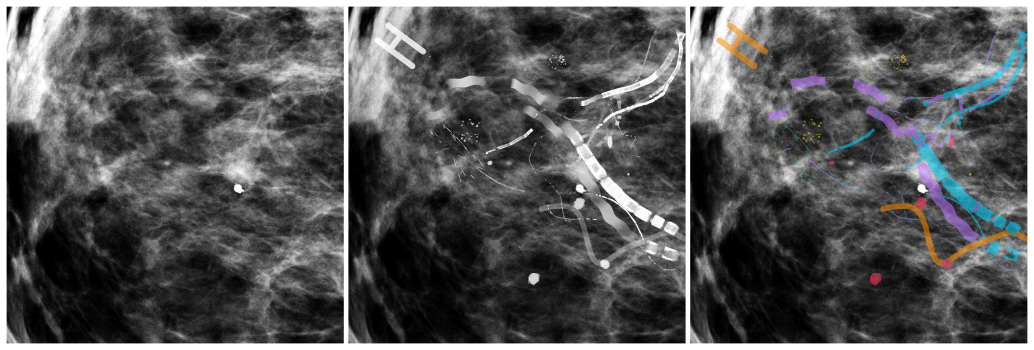
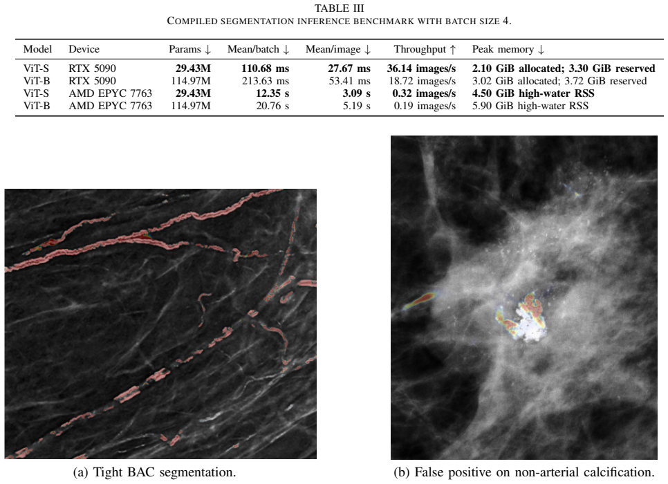
BAC-JEPA generates training data by selecting low-BAC real mammographic backgrounds and procedurally sampling arterial structure, disease burden, appearance parameters, and distractors to insert calcifications with perfect masks. These synthetic images train mammography self-supervised Vision Transformer encoders plus a convolutional decoder that outputs full-resolution segmentation maps. On synthetic validation the larger backbone reaches IoU 0.5325 and Dice 0.6357; on the BacSeg real dataset the segmentation-derived image-level scores reach AUROC 0.8719, showing that BAC-specific synthetic supervision produces useful transfer without any human pixel-level training masks.
What carries the argument
The procedural generator that samples arterial structure, disease burden, radiographic appearance, and hard-negative distractors to produce paired synthetic images and exact masks for supervision of the self-supervised ViT encoder and decoder.
If this is right
- Image-level BAC classification becomes feasible from segmentation maps without any pixel-level human labels during training.
- Four-view inference runs in 110-214 ms on current GPUs, supporting potential screening workflows.
- Synthetic generation at 2.7 seconds per severe-preset image allows scaling training data volume.
- Expert-reviewed real-mammogram segmentation remains required for clinical validation and calibration.
Where Pith is reading between the lines
- The same synthetic-insertion strategy could be tested on other sparse or costly-to-label calcifications such as coronary or aortic types if analogous procedural models are developed.
- Performance on real data may improve by increasing the diversity of background selection beyond the initial model-screened pool.
- The gap between synthetic IoU and real-image transfer suggests that hybrid fine-tuning on a small number of real masks could be a practical next calibration step.
Load-bearing premise
Backgrounds screened as low-BAC are free of actual calcifications and the synthetic radiographic appearances transfer to real human anatomy.
What would settle it
Apply the trained model to a new set of several thousand expert pixel-annotated real mammograms and measure whether pixel-level overlap or image-level AUROC falls below the levels reported on the 1,000 BacSeg cases.
Figures
read the original abstract
Breast arterial calcification (BAC) on screening mammograms is an emerging cardiovascular risk biomarker, but quantitative use requires reproducible segmentation and expert pixel-level labels are costly. We present BAC-JEPA, a label-efficient segmentation framework trained on procedurally generated arterial calcification inserted into real mammographic backgrounds with exact masks. Candidate backgrounds were selected from model-screened mammograms with low predicted BAC response; the generator samples arterial structure, disease burden, radiographic appearance, and hard-negative distractors including nonarterial calcifications and metallic objects. Synthetic masks are paired with mammography self-supervised Vision Transformer encoders and a high-resolution convolutional decoder to produce full-resolution segmentation maps. The study used 75,472 mammography studies from 34,956 patients for background selection and representation learning, trained on synthetic images from 10,000 backgrounds, selected checkpoints with 1,000 development backgrounds, and evaluated transfer on all 1,000 human-labeled BacSeg synthetic 2D mammograms. On held-out synthetic validation data, the larger backbone achieved IoU 0.5325 and Dice 0.6357. On BacSeg, image-level classification from segmentation probability maps reached AUROC 0.8719, with 0.8547 for the smaller backbone. Four-view inference required 110.68--213.63 ms on an RTX 5090 GPU, and severe-preset synthetic image generation averaged 2.7071 s per image on a multicore workstation. These results indicate that BAC-specific synthetic supervision can produce useful image-level transfer without human pixel-level training masks, while expert-reviewed real-mammogram segmentation remains necessary for clinical validation and calibration.
Editorial analysis
A structured set of objections, weighed in public.
Referee Report
Summary. The manuscript introduces BAC-JEPA, a label-efficient segmentation framework for breast arterial calcifications (BAC) trained exclusively on procedurally generated synthetic mammograms. Synthetic data are created by inserting generated arterial calcifications (with exact masks) into real mammographic backgrounds selected from a large corpus via model screening for low predicted BAC response; the generator also samples distractors. The architecture pairs mammography self-supervised Vision Transformer encoders with a high-resolution convolutional decoder. Reported results include IoU 0.5325 / Dice 0.6357 on held-out synthetic validation and AUROC 0.8719 (image-level classification from segmentation maps) on the real BacSeg dataset, with the claim that this yields useful synthetic-to-real transfer without human pixel-level training masks.
Significance. If the transfer claim holds, the work demonstrates a scalable route to training segmentation models for a clinically relevant biomarker when expert pixel annotations are scarce. Strengths include the scale of the representation-learning corpus (75,472 studies), the use of exact synthetic masks, concrete metrics on both synthetic and real held-out data, and reported inference/generation timings. These elements support the potential for reducing annotation burden while still requiring expert review for final clinical calibration.
major comments (1)
- [Abstract] Abstract: The central claim that synthetic supervision produces useful transfer (AUROC 0.8719 on BacSeg) rests on the assumption that model-screened low-response backgrounds contain negligible real calcifications. The manuscript reports only that candidates were 'model-screened' with 'low predicted BAC response' and provides no false-negative rate for the screening model on subtle BAC, no inter-rater agreement statistics, and no post-selection expert audit of the 10,000 training backgrounds. Undetected real BAC would introduce label noise into the 'exact' synthetic masks, directly threatening the validity of the learned features and the reported real-data performance.
minor comments (2)
- [Abstract] Abstract: No error bars, confidence intervals, or details on validation splits/runs are provided for the reported IoU 0.5325, Dice 0.6357, or AUROC 0.8719, limiting assessment of metric stability.
- [Abstract] Abstract: The description of 'severe-preset synthetic image generation' and 'procedural generator sampling parameters' lacks concrete specification of the sampling distributions or preset definitions used for arterial structure, disease burden, and distractors.
Simulated Author's Rebuttal
We thank the referee for the careful review and for identifying a key assumption in our background selection process. We respond to the major comment below.
read point-by-point responses
-
Referee: [Abstract] Abstract: The central claim that synthetic supervision produces useful transfer (AUROC 0.8719 on BacSeg) rests on the assumption that model-screened low-response backgrounds contain negligible real calcifications. The manuscript reports only that candidates were 'model-screened' with 'low predicted BAC response' and provides no false-negative rate for the screening model on subtle BAC, no inter-rater agreement statistics, and no post-selection expert audit of the 10,000 training backgrounds. Undetected real BAC would introduce label noise into the 'exact' synthetic masks, directly threatening the validity of the learned features and the reported real-data performance.
Authors: We agree that undetected real BAC in the selected backgrounds would constitute label noise and weaken the claim of exact synthetic masks. The screening step applied a model to a corpus of 75,472 studies to retain only low-response images before procedural insertion of synthetic calcifications. However, we did not compute a false-negative rate on subtle BAC, collect inter-rater statistics, or perform a post-selection expert audit on the final 10,000 backgrounds. The reported AUROC of 0.8719 on the independently labeled BacSeg set provides indirect support that any residual noise did not prevent useful transfer, yet this does not substitute for direct validation of the screening assumption. We will revise the manuscript to state this limitation explicitly in the methods and discussion sections. revision: partial
- false-negative rate for the screening model on subtle BAC
- inter-rater agreement statistics for the background screening
- post-selection expert audit results for the 10,000 training backgrounds
Circularity Check
No significant circularity; performance metrics derive from direct evaluation on held-out real data
full rationale
The paper generates synthetic training data via procedural insertion of calcifications into model-screened backgrounds and evaluates segmentation transfer via AUROC and IoU on a separate human-labeled BacSeg set of 1,000 real mammograms. No equations, predictions, or central claims reduce reported metrics to fitted parameters, self-defined quantities, or self-citation chains within the paper; the synthetic supervision and real-data evaluation remain independent inputs and outputs.
Axiom & Free-Parameter Ledger
free parameters (1)
- procedural generator sampling parameters for arterial structure, disease burden, and distractors
axioms (2)
- domain assumption Self-supervised Vision Transformer encoders pretrained on mammography learn representations transferable to pixel-level BAC segmentation when paired with synthetic masks.
- ad hoc to paper Model-screened low predicted BAC backgrounds contain negligible real calcifications suitable for clean synthetic insertion.
Reference graph
Works this paper leans on
-
[1]
Artificial intelligence-based quantification of breast arterial calcifications to predict cardiovascular morbidity and mortality,
T. Dapamede, A. Urooj, V . Joshi, G. Gershon, F. Li, M. Chavoshi, B. Brown-Mulry, R. S. Isaac, A. Mansuri, C. Robichaux, C. Ayoub, R. Arsanjani, L. Sperling, J. Gichoya, M. van Assen, W. C. O’Neill, I. Banerjee, and H. Trivedi, “Artificial intelligence-based quantification of breast arterial calcifications to predict cardiovascular morbidity and mortality...
2026
-
[2]
Canadian Society of Breast Imaging Position Statement on Breast Arterial Calcification Reporting on Mammography,
Canadian Society of Breast Imaging, “Canadian Society of Breast Imaging Position Statement on Breast Arterial Calcification Reporting on Mammography,” Jan. 2023, accessed: 2026-05-25. [Online]. Available: https://csbi.ca/canadian-society-of-breast-imaging-position- statement-on-breast-arterial-calcification-reporting-on-mammography/
2023
-
[3]
Breast arterial cal- cification: A novel cardiovascular risk enhancer among postmenopausal women,
C. Iribarren, M. Chandra, C. Lee, G. Sanchez, D. L. Sam, F. F. Azamian, H.-M. Cho, H. Ding, N. D. Wong, and S. Molloi, “Breast arterial cal- cification: A novel cardiovascular risk enhancer among postmenopausal women,”Circulation: Cardiovascular Imaging, vol. 15, no. 3, Mar. 2022
2022
-
[4]
Automated breast arterial calcification score is associated with cardiovascular outcomes and mortality,
T. S. Allen, Q. M. Bui, G. M. Petersen, R. Mantey, J. Wang, N. Nerlekar, M. Eghtedari, and L. B. Daniels, “Automated breast arterial calcification score is associated with cardiovascular outcomes and mortality,”JACC: Advances, vol. 3, no. 11, p. 101283, Nov. 2024
2024
-
[5]
SCU-Net: A deep learning method for segmentation and quantification of breast arterial calcifications on mammograms,
X. Guo, W. C. O’Neill, B. Vey, T. C. Yang, T. J. Kim, M. Ghassemi, I. Pan, J. W. Gichoya, H. Trivedi, and I. Banerjee, “SCU-Net: A deep learning method for segmentation and quantification of breast arterial calcifications on mammograms,”Medical Physics, vol. 48, no. 10, pp. 5851–5861, Aug. 2021
2021
-
[6]
Improving segmentation of breast arterial calcifications from digital mammography: Good annotation is all you need,
K. Wang, M. Hill, S. Knowles-Barley, A. Tikhonov, L. Litchfield, and J. C. Bare, “Improving segmentation of breast arterial calcifications from digital mammography: Good annotation is all you need,” inProceedings of the Asian Conference on Computer Vision Workshops, Dec. 2022, pp. 130–146
2022
-
[7]
Evaluation of digital breast tomosynthesis as replacement of full-field digital mammography using an in silico imaging trial,
A. Badano, C. G. Graff, A. Badal, D. Sharma, R. Zeng, F. W. Samuelson, S. J. Glick, and K. J. Myers, “Evaluation of digital breast tomosynthesis as replacement of full-field digital mammography using an in silico imaging trial,”JAMA Network Open, vol. 1, no. 7, p. e185474, Nov. 2018
2018
-
[8]
Technical note: In silico imaging tools from the VICTRE clinical trial,
D. Sharma, C. G. Graff, A. Badal, R. Zeng, P. Sawant, A. Sengupta, E. Dahal, and A. Badano, “Technical note: In silico imaging tools from the VICTRE clinical trial,”Medical Physics, vol. 46, no. 9, pp. 3924– 3928, Jul. 2019
2019
-
[9]
Knowledge-based in silico models and dataset for the comparative evaluation of mammography AI for a range of breast characteristics, lesion conspicuities and doses,
E. Sizikova, N. Saharkhiz, D. Sharma, M. Lago, B. Sahiner, J. Delfino, and A. Badano, “Knowledge-based in silico models and dataset for the comparative evaluation of mammography AI for a range of breast characteristics, lesion conspicuities and doses,” inAdvances in Neural Information Processing Systems, vol. 36, 2023
2023
-
[10]
A generative adversarial network for syntheti- zation of regions of interest based on digital mammograms,
O. N. Oyelade, A. E. Ezugwu, M. S. Almutairi, A. K. Saha, L. Abuali- gah, and H. Chiroma, “A generative adversarial network for syntheti- zation of regions of interest based on digital mammograms,”Scientific Reports, vol. 12, no. 1, Apr. 2022
2022
-
[11]
MAM-E: Mammographic synthetic image generation with diffusion models,
R. Montoya-del Angel, K. Sam-Millan, J. C. Vilanova, and R. Marti, “MAM-E: Mammographic synthetic image generation with diffusion models,”Sensors, vol. 24, no. 7, p. 2076, Mar. 2024
2076
-
[12]
An image is worth 16x16 words: Trans- formers for image recognition at scale,
A. Dosovitskiy, L. Beyer, A. Kolesnikov, D. Weissenborn, X. Zhai, T. Unterthiner, M. Dehghani, M. Minderer, G. Heigold, S. Gelly, J. Uszkoreit, and N. Houlsby, “An image is worth 16x16 words: Trans- formers for image recognition at scale,” inInternational Conference on Learning Representations, 2021
2021
-
[13]
Self-supervised learning from images with a joint-embedding predictive architecture,
M. Assran, Q. Duval, I. Misra, P. Bojanowski, P. Vincent, M. Rab- bat, Y . LeCun, and N. Ballas, “Self-supervised learning from images with a joint-embedding predictive architecture,” inProceedings of the IEEE/CVF Conference on Computer Vision and Pattern Recognition, 2023, pp. 15 619–15 629
2023
-
[14]
DU-Net: Convolutional network for the detection of arterial calcifications in mammograms,
M. AlGhamdi, M. Abdel-Mottaleb, and F. Collado-Mesa, “DU-Net: Convolutional network for the detection of arterial calcifications in mammograms,”IEEE Transactions on Medical Imaging, vol. 39, no. 10, pp. 3240–3249, Oct. 2020
2020
-
[15]
Difference-of-Gaussian generative adversarial network for segmenting breast arterial calcifications in mammograms,
M. Alamir, M. AlGhamdi, F. Collado-Mesa, and M. Abdel-Mottaleb, “Difference-of-Gaussian generative adversarial network for segmenting breast arterial calcifications in mammograms,”Expert Systems with Applications, vol. 217, p. 119506, May 2023
2023
-
[16]
Recurrent attention U-Net for segmentation and quantification of breast arterial calcifications on synthesized 2D mammograms,
M. AlJabri, M. Alghamdi, F. Collado-Mesa, and M. Abdel-Mottaleb, “Recurrent attention U-Net for segmentation and quantification of breast arterial calcifications on synthesized 2D mammograms,”PeerJ Computer Science, vol. 10, p. e2076, May 2024
2024
-
[17]
Quantification of breast arterial calcification in mammo- grams using a UNet-based deep learning for detecting cardiovascular disease,
W. Li, Q. Zhang, D. Black, H. Ding, C. Iribarren, A. Shojazadeh, and S. Molloi, “Quantification of breast arterial calcification in mammo- grams using a UNet-based deep learning for detecting cardiovascular disease,”Academic Radiology, vol. 32, no. 9, pp. 5028–5038, Sep. 2025
2025
-
[18]
MC-GenRef: Annotation-free mammography microcalcification segmentation with generative posterior refinement,
H. Cho, Y . Kwon, M. J. Kim, and Y . Yoo, “MC-GenRef: Annotation-free mammography microcalcification segmentation with generative posterior refinement,” 2026
2026
-
[19]
A curated mammography data set for use in computer-aided detection and diagnosis research,
R. S. Lee, F. Gimenez, A. Hoogi, K. K. Miyake, M. Gorovoy, and D. L. Rubin, “A curated mammography data set for use in computer-aided detection and diagnosis research,”Scientific Data, vol. 4, p. 170177, Dec. 2017
2017
-
[20]
OPTIMAM mammography image database: A large- scale resource of mammography images and clinical data,
M. D. Halling-Brown, L. M. Warren, D. Ward, E. Lewis, A. Mackenzie, M. G. Wallis, L. S. Wilkinson, R. M. Given-Wilson, R. McAvinchey, and K. C. Young, “OPTIMAM mammography image database: A large- scale resource of mammography images and clinical data,”Radiology: Artificial Intelligence, vol. 3, no. 1, p. e200103, Jan. 2021
2021
-
[21]
Simeoni, H
O. Simeoni, H. V . V o, M. Seitzer, F. Baldassarre, M. Oquab, C. Jose, V . Khalidov, M. Szafraniec, S. Yi, M. Ramamonjisoa, F. Massa, D. Haz- iza, L. Wehrstedt, J. Wang, T. Darcet, T. Moutakanni, L. Sentana, C. Roberts, A. Vedaldi, J. Tolan, J. Brandt, C. Couprie, J. Mairal, H. Jegou, P. Labatut, and P. Bojanowski, “DINOv3,” 2025
2025
-
[22]
A convnet for the 2020s,
Z. Liu, H. Mao, C.-Y . Wu, C. Feichtenhofer, T. Darrell, and S. Xie, “A convnet for the 2020s,” inProceedings of the IEEE/CVF Conference on Computer Vision and Pattern Recognition, 2022, pp. 11 976–11 986
2022
-
[23]
Focal loss for dense object detection,
T.-Y . Lin, P. Goyal, R. Girshick, K. He, and P. Dollar, “Focal loss for dense object detection,” inProceedings of the IEEE International Conference on Computer Vision, 2017, pp. 2980–2988
2017
-
[24]
V-Net: Fully convolutional neural networks for volumetric medical image segmentation,
F. Milletari, N. Navab, and S.-A. Ahmadi, “V-Net: Fully convolutional neural networks for volumetric medical image segmentation,” inFourth International Conference on 3D Vision, 2016, pp. 565–571
2016
discussion (0)
Sign in with ORCID, Apple, or X to comment. Anyone can read and Pith papers without signing in.