Intracranial Aneurysm Classification and Segmentation via Tri-Axial ROI and Multi-Task Learning
Pith reviewed 2026-06-26 05:42 UTC · model grok-4.3
The pith
Tri-axial ROI extraction with a dual-decoder multi-task network enables simultaneous location-specific classification and segmentation of intracranial aneurysms and vessels across modalities.
A machine-rendered reading of the paper's core claim, the machinery that carries it, and where it could break.
Core claim
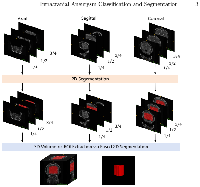
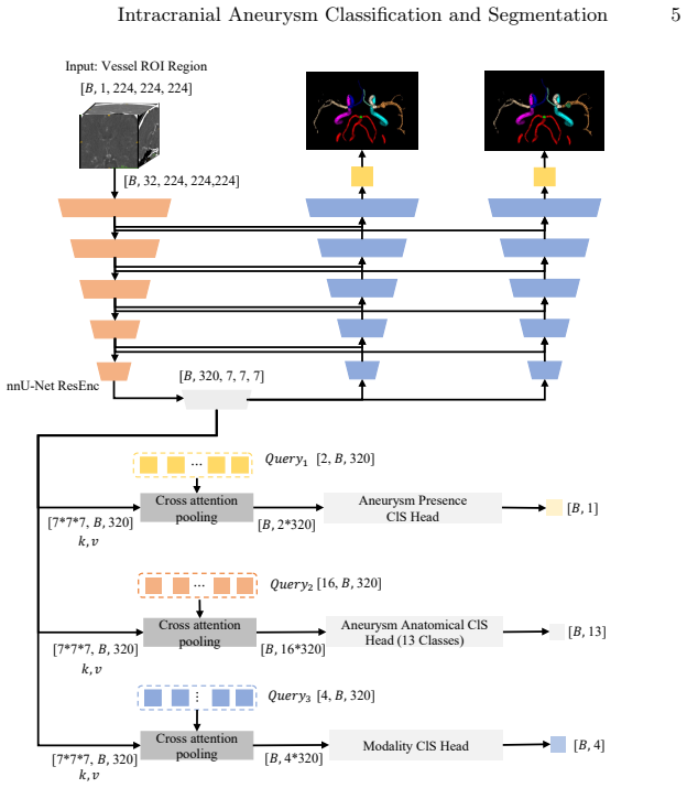
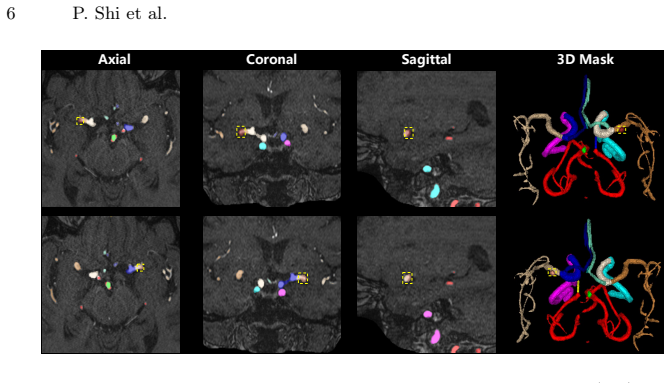
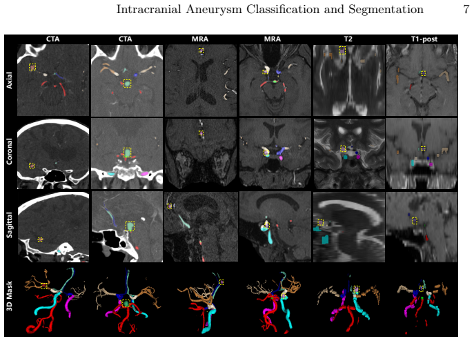
The framework simultaneously performs multi-label classification, multi-class aneurysm segmentation, and multi-class vessel segmentation across 13 anatomical locations and four imaging modalities by combining fast 2D tri-axial ROI extraction with a 3D multi-task nnU-Net backbone whose dual-decoder design, cross-attention pooling, and modality-specific auxiliary heads mitigate volume imbalance and improve feature learning from heterogeneous inputs.
What carries the argument
Tri-axial ROI extraction paired with a dual-decoder 3D multi-task network that uses cross-attention pooling to balance aneurysm and vessel segmentation.
If this is right
- The model outputs both location labels for 13 sites and separate masks for aneurysms and vessels in one forward pass.
- The dual-decoder structure reduces the effect of extreme class imbalance between small aneurysms and large vessels.
- Modality-specific auxiliary heads allow the same backbone to process CTA, MRA, T2, and T1-post scans without retraining.
- A two-fold ensemble of the trained models placed second in the RSNA 2025 Intracranial Aneurysm Detection challenge.
Where Pith is reading between the lines
- Public release of the code, weights, and 3D Slicer plugin lowers the barrier for clinical groups to test the pipeline on their own scans.
- Location-aware outputs could feed directly into existing rupture-risk calculators that already weight certain anatomical sites more heavily.
- The multi-modality design suggests a route to training on mixed or incomplete imaging datasets for other focal vascular lesions.
Load-bearing premise
The performance improvements result from the tri-axial ROI extraction and dual-decoder architecture rather than from the two-fold ensemble or tuning choices on the challenge test set.
What would settle it
An ablation experiment on an independent held-out dataset that removes the tri-axial ROI step or the dual-decoder while keeping the ensemble shows no measurable drop in classification or segmentation scores.
Figures
read the original abstract
Intracranial aneurysms are often asymptomatic until rupture, which carries high mortality. Rupture risk assessment and treatment planning depend on both aneurysm morphology and anatomical location, yet existing automated methods remain limited to binary detection without fine-grained anatomical classification or multi-class segmentation. We present a multi-task framework that simultaneously performs multi-label classification, multi-class aneurysm segmentation, and multi-class vessel segmentation across 13 anatomical locations and four imaging modalities (CTA, MRA, T2, T1-post). Our two-stage approach combines a fast 2D tri-axial Region of Interest (ROI) extraction method with a 3D multi-task nnU-Net backbone. A dual-decoder design mitigates the extreme volume imbalance between aneurysm and vessel classes, while cross-attention pooling and modality-specific auxiliary heads improve feature learning across heterogeneous inputs. Our two-fold ensemble achieved 2nd place in the RSNA 2025 Intracranial Aneurysm Detection challenge. Code, model weights, and a 3D Slicer plugin are publicly available.
Editorial analysis
A structured set of objections, weighed in public.
Referee Report
Summary. The manuscript presents a multi-task framework for intracranial aneurysm multi-label classification, multi-class aneurysm segmentation, and multi-class vessel segmentation across 13 anatomical locations and four modalities (CTA, MRA, T2, T1-post). The approach uses a two-stage pipeline consisting of fast 2D tri-axial ROI extraction followed by a 3D multi-task nnU-Net backbone with dual decoders to address class imbalance, cross-attention pooling, and modality-specific auxiliary heads. The central empirical claim is that a two-fold ensemble of this model achieved 2nd place in the RSNA 2025 Intracranial Aneurysm Detection challenge.
Significance. If the architectural modifications can be shown to drive the ranking beyond ensembling, the framework would advance automated aneurysm analysis by providing simultaneous classification and fine-grained segmentation that existing binary-detection methods lack. The public release of code, model weights, and a 3D Slicer plugin is a clear strength that supports reproducibility and downstream use.
major comments (1)
- [Abstract] Abstract: The 2nd-place ranking is reported for a two-fold ensemble, yet no ablation results, single-model baselines, or direct comparisons against plain nnU-Net on the same data splits are provided to establish that the tri-axial ROI extraction, dual-decoder design, cross-attention pooling, or modality-specific heads are the primary contributors rather than the ensemble procedure or challenge-specific tuning.
minor comments (1)
- [Abstract] Abstract: Dataset size, class distribution, exclusion criteria, and any error bars or statistical details around the challenge ranking are omitted, limiting assessment of result robustness.
Simulated Author's Rebuttal
We thank the referee for the detailed review and constructive feedback. We address the major comment below and commit to revisions that strengthen the empirical validation of our contributions.
read point-by-point responses
-
Referee: [Abstract] Abstract: The 2nd-place ranking is reported for a two-fold ensemble, yet no ablation results, single-model baselines, or direct comparisons against plain nnU-Net on the same data splits are provided to establish that the tri-axial ROI extraction, dual-decoder design, cross-attention pooling, or modality-specific heads are the primary contributors rather than the ensemble procedure or challenge-specific tuning.
Authors: We agree that the current manuscript does not include explicit ablation studies or single-model baselines on the challenge data splits, which limits the ability to isolate the contribution of each proposed component from the ensemble procedure. In the revised manuscript we will add (1) performance metrics for the single-model (non-ensembled) version, (2) a direct comparison against a plain nnU-Net baseline trained on the same splits, and (3) targeted ablations removing the tri-axial ROI extractor, dual-decoder, and modality-specific heads. These additions will be reported on the official validation set to clarify the source of the ranking improvement. revision: yes
Circularity Check
Empirical challenge result with no derivation chain or self-referential reductions
full rationale
The paper reports an empirical 2nd-place ranking on the external RSNA 2025 hidden test set using a two-fold ensemble of a multi-task nnU-Net variant. No equations, fitted parameters renamed as predictions, or load-bearing self-citations appear in the provided text. The central claim is a factual external ranking rather than a derived quantity that reduces to the method's own inputs by construction. The architecture descriptions (tri-axial ROI, dual-decoder, cross-attention) are presented as design choices whose value is assessed by challenge performance, not by internal redefinition or ansatz smuggling.
Axiom & Free-Parameter Ledger
free parameters (2)
- nnU-Net training hyperparameters
- tri-axial ROI extraction parameters
axioms (2)
- domain assumption nnU-Net provides a competitive baseline for volumetric medical image segmentation
- domain assumption Dual-decoder and cross-attention designs mitigate class imbalance and modality heterogeneity
Reference graph
Works this paper leans on
-
[1]
Patterns2(2), 100197 (2021) 2 Intracranial Aneurysm Classification and Segmentation 11
Bo, Z.H., Qiao, H., Tian, C., Guo, Y., Li, W., Liang, T., Li, D., Liao, D., Zeng, X., Mei, L., et al.: Toward human intervention-free clinical diagnosis of intracranial aneurysm via deep neural network. Patterns2(2), 100197 (2021) 2 Intracranial Aneurysm Classification and Segmentation 11
2021
-
[2]
Neu- roinformatics21(1), 21–34 (2023) 2
Di Noto, T., Marie, G., Tourbier, S., Aleman-Gomez, Y., Esteban, O., Saliou, G., Cuadra, M.B., Hagmann, P., Richiardi, J.: Towards automated brain aneurysm detection in TOF-MRA: open data, weak labels, and anatomical knowledge. Neu- roinformatics21(1), 21–34 (2023) 2
2023
-
[3]
Medical Image Analysis101, 103493 (2025) 1
Hsu, W.C., Meuschke, M., Frangi, A.F., Preim, B., Lawonn, K.: A survey of in- tracranial aneurysm detection and segmentation. Medical Image Analysis101, 103493 (2025) 1
2025
-
[4]
Nature methods18(2), 203–211 (2021) 2, 3, 5, 8
Isensee, F., Jaeger, P.F., Kohl, S.A., Petersen, J., Maier-Hein, K.H.: nnu-net: a self-configuring method for deep learning-based biomedical image segmentation. Nature methods18(2), 203–211 (2021) 2, 3, 5, 8
2021
-
[5]
In: International Conference on Medical Image Computing and Computer- Assisted Intervention
Isensee, F., Wald, T., Ulrich, C., Baumgartner, M., Roy, S., Maier-Hein, K., Jaeger, P.F.: nnu-net revisited: A call for rigorous validation in 3d medical image segmen- tation. In: International Conference on Medical Image Computing and Computer- Assisted Intervention. pp. 488–498. Springer (2024) 2
2024
-
[6]
Rudie, J., Calabrese, E., Ball, R., Chang, P., Chen, R., Colak, E., de Verdier, M.C., Prevedello, L., Richards, T., Saluja, R., Zaharchuk, G., Sho, J., Vazirabad,M.:RSNA2025intracranialaneurysmdetection.https://kaggle.com/ competitions/rsna-intracranial-aneurysm-detection(2025), kaggle 2, 8
2025
-
[7]
Nature communications11(1), 6090 (2020) 2
Shi, Z., Miao, C., Schoepf, U.J., Savage, R.H., Dargis, D.M., Pan, C., Chai, X., Li, X.L., Xia, S., Zhang, X., et al.: A clinically applicable deep-learning model for detecting intracranial aneurysm in computed tomography angiography images. Nature communications11(1), 6090 (2020) 2
2020
-
[8]
Neuroimage238, 118216 (2021) 1
Timmins, K.M., van der Schaaf, I.C., Bennink, E., Ruigrok, Y.M., An, X., Baum- gartner, M., Bourdon, P., De Feo, R., Di Noto, T., Dubost, F., et al.: Comparing methods of detecting and segmenting unruptured intracranial aneurysms on tof- mras: the adam challenge. Neuroimage238, 118216 (2021) 1
2021
-
[9]
Radiology290(1), 187–194 (2019) 2
Ueda, D., Yamamoto, A., Nishimori, M., Shimono, T., Doishita, S., Shimazaki, A., Katayama, Y., Fukumoto, S., Choppin, A., Shimahara, Y., et al.: Deep learning for mr angiography: automated detection of cerebral aneurysms. Radiology290(1), 187–194 (2019) 2
2019
-
[10]
Radiology: Artificial Intelligence 5(5), e230024 (2023) 7, 9
Wasserthal, J., Breit, H.C., Meyer, M.T., Pradella, M., Hinck, D., Sauter, A.W., Heye, T., Boll, D.T., Cyriac, J., Yang, S., et al.: Totalsegmentator: robust segmen- tation of 104 anatomic structures in ct images. Radiology: Artificial Intelligence 5(5), e230024 (2023) 7, 9
2023
-
[11]
Radiology312(2), e233197 (2024) 2
Wei, J., Song, X., Wei, X., Yang, Z., Dai, L., Wang, M., Sun, Z., Jin, Y., Ma, C., Hu, C., et al.: Knowledge-augmented deep learning for segmenting and detecting cerebral aneurysms with ct angiography: a multicenter study. Radiology312(2), e233197 (2024) 2
2024
-
[12]
ArXiv pp
Yang, K., Musio, F., Ma, Y., Juchler, N., Paetzold, J.C., Al-Maskari, R., Höher, L., Li, H.B., Hamamci, I.E., Sekuboyina, A., et al.: Benchmarking the cow with the topcow challenge: Topology-aware anatomical segmentation of the circle of willis for cta and mra. ArXiv pp. arXiv–2312 (2025) 4, 7
2025
-
[13]
medRxiv pp
Yang, K., Shi, P., Huang, H., Musio, F., Baazaoui, H., Aydin, O.U., Hilbert, A., Hamadache, R.E., Yalcin, C., Zhang, M., et al.: Topbrain segmentation challenge for whole brain vessel anatomy. medRxiv pp. 2026–05 (2026) 4, 7
2026
-
[14]
IEEE Transactions on Medical Imaging 44(3), 1273–1283 (2024) 2
Yao, L., Chen, D., Zhao, X., Fei, M., Song, Z., Xue, Z., Zhan, Y., Song, B., Shi, F., Wang, Q., et al.: Aaseg: Artery-aware global-to-local framework for aneurysm segmentation in head and neck cta images. IEEE Transactions on Medical Imaging 44(3), 1273–1283 (2024) 2
2024
discussion (0)
Sign in with ORCID, Apple, or X to comment. Anyone can read and Pith papers without signing in.