Self-Auditing Residual Drifting for Pathology-Preserving Accelerated Knee MRI
Pith reviewed 2026-07-03 03:35 UTC · model grok-4.3
The pith
SA-RDM-DC reconstructs accelerated knee MRI with highest SSIM while generating self-audit error maps and risk scores.
A machine-rendered reading of the paper's core claim, the machinery that carries it, and where it could break.
Core claim
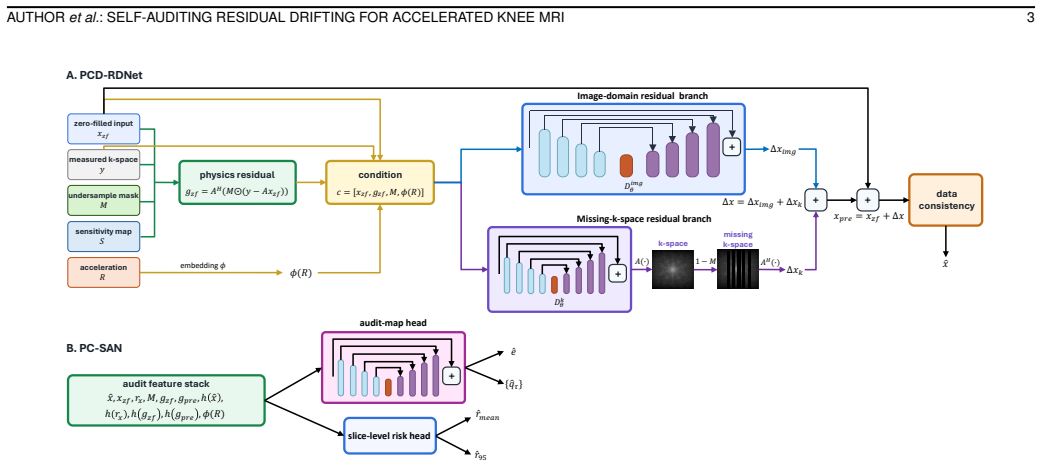
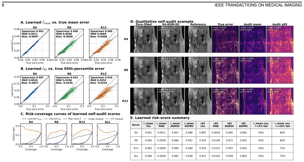
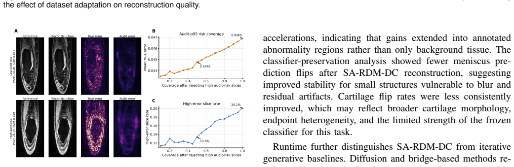
SA-RDM-DC adapts the generative drifting paradigm to accelerated MRI by training a physics-conditioned drift field from the zero-filled reconstruction toward the fully sampled residual correction. It predicts image- and missing-k-space residual corrections, enforces data consistency with acquired k-space, uses frequency-aware and residual drifting supervision to recover fine detail, and produces dense error maps and slice-level risk scores in the same inference pass.
What carries the argument
The physics-conditioned drift field trained from zero-filled reconstruction toward fully sampled residual correction, which predicts image- and k-space residual corrections while enforcing data consistency and producing self-audit scores.
If this is right
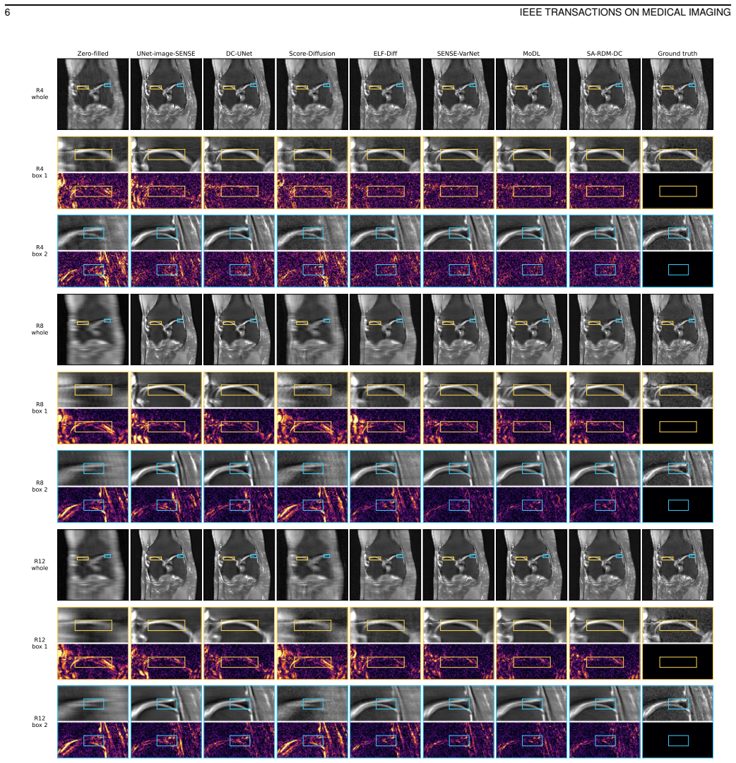
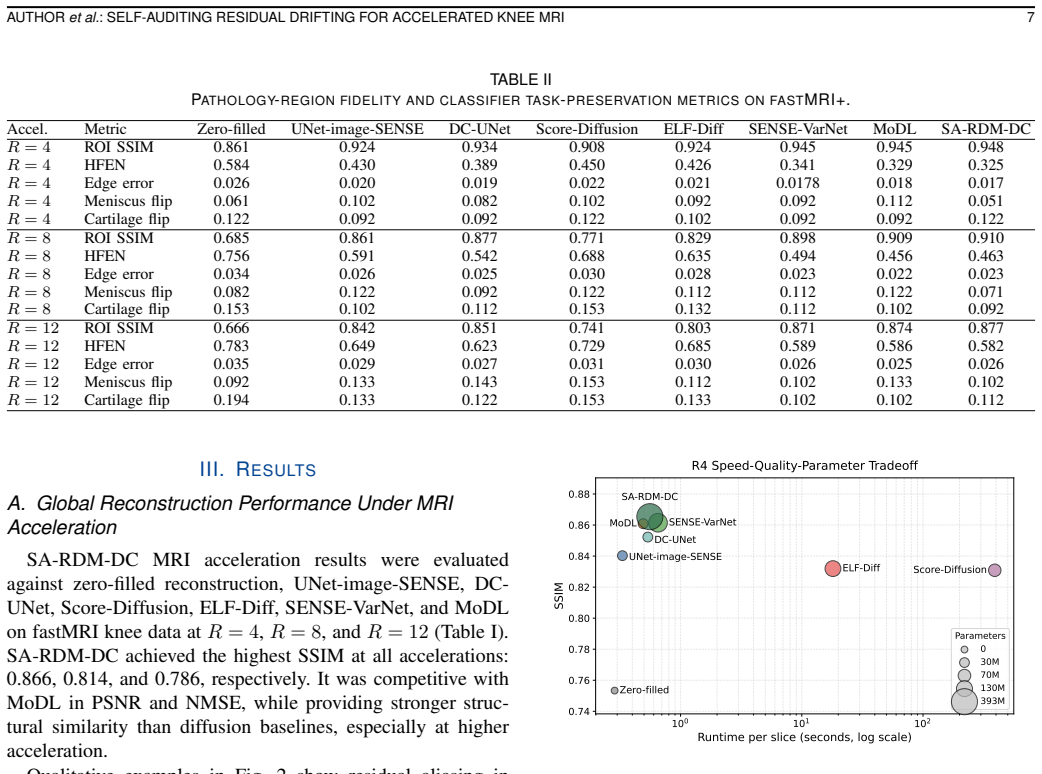
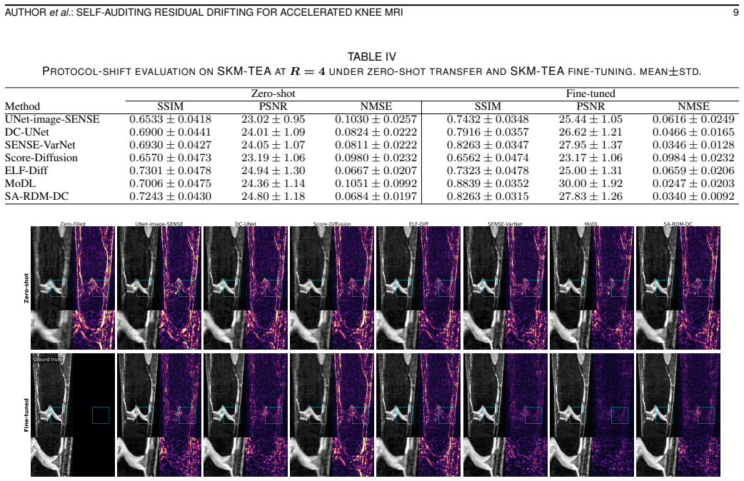
- SSIM exceeds UNet, diffusion, and variational network baselines at acceleration factors 4, 8, and 12.
- Lesion structural fidelity is retained and meniscus prediction instability is reduced compared with competing methods.
- Self-auditing scores strongly flag high-error reconstructions on fastMRI and partially transfer to SKM-TEA.
- Subsecond per-slice inference is retained without the sampling time cost of iterative diffusion approaches.
Where Pith is reading between the lines
- Risk scores could enable selective human review of cases likely to contain clinically relevant errors.
- Partial protocol transfer suggests the self-auditing signal may support deployment across varied scanner settings with limited retraining.
- The residual drifting approach might extend to other anatomies or modalities where both speed and case-specific reliability matter.
Load-bearing premise
A drift field trained on fastMRI knee data will generalize to preserve pathology-specific features and produce transferable risk scores on SKM-TEA without introducing artifacts missed by global metrics.
What would settle it
A demonstration that self-auditing scores fail to correlate with actual reconstruction errors on fastMRI or that lesion-region fidelity falls below baselines in pathology-annotated regions.
Figures
read the original abstract
Accelerated magnetic resonance imaging reduces acquisition time, but reconstruction from undersampled k-space can blur diagnostically relevant structures or introduce failures that are not captured by global image metrics. We propose SA-RDM-DC, a Self-Auditing Residual generative Drifting Model with Data Consistency for accelerated knee MRI. The method adapts the newly proposed generative drifting paradigm to accelerated MRI by training a physics-conditioned drift field from the zero-filled reconstruction toward the fully sampled residual correction. It predicts image- and missing-k-space residual corrections, enforces data consistency with acquired k-space, uses frequency-aware and residual drifting supervision to recover fine detail, and produces dense error maps and slice-level risk scores in the same inference pass. We evaluate SA-RDM-DC on multi-coil fastMRI knee data at acceleration factors of 4, 8, and 12, with fastMRI+ pathology annotations for region-level and classifier-based task preservation, and on SKM-TEA for zero-shot and fine-tuned protocol-shift evaluation. Compared with zero-filled reconstruction, UNet-image-SENSE, DC-UNet, Score-Diffusion, ELF-Diff, SENSE-VarNet, and MoDL baselines, SA-RDM-DC achieves the highest SSIM across fastMRI acceleration factors while retaining subsecond per-slice inference and avoiding the long sampling time of iterative diffusion baselines. In pathology-aware analysis, SA-RDM-DC preserves lesion-region structural fidelity and reduces meniscus prediction instability. Its self-auditing scores strongly identify high-error reconstructions on fastMRI and partially transfer as a selective-review signal under SKM-TEA protocol shift. These results support reconstruction evaluation that jointly considers image fidelity, pathology preservation, runtime, and case-specific reliability.
Editorial analysis
A structured set of objections, weighed in public.
Referee Report
Summary. The manuscript proposes SA-RDM-DC, a Self-Auditing Residual generative Drifting Model with Data Consistency for accelerated knee MRI. It trains a physics-conditioned drift field on zero-filled reconstructions to predict image- and k-space residual corrections, enforces data consistency, applies frequency-aware and residual supervision, and outputs dense error maps plus slice-level risk scores in one pass. On fastMRI knee data at 4x/8x/12x acceleration it reports the highest SSIM versus zero-filled, UNet-image-SENSE, DC-UNet, Score-Diffusion, ELF-Diff, SENSE-VarNet and MoDL baselines while retaining sub-second inference; pathology-aware tests on fastMRI+ annotations claim preserved lesion-region fidelity and reduced meniscus instability; risk scores are said to identify high-error cases on fastMRI and to partially transfer under SKM-TEA protocol shift.
Significance. If the quantitative claims are substantiated, the work would be significant for combining fast generative reconstruction, explicit data consistency, and built-in reliability scoring in a single model, thereby addressing the known mismatch between global image metrics and diagnostic utility in accelerated MRI. The self-auditing component, if shown to be informative under distribution shift, would constitute a practical advance over purely fidelity-driven methods.
major comments (2)
- [Pathology-aware analysis] Pathology-aware analysis (abstract and corresponding results section): the statements that SA-RDM-DC 'preserves lesion-region structural fidelity' and 'reduces meniscus prediction instability' rest on unspecified quantitative evidence; no per-region SSIM, lesion Dice scores, or downstream classifier AUC on the reconstructed images are reported, which is load-bearing for the pathology-preservation claim.
- [SKM-TEA protocol-shift evaluation] SKM-TEA protocol-shift evaluation (abstract and corresponding results section): the claim that self-auditing scores 'partially transfer as a selective-review signal' is unsupported by any reported correlation, AUC, or calibration metric under the shift; without these numbers the generalization argument for the risk scores cannot be evaluated and is central to the self-auditing contribution.
minor comments (1)
- A consolidated results table listing SSIM, PSNR and runtime for all methods and accelerations would improve readability; the current narrative description makes direct comparison difficult.
Simulated Author's Rebuttal
We thank the referee for the constructive comments. We address the two major points below and will strengthen the manuscript with the requested quantitative metrics.
read point-by-point responses
-
Referee: [Pathology-aware analysis] Pathology-aware analysis (abstract and corresponding results section): the statements that SA-RDM-DC 'preserves lesion-region structural fidelity' and 'reduces meniscus prediction instability' rest on unspecified quantitative evidence; no per-region SSIM, lesion Dice scores, or downstream classifier AUC on the reconstructed images are reported, which is load-bearing for the pathology-preservation claim.
Authors: We agree that the pathology-preservation claims require explicit quantitative backing. The manuscript performs region-level analysis on fastMRI+ annotations but reports only aggregate SSIM and qualitative observations on lesion fidelity and meniscus stability. We will add per-region SSIM, lesion Dice scores, and downstream classifier AUC to the pathology-aware results section. revision: yes
-
Referee: [SKM-TEA protocol-shift evaluation] SKM-TEA protocol-shift evaluation (abstract and corresponding results section): the claim that self-auditing scores 'partially transfer as a selective-review signal' is unsupported by any reported correlation, AUC, or calibration metric under the shift; without these numbers the generalization argument for the risk scores cannot be evaluated and is central to the self-auditing contribution.
Authors: We acknowledge that the SKM-TEA transfer claim is stated qualitatively without the supporting statistics. The manuscript notes that risk scores identify high-error cases on fastMRI and show partial transfer on SKM-TEA, but does not include correlation, AUC, or calibration metrics under the shift. We will compute and report these metrics in the revised protocol-shift evaluation. revision: yes
Circularity Check
No significant circularity; method and claims are self-contained against external benchmarks.
full rationale
The paper proposes SA-RDM-DC as a trained generative drifting model with data consistency, evaluated via SSIM, pathology-region metrics, and risk-score transfer on independent fastMRI and SKM-TEA datasets against listed external baselines. No equations, fitted parameters, or self-citations are shown that reduce any claimed performance metric or generalization result to a definition or input by construction. The central claims rest on empirical comparisons to held-out data and prior non-overlapping methods rather than self-referential definitions or load-bearing self-citations.
Axiom & Free-Parameter Ledger
Reference graph
Works this paper leans on
-
[1]
Sparse MRI: The application of compressed sensing for rapid MR imaging,
M. Lustig, D. Donoho, and J. M. Pauly, “Sparse MRI: The application of compressed sensing for rapid MR imaging,”Magnetic Resonance in Medicine, vol. 58, no. 6, pp. 1182–1195, 2007
2007
-
[2]
fastMRI: An Open Dataset and Benchmarks for Accelerated MRI
J. Zbontaret al., “fastMRI: An open dataset and benchmarks for accelerated MRI,”arXiv preprint arXiv:1811.08839, 2018
work page internal anchor Pith review Pith/arXiv arXiv 2018
-
[3]
SENSE: Sensitivity encoding for fast MRI,
K. P. Pruessmann, M. Weiger, M. B. Scheidegger, and P. Boesiger, “SENSE: Sensitivity encoding for fast MRI,”Magnetic Resonance in Medicine, vol. 42, no. 5, pp. 952–962, 1999
1999
-
[4]
Generalized autocalibrating partially parallel acquisitions (GRAPPA),
M. A. Griswoldet al., “Generalized autocalibrating partially parallel acquisitions (GRAPPA),”Magnetic Resonance in Medicine, vol. 47, no. 6, pp. 1202–1210, 2002
2002
-
[5]
U-Net: Convolutional net- works for biomedical image segmentation,
O. Ronneberger, P. Fischer, and T. Brox, “U-Net: Convolutional net- works for biomedical image segmentation,” inMedical Image Comput- ing and Computer-Assisted Intervention – MICCAI 2015, ser. Lecture Notes in Computer Science, vol. 9351. Springer, 2015, pp. 234–241
2015
-
[6]
A deep cascade of convolutional neural networks for dynamic MR image reconstruction,
J. Schlemper, J. Caballero, J. V . Hajnal, A. N. Price, and D. Rueckert, “A deep cascade of convolutional neural networks for dynamic MR image reconstruction,”IEEE Transactions on Medical Imaging, vol. 37, no. 2, pp. 491–503, 2018
2018
-
[7]
MoDL: Model-based deep learning architecture for inverse problems,
H. K. Aggarwal, M. P. Mani, and M. Jacob, “MoDL: Model-based deep learning architecture for inverse problems,”IEEE Transactions on Medical Imaging, vol. 38, no. 2, pp. 394–405, 2019
2019
-
[8]
Learning a variational network for reconstruction of accelerated MRI data,
K. Hammerniket al., “Learning a variational network for reconstruction of accelerated MRI data,”Magnetic Resonance in Medicine, vol. 79, no. 6, pp. 3055–3071, 2018
2018
-
[9]
End-to-end variational networks for accelerated MRI reconstruction,
A. Sriramet al., “End-to-end variational networks for accelerated MRI reconstruction,” inMedical Image Computing and Computer Assisted Intervention – MICCAI 2020, ser. Lecture Notes in Computer Science, vol. 12262. Springer, 2020, pp. 64–73
2020
-
[10]
fastMRI+, clinical pathology annotations for knee and brain fully sampled magnetic resonance imaging data,
R. Zhaoet al., “fastMRI+, clinical pathology annotations for knee and brain fully sampled magnetic resonance imaging data,”Scientific Data, vol. 9, no. 1, p. 152, 2022
2022
-
[11]
Estimating MRI image quality via image reconstruction uncertainty,
R. Shaw, C. H. Sudre, S. Ourselin, and M. J. Cardoso, “Estimating MRI image quality via image reconstruction uncertainty,”arXiv preprint arXiv:2106.10992, 2021
-
[12]
SKM-TEA: A dataset for accelerated MRI reconstruction with dense image labels for quantitative clinical evaluation,
A. D. Desaiet al., “SKM-TEA: A dataset for accelerated MRI reconstruction with dense image labels for quantitative clinical evaluation,” inProceedings of the Neural Information Processing Systems Track on Datasets and Benchmarks, 2021. [Online]. Avail- able: https://datasets-benchmarks-proceedings.neurips.cc/paper/2021/ hash/03c6b06952c750899bb03d998e6318...
2021
-
[13]
Score-based diffusion models for accelerated MRI,
H. Chung and J. C. Ye, “Score-based diffusion models for accelerated MRI,”Medical Image Analysis, vol. 80, p. 102479, 2022
2022
-
[14]
High-frequency space diffusion model for accelerated MRI,
C. Caoet al., “High-frequency space diffusion model for accelerated MRI,”IEEE Transactions on Medical Imaging, vol. 43, no. 5, pp. 1853– 1865, 2024
2024
-
[15]
Ensemble and low-frequency mixing with diffusion models for accelerated MRI reconstruction,
Y . Shin, G. Son, D. Hwang, and T. Eo, “Ensemble and low-frequency mixing with diffusion models for accelerated MRI reconstruction,” Medical Image Analysis, vol. 101, p. 103477, 2025
2025
-
[16]
SPIRiT-Diffusion: Self-consistency driven diffusion model for accelerated MRI,
Z.-X. Cuiet al., “SPIRiT-Diffusion: Self-consistency driven diffusion model for accelerated MRI,”IEEE Transactions on Medical Imaging, vol. 44, pp. 1019–1031, 2025
2025
-
[17]
Learning Fourier-constrained diffusion bridges for MRI reconstruction,
M. U. Mirzaet al., “Learning Fourier-constrained diffusion bridges for MRI reconstruction,”IEEE Transactions on Medical Imaging, 2026, early access
2026
-
[18]
Generative Modeling via Drifting
M. Deng, H. Li, T. Li, Y . Du, and K. He, “Generative modeling via drifting,”arXiv preprint arXiv:2602.04770, 2026. [Online]. Available: https://arxiv.org/abs/2602.04770
work page internal anchor Pith review Pith/arXiv arXiv 2026
-
[19]
RDDM: A Residual-Driven Drifting Model for High-Fidelity Low-Dose CT Denoising
J. Wang, Q. Lyu, and G. Wang, “RDDM: A residual-driven drifting model for high-fidelity low-dose CT denoising,”arXiv preprint arXiv:2605.17188, 2026. [Online]. Available: https://arxiv.org/abs/2605. 17188
work page internal anchor Pith review Pith/arXiv arXiv 2026
-
[20]
Uncertainty quantification in deep MRI reconstruction,
V . Edupuganti, M. Mardani, S. Vasanawala, and J. Pauly, “Uncertainty quantification in deep MRI reconstruction,”IEEE Transactions on Med- ical Imaging, vol. 40, no. 1, pp. 239–250, 2021
2021
-
[21]
Learning to predict error for MRI reconstruction,
S. Hu, N. Pezzotti, and M. Welling, “Learning to predict error for MRI reconstruction,” inMedical Image Computing and Computer Assisted Intervention – MICCAI 2021, ser. Lecture Notes in Computer Science, vol. 12903. Springer, 2021, pp. 604–613
2021
-
[22]
PixCUE: Joint uncertainty estimation and image reconstruction in MRI using deep pixel classification,
M. Ekanayake, K. Pawar, Z. Chen, G. Egan, and Z. Chen, “PixCUE: Joint uncertainty estimation and image reconstruction in MRI using deep pixel classification,”Journal of Imaging Informatics in Medicine, vol. 38, no. 4, pp. 2071–2084, 2025
2071
-
[23]
Automated quality control in image segmentation: Application to the UK Biobank cardiovascular magnetic resonance imaging study,
R. Robinsonet al., “Automated quality control in image segmentation: Application to the UK Biobank cardiovascular magnetic resonance imaging study,”Journal of Cardiovascular Magnetic Resonance, vol. 21, no. 1, p. 18, 2019
2019
-
[24]
Confidence calibration and predictive uncertainty estimation for deep medical image segmentation,
A. Mehrtash, W. M. Wells, C. M. Tempany, P. Abolmaesumi, and T. Kapur, “Confidence calibration and predictive uncertainty estimation for deep medical image segmentation,”IEEE Transactions on Medical Imaging, vol. 39, no. 12, pp. 3868–3878, 2020
2020
-
[25]
Medical image segmentation automatic quality control: A multi-dimensional approach,
J. Fournelet al., “Medical image segmentation automatic quality control: A multi-dimensional approach,”Medical Image Analysis, vol. 74, p. 102213, 2021
2021
-
[26]
Comparative benchmarking of failure detection meth- ods in medical image segmentation: Unveiling the role of confidence aggregation,
M. Zenket al., “Comparative benchmarking of failure detection meth- ods in medical image segmentation: Unveiling the role of confidence aggregation,”Medical Image Analysis, vol. 101, p. 103392, 2025
2025
-
[27]
Image quality assessment: From error visibility to structural similarity,
Z. Wang, A. C. Bovik, H. R. Sheikh, and E. P. Simoncelli, “Image quality assessment: From error visibility to structural similarity,”IEEE Transactions on Image Processing, vol. 13, no. 4, pp. 600–612, 2004
2004
-
[28]
Squeeze-and-excitation networks,
J. Hu, L. Shen, S. Albanie, G. Sun, and E. Wu, “Squeeze-and-excitation networks,” inProceedings of the IEEE Conference on Computer Vision and Pattern Recognition, 2018, pp. 7132–7141
2018
-
[29]
Robust estimation of a location parameter,
P. J. Huber, “Robust estimation of a location parameter,”The Annals of Mathematical Statistics, vol. 35, no. 1, pp. 73–101, 1964
1964
-
[30]
Regression quantiles,
R. Koenker and G. Bassett, Jr., “Regression quantiles,”Econometrica, vol. 46, no. 1, pp. 33–50, 1978
1978
-
[31]
Attention-based deep multiple instance learning,
M. Ilse, J. M. Tomczak, and M. Welling, “Attention-based deep multiple instance learning,” inProceedings of the 35th International Conference on Machine Learning, ser. Proceedings of Machine Learning Research, vol. 80. PMLR, 2018, pp. 2127–2136
2018
-
[32]
Selective Classification for Deep Neural Networks
Y . Geifman and R. El-Yaniv, “Selective classification for deep neural networks,”arXiv preprint arXiv:1705.08500, 2017
work page internal anchor Pith review Pith/arXiv arXiv 2017
-
[33]
Decoupled weight decay regularization,
I. Loshchilov and F. Hutter, “Decoupled weight decay regularization,” in International Conference on Learning Representations, 2019. [Online]. Available: https://openreview.net/forum?id=Bkg6RiCqY7
2019
-
[34]
PyTorch: An imperative style, high-performance deep learning library,
A. Paszkeet al., “PyTorch: An imperative style, high-performance deep learning library,” inAdvances in Neural Information Processing Systems, vol. 32, 2019, pp. 8024–8035
2019
discussion (0)
Sign in with ORCID, Apple, or X to comment. Anyone can read and Pith papers without signing in.