REVIEW 4 major objections 4 minor 24 references
Risk-Routed Implicit Boundary Refinement for Robust Ultrasound Image Segmentation
T0 review · 4 major / 4 minor · reviewed 2026-08-01 · deepseek-v4-flash
Pith's one-line read The paper claims that implicit neural networks should be used as risk-gated boundary residuals, not full mask predictors, in ultrasound segmentation, and that a 0.4M-parameter model built this way beats larger baselines on boundary error.
desk verdict Solid, well-scoped empirical paper: risk-routed implicit residual gives a compact model with consistent boundary-error gains across nine US datasets, but missing significance tests and baseline training details keep the exact margins unproven. read the letter →
The pith
A machine-rendered reading of the paper's core claim, the machinery that carries it, and where it could break.
The reading
What carries the argument
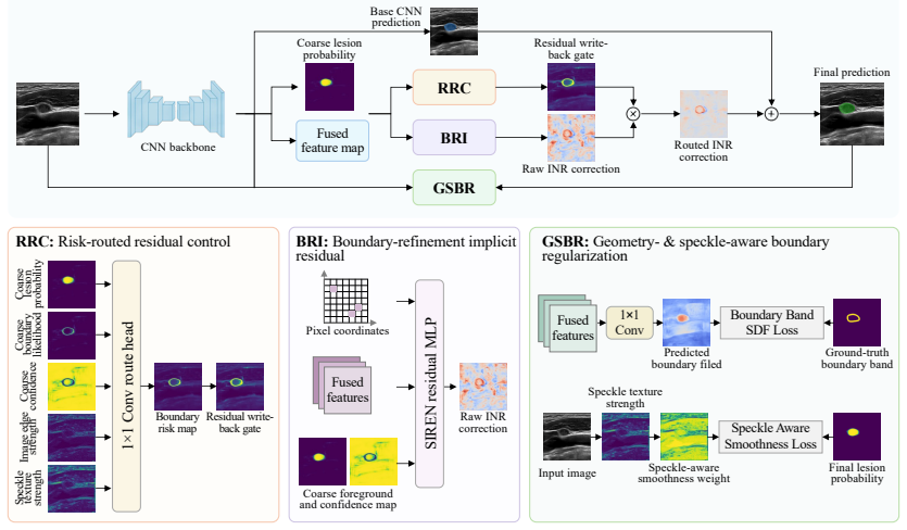
The load-bearing mechanism is the gated residual composition z_i = z^0_i + β α_i δ_i: the base logit z^0 from a convolutional predictor is modified by an implicit residual δ_i produced by a small sinusoidal MLP that takes pixel coordinates, object-relative coordinates, fused features, local intensity statistics, coarse probability, and confidence; α_i ∈ [0,1] is a risk-routing gate computed from a boundary-proximity score b_i = 1 - 2|p_i - 0.5| plus a detached risk-adjusted offset, passed through a sigmoid. This gate confines the implicit network's high-frequency capacity to ambiguous contour regions, preventing it from rewriting confident interiors or background. A signed-distance boundary
What would settle it
Train each baseline with its own best-known configuration (longer schedules, tuned augmentation, optimizer) on the same splits and external test sets, then compare the LN and breast macro-averages; if any larger transformer baseline—most plausibly SwinUNETR—overtakes RIBR on HD95, the paper's central boundary-accuracy claim collapses.
Extended reading notes
Core claim
On its own terms, the paper's central discovery is that the high-frequency capacity of implicit neural representation can be made useful in ultrasound segmentation by constraining it to a logit-space residual correction. A compact convolutional U-Net supplies stable coarse logits; a small sinusoidal (SIREN) implicit branch predicts coordinate-conditioned corrections; and a risk-routing module computes a per-pixel gate—based on boundary proximity plus detached cues like confidence, local intensity statistics, and gradient strength—so the correction is written back only near uncertain boundaries. Auxiliary geometry- and speckle-aware regularization supervises a narrow band around the true cont
Load-bearing premise
The comparison assumes all baselines were trained under conditions that suit them; if transformer or state-space models need more epochs, larger crops, or specialized augmentation to converge, the reported margins (0.84 Dice, 7.05 HD95) could be inflated.
Editorial extensions
If this is right
- If RIBR's results hold, a ~0.4M-parameter segmentation model can outperform much larger transformer and state-space models on ultrasound boundary metrics, making boundary-sensitive AI achievable on low-resource hardware.
- The design principle—implicit residual refinement gated by risk—can be attached to any stable segmentation baseline as a module, potentially improving boundary quality without retraining the base from scratch.
- The strongest gains on external LN and breast cohorts imply that risk-routed refinement transfers across acquisition centers better than simply raising backbone capacity.
- The ablation indicates that risk routing is necessary: without the gate, implicit residual refinement can degrade surface-distance metrics, which argues that uncontrolled high-frequency correction is the failure mode to avoid.
Reading between the lines
- Beyond the paper, the same risk-gated residual correction could be applied to other imaging modalities with speckle-like noise or weak boundaries (e.g., optical coherence tomography, low-dose CT), since the mechanism is not intrinsically ultrasound-specific.
- The routing gate is effectively an uncertainty signal; a future system could expose α_i as a calibrated boundary-uncertainty map for clinicians without changing the architecture.
- The paper fixes deterministic post-processing (hole filling, small-component removal); a natural test is to learn or calibrate these steps end-to-end, which might change the reported boundary-error margins.
- Given the 0.4M-parameter footprint, a plausible next step is combining RIBR with semi-supervised learning on unlabeled ultrasound data—something the paper does not explore—potentially improving external robustness further.
Editorial analysis
A structured set of objections, weighed in public.
Referee Report
Summary. The paper proposes RIBR, a compact ultrasound segmentation framework that keeps a convolutional encoder-decoder as the base predictor and adds a boundary-refinement implicit residual (BRI) modulated by a risk-routed residual control (RRC), with geometry- and speckle-aware boundary regularization (GSBR) during training. The final logits are the base logits plus a gated, coordinate-conditioned SIREN residual. The method is evaluated on nine ultrasound datasets covering lymph nodes, breast, thyroid, and prostate, including external test cohorts, and compared against eleven baselines. The central claims are that RIBR achieves the best overall macro-average Dice and HD95 (76.67±0.42% Dice, 29.55±0.55 HD95 with 0.4M parameters), has the lowest HD95 on every one of the nine datasets, and that module ablations on the lymph-node task show all three modules are complementary.
Significance. If the empirical claims hold, RIBR makes a useful contribution to medical ultrasound segmentation by demonstrating that implicit neural representations are most effective as controlled, risk-routed residuals rather than as unconstrained full-mask predictors. The compact parameter budget (0.4M) and consistent point-estimate improvements in boundary metrics across diverse ultrasound datasets, including external cohorts, are practically relevant. The paper also releases source code, which supports reproducibility, and the ablation design isolates the contribution of each module. However, the strength of the central claim depends on the fairness of the synchronized baseline comparison and on whether the reported differences are statistically reliable; both points are not yet adequately demonstrated in the manuscript.
major comments (4)
- [§IV-B, Table II] The synchronized baseline comparison is not yet shown to be fair. The text states that 'RIBR uses the same split files and synchronized evaluation pipeline as the supervised baselines' but gives no training budget, optimizer, learning rate, batch size, augmentation, loss schedule, or convergence checks for the eleven methods. These methods have very different training requirements: DynUNet relies on self-configuration, UNETR/SwinUNETR/TransUNet/Swin-Unet are large transformers, and VM-UNet is a state-space model. If all were trained with a one-size-fits-all budget, some may be undertrained. The anomalously low results for MetaSeg (e.g., 41.64±1.08 LN average Dice) and VM-UNet (54.88±2.01 LN average Dice) relative to their published operating ranges raise this concern. The authors should report per-method training configurations, show that each baseline converged (e.g., validation-loss cu
- [§IV-C, Table II] No statistical significance tests or confidence intervals are reported beyond mean ± std over three seeds. The claims that RIBR 'consistently reduces boundary error' and achieves the 'best' results on individual datasets are not supportable from overlapping distributions. For example, on BUSI the Dice values are RIBR 74.56±4.62 vs. SwinUNETR 77.02±2.93, and the HD95 values are 28.13±6.64 vs. 29.64±7.58; on several other datasets the standard deviations overlap. A paired test over the three seeds, or preferably a bootstrap or Wilcoxon test over test samples, should be reported for the key comparisons (overall macro-average, grouped averages, and dataset-level HD95). This is particularly important because the headline advantage is a boundary-metric gain, which is known to be sensitive to small numbers of outlier pixels.
- [§IV-D, Table II] The paper's wording 'achieves the best overall macro-average and consistently reduces boundary error across grouped and organ-specific comparisons' is accurate only as a point-estimate statement. It is not shown statistically, and for Dice, RIBR is not the best on all datasets (e.g., BUSI, TN3K, LN-INT). The authors should either add the appropriate significance testing or soften the claims to 'the point estimates suggest...' In particular, the 'all nine datasets' HD95 claim should be accompanied by tests of whether the per-dataset differences (e.g., the small BUSI HD95 difference of 1.5) are meaningful given the reported variability.
- [§IV-H] The limitations section acknowledges that the evaluation is retrospective and that post-processing is deterministic, but it does not address the comparison-fairness limitation raised above. Since the paper's main contribution is empirical, a statement about the lack of per-baseline tuning and the absence of significance tests should be added to the limitations, or the claims should be correspondingly limited.
minor comments (4)
- [§III-C] The post-processing threshold and the specific values for hole filling and small-component removal are not given. Since the paper says 'deterministic post-processing used in all synchronized experiments,' the threshold and component-size parameters should be reported in the implementation details for reproducibility.
- [§IV-A] The internal LN dataset (LN-INT) is not publicly described in detail. Please include scanner, annotation protocol, and any ethics/IRB information, consistent with the reporting for the public datasets.
- [Fig. 1] There is a typo in the figure: 'Predicted boundary filed' should be 'field'.
- [Table II] The table spans many columns and the grouping by organ is helpful, but the visual structure could be improved by separating the organ blocks with vertical lines or subheaders to reduce the chance of misreading grouped macro-averages as dataset-level results.
Circularity Check
No significant circularity: RIBR's reported gains come from standard supervised training with fixed hyperparameters and held-out external tests; self-citations are related work, not load-bearing evidence.
full rationale
The derivation is an empirical architecture-and-loss specification, not a reduction of its outputs to fitted inputs. The final prediction is z_i = z0_i + β α_i δ_i (Eq. 1), with δ_i from the SIREN residual MLP (Eq. 2), α_i from the clipped risk score q_i = clip(b_i + Δq_i, 0, 1) and σ(γ(q_i - τ)) (Eqs. 3-6), and the total loss L = λ_f L_focal + λ_d L_dice + λ_c L_coarse + ... (Eq. 8). These are architectural and training objectives; all weighting constants (τ=0.5, γ=10, λ=0.5, and the λ losses) are stated as fixed implementation choices, so no headline number is obtained by fitting constants to the test tables. External cohorts are explicitly used only for testing and never for training, validation, or hyperparameter selection (Section IV-A), making the cross-center claims genuinely predictive rather than constructed. The self-citations [15], [16] are related-work references to the authors' prior US segmentation methods and are not invoked to justify the residual-routing mechanism or to exclude alternatives. The module ablation (Table III) is a standard ablation, and the limitations in Section IV-H (retrospective design, binary-only, fixed post-processing) are external-validity caveats, not circular steps. The skepticism about baseline training budgets and convergence fairness (no per-method training schedule reported) is a legitimate correctness/fairness risk in the synchronized comparison, but it does not show that any RIBR result is equivalent by construction to its inputs. Therefore no specific circular reduction can be quoted, and the appropriate finding is no circularity.
Assumptions & free parameters
free parameters (13)
- residual_scale_beta
- risk_gate_tau =
0.5
- risk_gate_gamma =
10
- risk_gate_lambda =
0.5
- loss_weight_lambda_focal =
1.0
- loss_weight_lambda_dice =
1.0
- loss_weight_lambda_coarse =
0.5
- loss_weight_lambda_bce =
0.25
- loss_weight_lambda_sdf =
0.1
- loss_weight_lambda_speckle =
0.05
- loss_weight_lambda_residual =
0.01
- sdf_boundary_band_width
- post_process_threshold
assumptions (5)
- domain assumption Ground-truth annotations in all nine datasets are accurate and consistent enough for fair comparison.
- domain assumption The compact U-Net base provides coarse logits that are a stable starting point; if the base fails completely, the residual cannot recover.
- domain assumption The synchronized training recipe is equally appropriate for all baseline architectures.
- domain assumption Detached risk features in RRC are sufficient to distinguish boundary uncertainty from speckle-induced uncertainty.
- domain assumption Three random seeds are sufficient to characterize performance variability.
Cite this review
Pith. "Pith review of Risk-Routed Implicit Boundary Refinement for Robust Ultrasound Image Segmentation." pith.science (2026). https://pith.science/paper/2KXZYKU3
@misc{pith2026260721787,
  author       = {Pith},
  title        = {Pith review of: Risk-Routed Implicit Boundary Refinement for Robust Ultrasound Image Segmentation},
  year         = {2026},
  howpublished = {\url{https://pith.science/paper/2KXZYKU3}},
  note         = {Machine review of arXiv:2607.21787}
}
read the original abstract
Medical ultrasound (US) image segmentation faces significant challenges due to speckle noise, low-contrast boundaries, acoustic shadowing, and acquisition variation across operators and clinical centers. Although encoder-decoder and transformer-based networks have achieved strong performance, many methods recover boundary details through dense decoders or larger backbones, which may still produce over-smoothed contours or unstable predictions under external distribution shifts. In this article, we propose Risk-routed Implicit Boundary Refinement (RIBR), a compact segmentation framework that uses implicit neural representation as a risk-routed residual correction rather than an unconstrained full-mask predictor. RIBR combines boundary-refinement implicit residuals, risk-routed residual control, and geometry- and speckle-aware boundary regularization to refine uncertain contours while suppressing non-boundary oscillations. Evaluation on nine US datasets covering lymph nodes, breast lesions, thyroid nodules, and prostate shows that RIBR achieves the best overall macro-average and consistently reduces boundary error across grouped and organ-specific comparisons under a compact parameter budget. These findings suggest that controlled implicit residual learning is a practical strategy for resource-constrained and boundary-sensitive US segmentation. Source code is available at https://github.com/jinggqu/ribr.
Figures


Reference graph
Works this paper leans on
-
[1]
U-net: Convolutional networks for biomedical image segmentation,
O. Ronneberger, P. Fischer, and T. Brox, “U-net: Convolutional networks for biomedical image segmentation,” inMedical Image Computing and Computer-Assisted Intervention. Springer, 2015, pp. 234–241
2015
-
[2]
Unet++: A nested u-net architecture for medical image segmentation,
Z. Zhou, M. M. R. Siddiquee, N. Tajbakhsh, and J. Liang, “Unet++: A nested u-net architecture for medical image segmentation,” inDeep Learning in Medical Image Analysis and Multimodal Learning for Clinical Decision Support. Springer, 2018, pp. 3–11
2018
-
[3]
Attention u-net: Learning where to look for the pancreas,
O. Oktay, J. Schlemper, L. L. Folgoc, M. Lee, M. Heinrich, K. Misawa, K. Mori, S. McDonagh, N. Y . Hammerla, B. Kainz, B. Glocker, and D. Rueckert, “Attention u-net: Learning where to look for the pancreas,” inMedical Imaging with Deep Learning, 2018
2018
-
[4]
nnu-net: A self-configuring method for deep learning-based biomedical image segmentation,
F. Isensee, P. F. Jaeger, S. A. A. Kohl, J. Petersen, and K. H. Maier-Hein, “nnu-net: A self-configuring method for deep learning-based biomedical image segmentation,”Nature Methods, vol. 18, no. 2, pp. 203–211, 2021
2021
-
[5]
Unetr: Transformers for 3d medical image segmentation,
A. Hatamizadeh, Y . Tang, V . Nath, D. Yang, A. Myronenko, B. Landman, H. R. Roth, and D. Xu, “Unetr: Transformers for 3d medical image segmentation,” inIEEE/CVF Winter Conference on Applications of Computer Vision, 2022, pp. 1748–1758
2022
-
[6]
Swin unetr: Swin transformers for semantic segmentation of brain tumors in mri images,
A. Hatamizadeh, V . Nath, Y . Tang, D. Yang, H. R. Roth, and D. Xu, “Swin unetr: Swin transformers for semantic segmentation of brain tumors in mri images,” inBrainlesion: Glioma, Multiple Sclerosis, Stroke and Traumatic Brain Injuries. Springer, 2022, pp. 272–284
2022
-
[7]
Vm-unet: Vision mamba unet for medical image segmentation,
J. Ruan, J. Li, and S. Xiang, “Vm-unet: Vision mamba unet for medical image segmentation,”ACM Transactions on Multimedia Computing, Communications, and Applications, 2025
2025
-
[8]
V-net: Fully convolutional neural networks for volumetric medical image segmentation,
F. Milletari, N. Navab, and S.-A. Ahmadi, “V-net: Fully convolutional neural networks for volumetric medical image segmentation,” inInterna- tional Conference on 3D Vision, 2016, pp. 565–571
2016
Show all 24 references
-
[9]
Focal loss for dense object detection,
T.-Y . Lin, P. Goyal, R. Girshick, K. He, and P. Doll ´ar, “Focal loss for dense object detection,” inIEEE International Conference on Computer Vision, 2017, pp. 2980–2988
2017
-
[10]
Boundary loss for highly unbalanced segmentation,
H. Kervadec, J. Bouchtiba, C. Desrosiers, E. Granger, J. Dolz, and I. Ben Ayed, “Boundary loss for highly unbalanced segmentation,” Medical Image Analysis, vol. 67, p. 101851, 2021
2021
-
[11]
Reducing the hausdorff distance in medical image segmentation with convolutional neural networks,
D. Karimi and S. E. Salcudean, “Reducing the hausdorff distance in medical image segmentation with convolutional neural networks,”IEEE Transactions on Medical Imaging, vol. 39, no. 2, pp. 499–513, 2020
2020
-
[12]
Implicit neural representations with periodic activation functions,
V . Sitzmann, J. N. P. Martel, A. W. Bergman, D. B. Lindell, and G. Wetzstein, “Implicit neural representations with periodic activation functions,” inAdvances in Neural Information Processing Systems, vol. 33, 2020, pp. 7462–7473
2020
-
[13]
Transunet: Transformers make strong encoders for medical image segmentation,
J. Chen, Y . Lu, Q. Yu, X. Luo, E. Adeli, Y . Wang, L. Lu, A. L. Yuille, and Y . Zhou, “Transunet: Transformers make strong encoders for medical image segmentation,” 2021
2021
-
[14]
Swin-unet: Unet-like pure transformer for medical image segmentation,
H. Cao, Y . Wang, J. Chen, D. Jiang, X. Zhang, Q. Tian, and M. Wang, “Swin-unet: Unet-like pure transformer for medical image segmentation,” inEuropean Conference on Computer Vision Workshops. Springer, 2023, pp. 205–218
2023
-
[15]
Multiscale switch for semi-supervised and contrastive learning in medical ultrasound image segmentation,
J. Qu, X. Han, Y . Pu, M.-L. Chui, S. T. Gunda, Z. Chen, J. Qin, A. D. King, W. C.-W. Chu, J. Caiet al., “Multiscale switch for semi-supervised and contrastive learning in medical ultrasound image segmentation,”IEEE Transactions on Neural Networks and Learning Systems, pp. 1–15, 2026
2026
-
[16]
Adapting vision-language foundation model for next generation medical ultrasound image analysis,
J. Qu, X. Han, J. Ai, J. Wu, T. Zhao, T. Xiao, S. Ning, Y . Yang, J. Qin, A. D. Kinget al., “Adapting vision-language foundation model for next generation medical ultrasound image analysis,”Expert Systems with Applications, vol. 325, p. 132560, 2026
2026
-
[17]
Dataset of breast ultrasound images,
W. Al-Dhabyani, M. Gomaa, H. Khaled, and A. Fahmy, “Dataset of breast ultrasound images,”Data in Brief, vol. 28, p. 104863, 2020
2020
-
[18]
Lymphus: Multicenter open-access database of lymph node ultrasound images in patients with papillary thyroid carcinoma for clinical and artificial intelligence research,
A. Mohammadi, A. Mohebbi, M. Mirza-Aghazadeh-Attari, S. Moham- madzadeh, U. R. Acharya, R.-S. Tan, M. Salvi, S. Hatamikia, and A. Abbasian Ardakani, “Lymphus: Multicenter open-access database of lymph node ultrasound images in patients with papillary thyroid carcinoma for clin...
2026
-
[19]
Curated benchmark dataset for ultrasound based breast lesion analysis,
A. Pawłowska, A. ´Cwierz-Pie´nkowska, A. Domalik, D. Jagu ´s, P. Kasprzak, R. Matkowski, Ł. Fura, A. Nowicki, and N. ˙Zołek, “Curated benchmark dataset for ultrasound based breast lesion analysis,”Scientific Data, vol. 11, no. 1, p. 148, 2024
2024
-
[20]
Bus-uclm: Breast ultrasound lesion segmentation dataset,
N. Vallez, G. Bueno, O. Deniz, M. A. Rienda, and C. Pastor, “Bus-uclm: Breast ultrasound lesion segmentation dataset,”Scientific Data, vol. 12, no. 1, p. 242, 2025
2025
-
[21]
An open access thyroid ultrasound image database,
L. Pedraza, C. Vargas, F. Narv ´aez, O. Dur ´an, E. Mu ˜noz, and E. Romero, “An open access thyroid ultrasound image database,” inProceedings of SPIE Medical Imaging, vol. 9287, 2015, p. 92870W
2015
-
[22]
Multi-task learning for thyroid nodule segmentation with thyroid region prior,
H. Gong, G. Chen, R. Wang, X. Xie, M. Mao, Y . Yu, F. Chen, and G. Li, “Multi-task learning for thyroid nodule segmentation with thyroid region prior,” inIEEE International Symposium on Biomedical Imaging, 2021, pp. 257–261
2021
-
[23]
Microsegnet: A deep learning approach for prostate segmentation on micro-ultrasound images,
H. Jiang, M. Imran, P. Muralidharan, A. Patel, J. Pensa, M. Liang, T. Benidir, J. R. Grajo, J. P. Joseph, R. Terryet al., “Microsegnet: A deep learning approach for prostate segmentation on micro-ultrasound images,”Computerized Medical Imaging and Graphics, vol. 112, p. 102326, 2024
2024
-
[24]
Fit pixels, get labels: Meta-learned implicit networks for image segmentation,
K. Vyas, A. Veeraraghavan, and G. Balakrishnan, “Fit pixels, get labels: Meta-learned implicit networks for image segmentation,” inMedical Image Computing and Computer Assisted Intervention. Springer Nature Switzerland, 2025, pp. 194–203
2025
Reviewed August 1, 2026 · model on record in the stance chip above.
Discussion (0). Continue with ORCID to comment.