REVIEW 4 major objections 6 minor 44 references
$\texttt{DisMorph}$: learning to disentangle technical distortions from true biological change
T0 review · 4 major / 6 minor · reviewed 2026-08-12 · deepseek-v4-flash
Pith's one-line read DisMorph trains a registration network on synthetic images to factor each longitudinal brain scan pair into a scanner-distortion field and a biological-change field, and using only the anatomical field lowers atrophy-estimation error.
desk verdict New disentanglement idea with a solid synthetic check; real-data evidence for the split is still thin, but it deserves refereeing. read the letter →
The pith
A machine-rendered reading of the paper's core claim, the machinery that carries it, and where it could break.
The reading
What carries the argument
The central mechanism is the factored deformation model φ_da = φ_d ∘ φ_a, in which a dual-stream encoder with a pyramid-warping decoder predicts two stationary velocity fields that are integrated via scaling and squaring into diffeomorphic transforms. The network predicts the distortion field at quarter resolution and the anatomical field at half resolution, encoding the prior that GNL distortion is smoother than atrophy. Training supervises each field separately with the synthetic ground truth: the loss is the sum of a label-matching term for the distortion field, a label-matching term for the composed deformation, and a smoothness regularizer. The synthetic generator provides the disentanglement supervision: an elastic lattice deformation models GNL distortion, and iterative boundary reassignment between adjacent labels models local atrophy or growth, followed by an intensity generator that adds bias fields, blurring, noise, and contrast variation for domain randomization.
What would settle it
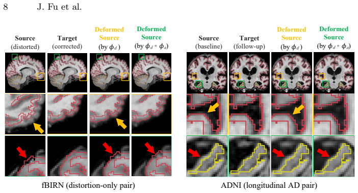
Construct a distortion-only pair by applying mri_gradunwarp to one scan and leaving the other original, as in the paper's fBIRN setup; if the predicted anatomical field then produces statistically significant volume change in structures, the split is misattributing technical distortion as biology.
Extended reading notes
Core claim
The paper's central claim is that the longitudinal deformation between two scans can be factorized into a technical transform and an anatomical transform, and that a network trained purely on synthetic data can recover both components separately. DisMorph outputs two stationary velocity fields, integrated by scaling and squaring into diffeomorphisms φ_d and φ_a, whose composition φ_da = φ_d ∘ φ_a aligns the moving image with the fixed image. The synthetic training pipeline constructs each effect independently—smooth random elastic fields for GNL distortion and boundary-reassignment for atrophy—so each predicted field has a direct ground-truth supervision target. With this split, the anatomical field alone yields lower absolute atrophy estimation error than the composite field on simulated data across low, moderate, and high atrophy levels, and on real fBIRN pairs that differ only by known GNL distortion the model attributes the majority of geometric change to the distortion field. On two-year ADNI Alzheimer's disease pairs, the anatomical field captures the expected subcortical volume loss while the distortion field absorbs remaining scanner-induced misalignment.
Load-bearing premise
Real residual GNL distortion is smooth enough to be captured by the quarter-resolution random elastic fields used in training, and longitudinal deformation factorizes exactly as a distortion step followed by an anatomical step.
Editorial extensions
If this is right
- Atrophy estimates can be computed from the predicted anatomical field alone, removing distortion-related bias without modifying the scanner or acquisition protocol.
- The distortion field itself can serve as a quality-control signal, flagging residual GNL distortion in datasets that were assumed corrected, as the method does for ADNI.
- Because training needs only synthetic data, the approach can be extended to other distortion sources such as B0 inhomogeneity or chemical shift by adding corresponding generative models.
- The largest relative improvement appears at low atrophy levels, meaning early-disease or slow-progressing studies benefit most from the decomposition.
- The two-field output preserves diffeomorphic smoothness for both components, so downstream morphometric measures remain well-defined.
Reading between the lines
- If the scale-separation prior is correct, one testable extension is to compare DisMorph's distortion field against direct phantom-based GNL measurements on the same scans; agreement would confirm that the smooth-field prior captures the true physical distortion.
- The factorization assumes distortion and atrophy compose sequentially; a natural extension would be to allow the distortion field to depend on tissue geometry, which the current generative model does not represent.
- The method could be applied across sites as a harmonization step: estimating each site's distortion signature from pairs of repeated scans and removing it before pooling longitudinal data, a use the paper does not explicitly discuss.
Signed reviews
Editorial analysis
A structured set of objections, weighed in public.
Referee Report
Summary. The paper proposes DisMorph, a learning-based deformable registration framework trained entirely on synthetic data that predicts two dense deformation fields—one intended to capture scanner-induced technical distortion (notably gradient non-linearity, GNL) and one intended to capture anatomical change. Training uses a procedural generator that applies a smooth random elastic transform to model distortion and label-boundary reassignment to model atrophy, with domain randomization over image intensities and corruptions. The method is evaluated in three settings: synthetic data with known atrophy generated by an external biophysical model; real fBIRN image pairs that differ only by GNL distortion; and real ADNI longitudinal pairs from Alzheimer's disease subjects. The results suggest that using only the anatomical field reduces atrophy estimation error relative to a single composite field in simulation, and that most distortion-only real pairs are attributed to the distortion field, while ADNI pairs show plausible atrophy in subcortical structures.
Significance. If the two-field decomposition is reliable, DisMorph would be a practically valuable tool for longitudinal morphometry in multi-site, retrospective, or protocol-changing studies where residual GNL distortion confounds atrophy estimates. The paper has notable strengths: the simulated atrophy ground truth comes from an independent biophysical model rather than the training generator; the fBIRN distortion-only experiment is a genuine out-of-distribution specificity test on real data; and the synthetic training strategy with domain randomization is a reasonable way to obtain dense supervision for two deformation fields. However, the central claim—that DisMorph separates true biological change from technical distortion on real images containing both effects—is not yet established. The current real-data evaluation lacks ground truth for the decomposition, and the simulation uses a distortion generator that appears to match the training distribution. The contribution is promising but needs additional validation before the claimed separation can be considered supported.
major comments (4)
- [Sec. 3.2, Fig. 5] The central claim that DisMorph separates technical distortion from biological change in real longitudinal pairs containing both is not established. The ADNI experiment (Fig. 5B) has no ground truth for either the distortion field or the atrophy field, so the reported decomposition cannot be verified. The fBIRN experiment (Fig. 5A) uses real pairs that differ only by GNL distortion, but because no atrophy is present, it cannot test whether the model misattributes real anatomical change to the distortion field when both effects coexist. A real-data mixed test is needed, for example by comparing the predicted distortion field against a known mri_gradunwarp correction on fBIRN pairs while simultaneously measuring atrophy on images with known disease-related changes, or by constructing a synthetic mixed test with a held-out distortion generator.
- [Sec. 2.1 vs. Sec. 3.1] The simulated experiment in Sec. 3.1 is likely in-distribution for the distortion prior. The training data in Sec. 2.1 synthesize GNL distortion with smooth random elastic transforms sampled on a coarse control lattice, and the simulation in Sec. 3.1 is described only as 'adding distortion' without stating whether it uses a different generator. If the same generator is used, the experiment cannot detect a miscalibrated synthetic prior: the model may have memorized the specific smoothness and magnitude distribution of the training distortions. The paper should explicitly state the relationship between the training and simulation distortion generators and should add out-of-distribution tests, such as distortion fields derived from a measured or biophysical GNL model, or with smoothness and magnitude ranges outside the training support, to demonstrate that the decomposition generalizes beyond the training distribution.
- [Sec. 2.2, Eq. (2)] The training objective supervises the distortion field through L_d and the composed deformation through L_a, but it does not supervise the anatomical field independently. Because the only label losses are on the intermediate distorted label map and the final distorted-and-atrophied label map, any pair (phi_d, phi_a) that preserves the composition phi_da is equally consistent with the label losses. The smoothness regularizer is applied to the composed displacement u, not to the individual fields, and the quarter-resolution distortion stream versus half-resolution anatomical stream is an architectural prior rather than a validated physical constraint. As a result, the split between v_d and v_a may be driven by inductive biases rather than by the data. The paper should provide a sensitivity analysis varying distortion smoothness and magnitude and should compare the predicted distortion field against a known reference such as mri_gradunwarp output to demonstrate that the decomposition is not arbitrary.
- [Sec. 3.2, Fig. 6 (left)] The skull-stripping inconsistency in the fBIRN example is explicitly assigned to the atrophy field and described as 'apparent atrophy.' This is a concrete case of an out-of-distribution technical artifact being misattributed to biological change, which undermines the claim of specificity beyond the training prior. The manuscript should quantify how frequently such misassignments occur across the fBIRN pairs and discuss the implications for analyses of cortical or boundary-adjacent structures where preprocessing inconsistencies are common. Without such quantification, the example suggests a known failure mode of the proposed decomposition rather than an isolated anecdote.
minor comments (6)
- [Sec. 2.2, Eq. (1)] The notation in Eq. (1) is slightly confusing: the text introduces intensity images I_F and I_M, but the loss is defined on one-hot encoded label maps. Clarify that Eq. (1) is computed on label representations, not on intensity images.
- [Fig. 4 and Fig. 5] The results in Figs. 4 and 5 are presented without confidence intervals or statistical tests. Given the small sample sizes (18 fBIRN pairs and 40 ADNI pairs), error bars or a paired statistical comparison would strengthen the claims of consistent improvement and specificity.
- [Sec. 3.1, Metrics] The metric AAE is used in the text and Fig. 4 but is never explicitly defined. Please provide the formula relating AAE to the prescribed and estimated ASPC values.
- [Sec. 2.1, Geometry generator] The phrase 'lattice resolutions between 2 and 10 control points for each dimension' is ambiguous; clarify whether the number of control points is sampled independently per dimension and whether this refers to the spacing or the count.
- [Conclusion, Sec. 4] The claim that DisMorph is 'the first registration framework' that explicitly decomposes longitudinal deformation into technical and anatomical transforms is strong and is not substantiated by a literature survey. Soften the claim or provide a focused comparison with prior works that estimate separate deformation components.
- [Sec. 3.2, Data] The fBIRN experiment uses mri_gradunwarp with the manufacturer's coefficient files, but no details are given about the scanner model, version of mri_gradunwarp, or whether the same coefficients were used across subjects. These details would help readers assess the reliability of the distortion reference.
Circularity Check
No load-bearing circularity: DisMorph's decomposition is supervised from synthetic labels and tested on external biophysical and real data; the main limitation is a possibly in-distribution simulated distortion test, which is not a circular derivation.
full rationale
The paper's derivation is a supervised learning pipeline, not a claimed first-principles result. The training objective (Eq. 2) supervises the two predicted fields against distortion and atrophy label maps produced by the geometry generator, which is standard label-based training rather than a circular reduction. The simulated evaluation uses a biophysical atrophy model (Khanal et al.) as an independent source of ground-truth anatomical change, and the real-data experiments (fBIRN distortion-only pairs and ADNI longitudinal pairs) are out-of-distribution with respect to the synthetic training data. The composition phi_da = phi_d ∘ phi_a is an explicit modeling assumption, stated in Sec. 2.2, not a hidden equivalence. The paper cites several prior works by the same authors (e.g., SynthMorph, Fu et al. synthetic longitudinal registration), but these are used for synthetic data generation and backbone choices, not as the load-bearing justification for the disentanglement claim. No uniqueness theorem or fitted parameter is renamed as a prediction. The only substantive concern is that the simulated test pairs are said to have 'distortion added' without specifying whether the generator matches training; if it does, the distortion-recovery part of the simulation is in-distribution and limited as evidence of generalization. That is an evaluation weakness, not circularity, because the central atrophy ground truth and the real-data results remain independent.
Assumptions & free parameters
free parameters (5)
- Smoothness regularization weight lambda =
0.01
- Distortion training magnitude cap =
2.5% of field of view
- Distortion control lattice resolution =
2 to 10 control points per dimension
- Training atrophy change ratio r_k =
U(-0.5, 0.5)
- Deformation field output resolutions =
distortion quarter resolution, atrophy half resolution
assumptions (5)
- domain assumption Real GNL distortion is statistically similar to the smooth random elastic fields used in training
- domain assumption Longitudinal deformation factorizes as phi_da = phi_d composed with phi_a
- domain assumption The two effects are separable by smoothness: distortion is low-frequency, atrophy is localized
- standard math Scaling and squaring integration yields diffeomorphic deformations
- domain assumption Label-map-based synthetic training transfers to real image contrast
Cite this review
Pith. "Pith review of $\texttt{DisMorph}$: learning to disentangle technical distortions from true biological change." pith.science (2026). https://pith.science/paper/2BVBMX2F
@misc{pith2026260808173,
  author       = {Pith},
  title        = {Pith review of: $\textttDisMorph$: learning to disentangle technical distortions from true biological change},
  year         = {2026},
  howpublished = {\url{https://pith.science/paper/2BVBMX2F}},
  note         = {Machine review of arXiv:2608.08173}
}
abstract
Longitudinal MRI enables sensitive measurement of structural brain change for studying aging and neurodegenerative disease. Deformable image registration is a key tool for estimating such change by computing a dense deformation that captures geometric differences between longitudinal scans. However, MRI scanners introduce geometric distortions that vary across acquisition systems and protocols, such as gradient non-linearity (GNL) distortion. Existing registration methods estimate a single field that conflates biological and technical effects, potentially biasing downstream morphometric measurements if distortions remain (partially) uncorrected. We propose $\texttt{DisMorph}$, a registration framework trained entirely on synthetic data that explicitly decomposes longitudinal deformation into technical and anatomical transforms. It predicts two dense deformations, each encoding one effect. During training, a novel generative model synthesizes both effects separately to provide disentanglement supervision, while domain randomization promotes generalization across imaging protocols. We evaluate our method in three complementary settings. On simulated data with known ground truth, our method detects anatomical change more accurately and consistently than conventional registration. On real image pairs that differ only by GNL distortion, our method assigns most geometric change to the distortion field, demonstrating specificity in the absence of anatomical change. On longitudinal Alzheimer's disease (AD) pairs, our method detects anatomical change in AD-related brain structures while identifying residual distortion left after standard correction. By disentangling MRI-induced distortion from biological change in the longitudinal deformation, our method paves the way for more accurate longitudinal morphometry in clinical settings where maintaining acquisition consistency is challenging.
Figures


Figures from the paper (3 more)


Reference graph
Works this paper leans on
-
[1]
In: 2025 IEEE/CVF Conference on Computer Vision and Pattern Recog- nition (CVPR)
Abulnaga, S.M., Hoopes, A., Dey, N., et al.: MultiMorph: on-demand atlas con- struction. In: 2025 IEEE/CVF Conference on Computer Vision and Pattern Recog- nition (CVPR). pp. 30906–30917. IEEE (2025)
work page 2025
-
[2]
Phys Imaging Radiat Oncol15, 80–84 (2020)
Alzahrani, M., et al.: Audit feasibility for geometric distortion in magnetic reso- nance imaging for radiotherapy. Phys Imaging Radiat Oncol15, 80–84 (2020)
work page 2020
-
[3]
Arsigny, V., et al.: A Log-Euclidean Framework for Statistics on Diffeomorphisms. In: MICCAI 2006. pp. 924–931 (2006)
work page 2006
-
[4]
Ashburner,J.,Friston,K.J.:Voxel-basedmorphometry—themethods.NeuroImage 11(6), 805–821 (2000)
work page 2000
-
[5]
Human Brain Mapping9, 212–225 (2000)
Ashburner, J., et al.: Image Registration Using a Symmetric Prior-in Three Di- mensions. Human Brain Mapping9, 212–225 (2000)
work page 2000
-
[6]
Medical Image Analysis12(1), 26–41 (2008)
Avants, B.B., et al.: Symmetric diffeomorphic image registration with cross- correlation: Evaluating automated labeling of elderly and neurodegenerative brain. Medical Image Analysis12(1), 26–41 (2008)
work page 2008
-
[7]
Balakrishnan, G., et al.: VoxelMorph: A Learning Framework for Deformable Med- ical Image Registration. IEEE Trans. on Medical Imaging38(8), 1788–1800 (2019)
work page 2019
-
[8]
NeuroImage23(2), 574–581 (2004)
Barnes,J.,etal.:DifferentiatingADfromagingusingsemiautomatedmeasurement of hippocampal atrophy rates. NeuroImage23(2), 574–581 (2004)
work page 2004
Show all 44 references
-
[9]
Medical Image Analysis86, 102789 (2023)
Billot, B., et al.: SynthSeg: Segmentation of brain MRI scans of any contrast and resolution without retraining. Medical Image Analysis86, 102789 (2023)
2023
-
[10]
Medical Image Analysis57, 226–236 (2019)
Dalca, A.V., et al.: Unsupervised learning of probabilistic diffeomorphic registra- tion for images and surfaces. Medical Image Analysis57, 226–236 (2019)
2019
-
[11]
Medical Image Analysis52, 128–143 (2019)
De Vos, B.D., et al.: A deep learning framework for unsupervised affine and de- formable image registration. Medical Image Analysis52, 128–143 (2019)
2019
-
[12]
NeuroImage62(2), 774–781 (2012)
Fischl, B.: FreeSurfer. NeuroImage62(2), 774–781 (2012)
2012
-
[13]
Neuron33(3), 341–355 (2002)
Fischl, B., et al.: Whole brain segmentation: automated labeling of neuroanatom- ical structures in the human brain. Neuron33(3), 341–355 (2002)
2002
-
[14]
Sci Rep15(1), 11813 (2025)
Fu, J., et al.: Decomposing the effect of normal aging and Alzheimer’s disease in brain morphological changes via learned aging templates. Sci Rep15(1), 11813 (2025)
2025
-
[15]
In: ISBI 2025
Fu, J., et al.: Learning Accurate Rigid Registration for Longitudinal Brain MRI from Synthetic Data. In: ISBI 2025. pp. 1–5 (2025)
2025
-
[16]
Medical Image Analysis105, 103669 (2025)
Fu, J., et al.: Synthesizing individualized aging brains in health and disease with generative models and parallel transport. Medical Image Analysis105, 103669 (2025)
2025
-
[17]
NeuroImage14(1), 21–36 (2001)
Good, C.D., et al.: A voxel-based morphometric study of ageing in 465 normal adult human brains. NeuroImage14(1), 21–36 (2001)
2001
-
[18]
In: WBIR
Gopinath, K., et al.: Registration by regression (RbR): A framework for inter- pretable and flexible atlas registration. In: WBIR. pp. 205–215 (2024)
2024
-
[19]
OpenNeuro (2024)
Greve, D., Fischl, B.: The FreeSurfer Maintenance Dataset. OpenNeuro (2024)
2024
-
[20]
Med Phys36(6Part1), 2193–2205 (2009)
Gunter, J.L., et al.: Measurement of MRI scanner performance with the ADNI phantom. Med Phys36(6Part1), 2193–2205 (2009)
2009
-
[21]
IEEE Signal Process Mag42(4), 78–90 (2025)
Hoffmann, M.: Domain-Randomized Deep Learning for Neuroimage Analysis. IEEE Signal Process Mag42(4), 78–90 (2025)
2025
-
[22]
IEEE Trans
Hoffmann,M.,etal.:SynthMorph:LearningContrast-InvariantRegistrationWith- out Acquired Images. IEEE Trans. on Medical Imaging41(3), 543–558 (2022)
2022
-
[23]
Imaging Neurosci2, imag–2–00197 (2024) DisMorph: disentangling technical distortions from biological change 11
Hoffmann, M., et al.: Anatomy-aware and acquisition-agnostic joint registration with SynthMorph. Imaging Neurosci2, imag–2–00197 (2024) DisMorph: disentangling technical distortions from biological change 11
2024
-
[24]
NeuroImage 260, 119474 (2022)
Hoopes, A., et al.: SynthStrip: skull-stripping for any brain image. NeuroImage 260, 119474 (2022)
2022
-
[25]
arXiv preprint arXiv:2410.08397 (2024)
Hoopes, A., et al.: VoxelPrompt: A Vision Agent for End-to-End Medical Image Analysis. arXiv preprint arXiv:2410.08397 (2024)
2024
-
[26]
Sci Adv9(5), eadd3607 (2023)
Iglesias, J.E., et al.: SynthSR: A public AI tool to turn heterogeneous clinical brain scans into high-resolution T1-weighted images for 3D morphometry. Sci Adv9(5), eadd3607 (2023)
2023
-
[27]
Information eXtraction from Images (IXI) Project: IXI Dataset: Nearly 600 MR images from normal, healthy subjects.https://brain-development.org/ ixi-dataset/(2015)
2015
-
[28]
J Magn Reson Imaging27(4), 685–691 (2008)
Jack Jr., C.R., et al.: The Alzheimer’s disease neuroimaging initiative (ADNI): MRI methods. J Magn Reson Imaging27(4), 685–691 (2008)
2008
-
[29]
arXiv preprint arXiv:2512.01913 (2025)
Jian, B., et al.: Disentangling progress in medical image registration: Beyond trend-driven architectures towards domain-specific strategies. arXiv preprint arXiv:2512.01913 (2025)
2025
-
[30]
NeuroImage30(2), 436–443 (2006)
Jovicich, J., et al.: Reliability in multi-site structural MRI studies: effects of gra- dient non-linearity correction on phantom and human data. NeuroImage30(2), 436–443 (2006)
2006
-
[31]
NeuroImage124, 1074–1079 (2016)
Keator, D.B., et al.: The Function Biomedical Informatics Research Network Data Repository. NeuroImage124, 1074–1079 (2016)
2016
-
[32]
NeuroImage134, 35–52 (2016)
Khanal, B., et al.: A biophysical model of brain deformation to simulate and an- alyze longitudinal MRIs of patients with Alzheimer’s disease. NeuroImage134, 35–52 (2016)
2016
-
[33]
NeuroImage40(2), 559–569 (2008)
Van der Kouwe, A.J.W., et al.: Brain morphometry with multiecho MPRAGE. NeuroImage40(2), 559–569 (2008)
2008
-
[34]
NeuroImage70, 33–36 (2013)
Malone, I.B., et al.: MIRIAD—Public release of a multiple time point Alzheimer’s MR imaging dataset. NeuroImage70, 33–36 (2013)
2013
-
[35]
NeuroImage23, S19–S33 (2004)
Miller, M.I.: Computational anatomy: shape, growth, and atrophy comparison via diffeomorphisms. NeuroImage23, S19–S33 (2004)
2004
-
[36]
MAGMA 33(3), 401–410 (2020)
Nousiainen, K., Mäkelä, T.: Measuring geometric accuracy in magnetic resonance imaging with 3D-printed phantom and nonrigid image registration. MAGMA 33(3), 401–410 (2020)
2020
-
[37]
NeuroImage53(4), 1181–1196 (2010)
Reuter, M., et al.: Highly accurate inverse consistent registration: A robust ap- proach. NeuroImage53(4), 1181–1196 (2010)
2010
-
[38]
NeuroImage61(4), 1402–1418 (2012)
Reuter, M., et al.: Within-subject template estimation for unbiased longitudinal image analysis. NeuroImage61(4), 1402–1418 (2012)
2012
-
[39]
Neurobiol Aging31(8), 1401–1418 (2010)
Risacher, S.L., et al.: Longitudinal MRI atrophy biomarkers: relationship to con- version in the ADNI cohort. Neurobiol Aging31(8), 1401–1418 (2010)
2010
-
[40]
Neuroradiol52(8), 691–698 (2010)
Takao, H., et al.: Computational analysis of cerebral cortex. Neuroradiol52(8), 691–698 (2010)
2010
-
[41]
Human Brain Mapping36(6), 2093–2117 (2015)
Tang, X., et al.: The diffeomorphometry of regional shape change rates and its relevance to cognitive deterioration in mild cognitive impairment and Alzheimer’s disease. Human Brain Mapping36(6), 2093–2117 (2015)
2015
-
[42]
Medical Image Analysis2(3), 243–260 (1998)
Thirion, J.P.: Image matching as a diffusion process: an analogy with Maxwell’s demons. Medical Image Analysis2(3), 243–260 (1998)
1998
-
[43]
In: MICCAI
Tian, L., et al.: uniGradICON: A foundation model for medical image registration. In: MICCAI. pp. 749–760 (2024)
2024
-
[44]
NeuroImage50(2), 434–445 (2010)
Yushkevich, P.A., et al.: Bias in estimation of hippocampal atrophy using deformation-based morphometry arises from asymmetric global normalization: an illustration in ADNI 3 T MRI data. NeuroImage50(2), 434–445 (2010)
2010
Reviewed August 12, 2026 · model on record in the stance chip above.
Discussion (0). Continue with ORCID to comment.