DuDoNet: Dual Domain Network for CT Metal Artifact Reduction
Pith reviewed 2026-05-25 12:23 UTC · model grok-4.3
The pith
A dual-domain network with a Radon inversion layer corrects both sinograms and CT images to reduce metal artifacts.
A machine-rendered reading of the paper's core claim, the machinery that carries it, and where it could break.
Core claim
The authors claim that simultaneously operating in the sinogram and image domains via an end-to-end network, connected by a novel Radon inversion layer that permits gradient flow from image to sinogram, produces superior metal artifact reduction compared with prior single-domain approaches and constitutes the first such dual-domain architecture for this task.
What carries the argument
The Radon inversion layer, which models the forward and backward projection processes to link the sinogram domain and image domain while allowing end-to-end gradient back-propagation.
If this is right
- Joint optimization across domains removes both the original metal streaks and the secondary artifacts that arise from inconsistent sinogram edits.
- Gradient signals from the final image quality can directly improve the sinogram restoration step.
- The same architecture can in principle be retrained on different scanner geometries or implant materials.
- Clinical CT workflows could incorporate the network as a post-processing step without separate sinogram and image pipelines.
Where Pith is reading between the lines
- The dual-domain linkage may extend to other tomography problems where projection data and reconstructed images must stay consistent, such as limited-angle reconstruction.
- Replacing the analytic Radon layer with a learned projection operator could test whether the performance gain comes from the domain coupling itself or from the specific inversion formula.
- If the method scales to cone-beam CT, it would directly affect dental and interventional imaging where metal artifacts are frequent.
Load-bearing premise
The Radon inversion layer must accurately represent the actual CT projection mathematics so that gradients can flow usefully between domains without creating new inconsistencies.
What would settle it
Training the network on a dataset of real CT scans containing known metallic implants and then measuring whether residual artifacts remain larger than those produced by the best single-domain baseline on the same scans.
Figures


read the original abstract
Computed tomography (CT) is an imaging modality widely used for medical diagnosis and treatment. CT images are often corrupted by undesirable artifacts when metallic implants are carried by patients, which creates the problem of metal artifact reduction (MAR). Existing methods for reducing the artifacts due to metallic implants are inadequate for two main reasons. First, metal artifacts are structured and non-local so that simple image domain enhancement approaches would not suffice. Second, the MAR approaches which attempt to reduce metal artifacts in the X-ray projection (sinogram) domain inevitably lead to severe secondary artifact due to sinogram inconsistency. To overcome these difficulties, we propose an end-to-end trainable Dual Domain Network (DuDoNet) to simultaneously restore sinogram consistency and enhance CT images. The linkage between the sigogram and image domains is a novel Radon inversion layer that allows the gradients to back-propagate from the image domain to the sinogram domain during training. Extensive experiments show that our method achieves significant improvements over other single domain MAR approaches. To the best of our knowledge, it is the first end-to-end dual-domain network for MAR.
Editorial analysis
A structured set of objections, weighed in public.
Referee Report
Summary. The paper proposes DuDoNet, an end-to-end trainable dual-domain neural network for metal artifact reduction (MAR) in CT. It uses a novel Radon inversion layer to link the sinogram and image domains, enabling simultaneous sinogram consistency restoration and image enhancement via gradient back-propagation. The central claim is that this yields significant improvements over single-domain MAR methods and is the first such end-to-end dual-domain network.
Significance. If the Radon inversion layer provides a consistent differentiable linkage, the work would represent a meaningful advance in MAR by moving beyond single-domain limitations to joint optimization. The introduction of the layer itself could have broader utility in differentiable tomography pipelines. The paper explicitly positions the contribution as the first end-to-end dual-domain approach.
major comments (1)
- [Abstract] Abstract (Radon inversion layer paragraph): The central claim that the layer 'allows the gradients to back-propagate from the image domain to the sinogram domain during training' without introducing new inconsistencies rests on an unverified assumption that the discrete implementation exactly matches the physical projection geometry and forms a consistent (adjoint) pair. No explicit verification (e.g., adjoint test, gradient consistency ablation, or comparison to the data-generation forward model) is referenced; if the pair is approximate, dual-domain training optimizes an inconsistent objective, which could undermine the reported gains over single-domain baselines.
minor comments (1)
- [Abstract] Abstract: 'sigogram' is a typo and should read 'sinogram'.
Simulated Author's Rebuttal
We thank the referee for the constructive comment on the Radon inversion layer. We address the concern point-by-point below and agree that additional verification will strengthen the manuscript.
read point-by-point responses
-
Referee: [Abstract] Abstract (Radon inversion layer paragraph): The central claim that the layer 'allows the gradients to back-propagate from the image domain to the sinogram domain during training' without introducing new inconsistencies rests on an unverified assumption that the discrete implementation exactly matches the physical projection geometry and forms a consistent (adjoint) pair. No explicit verification (e.g., adjoint test, gradient consistency ablation, or comparison to the data-generation forward model) is referenced; if the pair is approximate, dual-domain training optimizes an inconsistent objective, which could undermine the reported gains over single-domain baselines.
Authors: We agree that the manuscript does not reference explicit verification (adjoint test, gradient consistency ablation, or direct comparison to the forward model used for data generation). The layer is implemented as a differentiable approximation to the inverse Radon transform to enable gradient flow, but without the requested checks it is not demonstrated that the discrete pair is exactly consistent. In the revision we will add (i) an adjoint test for the layer, (ii) a gradient-consistency ablation, and (iii) a comparison against the simulation forward projector, together with a brief discussion of any residual approximation error. These additions will be placed in the Methods section and referenced from the abstract. revision: yes
Circularity Check
No circularity: data-driven network with experimental validation
full rationale
The paper introduces DuDoNet as an end-to-end trainable dual-domain CNN for metal artifact reduction, with a novel but explicitly constructed Radon inversion layer to enable gradient flow between sinogram and image domains. All performance claims rest on comparative experiments against single-domain baselines rather than any self-referential definition, fitted parameter renamed as prediction, or load-bearing self-citation. The linkage between domains is presented as an engineering choice whose correctness is tested empirically, not derived tautologically from the inputs. No equations or steps reduce by construction to the training data or prior outputs of the same model.
Axiom & Free-Parameter Ledger
free parameters (1)
- network hyperparameters and loss weights
axioms (1)
- domain assumption The CT imaging process can be accurately modeled by the Radon transform for the purpose of inversion and gradient flow.
invented entities (1)
-
Radon inversion layer
no independent evidence
Reference graph
Works this paper leans on
-
[1]
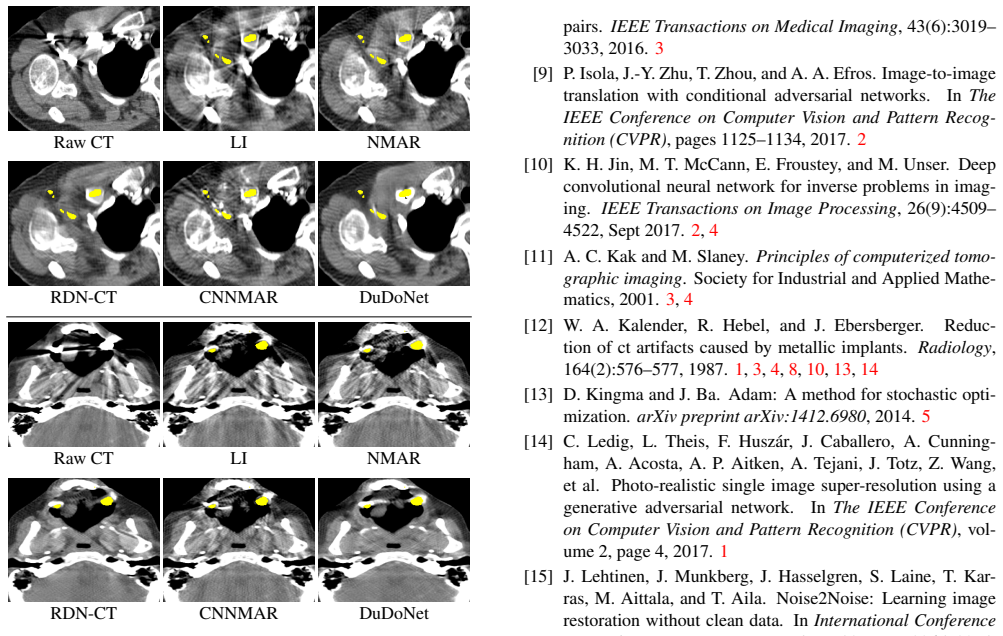
J. Adler and O. ¨Oktem. Learned primal-dual reconstruction. IEEE Transactions on Medical Imaging , 37(6):1322–1332, June 2018. 2, 4 4The domain of CT images with simulated metal artifacts. Raw CT LI NMAR RDN-CT CNNMAR DuDoNet Raw CT LI NMAR RDN-CT CNNMAR DuDoNet Figure 10: Evaluations on real data. All models are exactly the same as in the main paper (no ...
work page 2018
-
[2]
Bestimmung der absorption des rothen lichts in farbigen fl ¨ussigkeiten
Beer. Bestimmung der absorption des rothen lichts in farbigen fl ¨ussigkeiten. Annalen der Physik und Chemie , 162(5):78–88, 1852. 3
- [3]
-
[4]
X. Duan, L. Zhang, Y . Xiao, J. Cheng, Z. Chen, and Y . Xing. Metal artifact reduction in ct images by sinogram tv inpaint- ing. In Nuclear Science Symposium Conference Record,
- [5]
-
[6]
L. Gjesteby, Q. Yang, Y . Xi, B. Claus, Y . Jin, B. De Man, and G. Wang. Reducing metal streak artifacts in ct images via deep learning: Pilot results. In The 14th International Meeting on Fully Three-Dimensional Image Reconstruction in Radiology and Nuclear Medicine , pages 611–614, 2017. 3
work page 2017
-
[7]
L. Gjesteby, Q. Yang, Y . Xi, Y . Zhou, J. Zhang, and G. Wang. Deep learning methods to guide ct image reconstruction and reduce metal artifacts. In SPIE Medical Imaging, 2017. 3
work page 2017
- [8]
- [9]
-
[10]
P. Isola, J.-Y . Zhu, T. Zhou, and A. A. Efros. Image-to-image translation with conditional adversarial networks. In The IEEE Conference on Computer Vision and Pattern Recog- nition (CVPR), pages 1125–1134, 2017. 2
work page 2017
-
[11]
K. H. Jin, M. T. McCann, E. Froustey, and M. Unser. Deep convolutional neural network for inverse problems in imag- ing. IEEE Transactions on Image Processing , 26(9):4509– 4522, Sept 2017. 2, 4
work page 2017
-
[12]
A. C. Kak and M. Slaney. Principles of computerized tomo- graphic imaging. Society for Industrial and Applied Mathe- matics, 2001. 3, 4
work page 2001
-
[13]
W. A. Kalender, R. Hebel, and J. Ebersberger. Reduc- tion of ct artifacts caused by metallic implants. Radiology, 164(2):576–577, 1987. 1, 3, 4, 8, 10, 13, 14
work page 1987
-
[14]
Adam: A Method for Stochastic Optimization
D. Kingma and J. Ba. Adam: A method for stochastic opti- mization. arXiv preprint arXiv:1412.6980, 2014. 5
work page internal anchor Pith review Pith/arXiv arXiv 2014
-
[15]
C. Ledig, L. Theis, F. Husz ´ar, J. Caballero, A. Cunning- ham, A. Acosta, A. P. Aitken, A. Tejani, J. Totz, Z. Wang, et al. Photo-realistic single image super-resolution using a generative adversarial network. In The IEEE Conference on Computer Vision and Pattern Recognition (CVPR) , vol- ume 2, page 4, 2017. 1
work page 2017
-
[16]
J. Lehtinen, J. Munkberg, J. Hasselgren, S. Laine, T. Kar- ras, M. Aittala, and T. Aila. Noise2Noise: Learning image restoration without clean data. In International Conference on Machine Learning (ICML), volume 80, pages 2965–2974,
-
[17]
M. Makitalo and A. Foi. Optimal inversion of the anscombe transformation in low-count poisson image denoising. IEEE transactions on Image Processing, 20(1):99–109, 2011. 1
work page 2011
-
[18]
A. Mehranian, M. R. Ay, A. Rahmim, and H. Zaidi. X-ray ct metal artifact reduction using wavelet domain l0 sparse regularization. IEEE Transactions on Medical Imaging , 32:1707–1722, 2013. 1, 3
work page 2013
- [19]
-
[20]
J. Pan, W. Ren, Z. Hu, and M. Yang. Learning to deblur im- ages with exemplars. IEEE Transactions on Pattern Analysis and Machine Intelligence, 2018. 2
work page 2018
-
[21]
H. S. Park, Y . E. Chung, S. M. Lee, H. P. Kim, and J. K. Seo. Sinogram-consistency learning in ct for metal artifact reduction. arXiv preprint arXiv:1708.00607, 2017. 2, 3
work page internal anchor Pith review Pith/arXiv arXiv 2017
- [22]
-
[23]
O. Ronneberger, P. Fischer, and T. Brox. U-net: Convolu- tional networks for biomedical image segmentation. InMed- ical Image Computing and Computer Assisted Intervention (MICCAI), pages 234–241. Springer, 2015. 2, 9
work page 2015
-
[24]
D. Ulyanov, A. Vedaldi, and V . Lempitsky. Deep image prior. In The IEEE Conference on Computer Vision and Pattern Recognition (CVPR), June 2018. 2
work page 2018
-
[25]
J. Wang, Y . Zhao, J. H. Noble, and B. M. Dawant. Condi- tional generative adversarial networks for metal artifact re- duction in ct images of the ear. InMedical Image Computing and Computer Assisted Intervention (MICCAI) , 2018. 2, 3, 8, 10, 13, 14
work page 2018
-
[26]
X. Wang, K. Yu, S. Wu, J. Gu, Y . Liu, C. Dong, Y . Qiao, and C. C. Loy. Esrgan: Enhanced super-resolution genera- tive adversarial networks. In The European Conference on Computer Vision Workshops (ECCVW), September 2018. 1, 2, 8
work page 2018
- [27]
- [28]
-
[29]
K. Yan, X. Wang, L. Lu, L. Zhang, A. P. Harrison, M. Bagheri, and R. M. Summers. Deep lesion graphs in the wild: Relationship learning and organization of signifi- cant radiology image findings in a diverse large-scale lesion database. In The IEEE Conference on Computer Vision and Pattern Recognition (CVPR), June 2018. 5, 10
work page 2018
- [30]
-
[31]
H. Zhang and V . M. Patel. Densely connected pyramid de- hazing network. In The IEEE Conference on Computer Vi- sion and Pattern Recognition (CVPR), 2018. 2
work page 2018
- [32]
- [33]
-
[34]
Y . Zhang and H. Yu. Convolutional neural network based metal artifact reduction in x-ray computed tomography. IEEE Transactions on Medical Imaging , 2018. 1, 2, 3, 5, 8, 10, 13, 14
work page 2018
- [35]
-
[36]
Joint Sub-bands Learning with Clique Structures for Wavelet Domain Super-Resolution
Z. Zhong, T. Shen, Y . Yang, Z. Lin, and C. Zhang. Joint sub-bands learning with clique structures for wavelet domain super-resolution. arXiv preprint arXiv:1809.04508, 2018. 1 Ground Truth With Metal Artifact LI [12] NMAR [18] cGAN-CT [24] RDN-CT [32] CNNMAR [33] DuDoNet Ground Truth With Metal Artifact LI [12] NMAR [18] cGAN-CT [24] RDN-CT [32] CNNMAR [...
work page internal anchor Pith review Pith/arXiv arXiv 2018
discussion (0)
Sign in with ORCID, Apple, or X to comment. Anyone can read and Pith papers without signing in.