REVIEW 3 major objections 5 minor 33 references
ADN: Artifact Disentanglement Network for Unsupervised Metal Artifact Reduction
T0 review · 3 major / 5 minor · reviewed 2026-08-14 · deepseek-v4-flash
Pith's one-line read First unsupervised CT metal artifact reduction matches supervised models on synthetic data and outperforms them on clinical scans.
desk verdict A credible unsupervised MAR method with a clear disentanglement formulation, but the clinical generalization claim rests on qualitative images and an additivity assumption the paper itself concedes may transfer lesion-like anatomy. read the letter →
The pith
A machine-rendered reading of the paper's core claim, the machinery that carries it, and where it could break.
The reading
What carries the argument
The load-bearing object is the artifact consistency loss, Eq. (8): $\mathcal{L}_{\mathrm{art}} = \mathbb{E}[\|(x_a - \hat{x}) - (\hat{y}_a - y)\|_1]$, which forces the artifact subtracted from an artifact-affected image to equal the artifact added to an artifact-free image under the same artifact code. This is what lets the network learn anatomical precision without paired images. It is supported by an artifact pyramid decoding design that merges the artifact code into the content decoder at multiple scales, and by the full set of competing losses (adversarial, reconstruction, self-reduction) that keep the separated codes meaningful.
What would settle it
Take a set of clinical metal-affected CT images and their metal-free counterparts reconstructed from the same patient (e.g., before and after implant removal); run ADN trained only on unpaired images and check in metal-free tissue whether the difference between the network's output and the true clean image is near zero, and if the artifact code from one anatomy reproduces the true artifact when applied to a different anatomy.
Extended reading notes
Core claim
The paper's central claim is that metal artifact reduction can be learned without any anatomically paired artifact-free ground truth by treating it as artifact disentanglement. The artifact disentanglement network (ADN) maps an artifact-affected image through a content encoder and an artifact encoder into separate latent spaces, and an artifact-free image through its own content encoder. Artifact removal is decoding the content code alone; artifact synthesis is decoding a content code together with an artifact code. Four losses drive the separation: adversarial losses make each output look like its target domain, a reconstruction loss keeps the autoencoding faithful, an artifact consistency loss requires the artifact stripped from an artifact image to equal the artifact added to a clean image, and a self-reduction loss requires that removing the synthesized artifact from a transferred image recovers the clean original. The paper reports that on synthesized data ADN is on par with supervised methods, while on clinical scans—where supervised models trained on synthetic artifacts degrade—ADN removes streaks and shading without introducing secondary artifacts.
Load-bearing premise
The method assumes that the metal artifact in an image is an additive component that can be lifted out and re-attached to another image unchanged; if real artifacts depend on the underlying anatomy, the network may erase anatomical structures instead of artifacts.
Editorial extensions
If this is right
- Clinics could reduce metal artifacts without needing paired scans or access to raw projection data, since ADN works directly on reconstructed CT images.
- Supervised MAR models trained on synthetic artifacts fail under domain shift; an unsupervised model trained on the target domain's own unpaired images is a more robust route, as demonstrated by the clinical results.
- The same architecture should transfer to other artifact types (noise, streaks, blur) because the formulation does not assume a specific artifact mechanism.
- Artifact synthesis from one patient applied to another's artifact-free image could supply realistic metal-affected training data for downstream tasks like segmentation.
- The self-reduction loop provides a general recipe for adding self-supervision to image translation when paired data is unavailable.
Reading between the lines
- The artifact consistency loss is the likeliest failure point: if real metal artifacts are non-additive and anatomy-dependent (beam hardening, scatter), the equality in Eq. (8) can be satisfied by transferring anatomy-dependent residue into the artifact code, letting the network delete real structures.
- A testable extension: apply ADN to a dataset with known non-linear artifacts and compare the artifact code's transferability across anatomies; if the code carries anatomy-specific content, artifact transfer quality will drop when source and target anatomies differ greatly.
- Another extension: measure whether the model's clinical generalization comes from the unsupervised objective or from the absence of synthetic-data bias; training a supervised model directly on clinical artifact-affected images with noisy pseudo-labels could isolate the cause.
- If artifact synthesis is realistic enough, it could serve as data augmentation for supervised models, which would be a practical bridge between the two paradigms.
Signed reviews
Editorial analysis
A structured set of objections, weighed in public.
Referee Report
Summary. The paper proposes ADN, an unsupervised metal artifact reduction (MAR) method for CT images. The method is based on disentangling an artifact code and a content code in latent space, using specialized encoders/decoders and a combination of adversarial, reconstruction, artifact consistency, and self-reduction losses to avoid the need for paired training data. The authors evaluate ADN on one synthesized dataset (SYN) with quantitative PSNR/SSIM and on two clinical datasets (CL1, CL2) with qualitative comparisons. They report that ADN outperforms existing unsupervised baselines on SYN, attains performance comparable to supervised models, and generalizes better to clinical data than supervised models. The source code is released publicly.
Significance. If the claims hold, this would be the first unsupervised MAR method that avoids paired synthetic training and may generalize better to clinical data. The architecture is well motivated, and the ablation study (M1-M4) shows that each loss component contributes meaningfully to the quantitative results on SYN. The public implementation is a strength for reproducibility. The central limitation is that the key clinical-generalization claim rests on qualitative evaluation alone, and the artifact consistency loss embodies a potentially unjustified additivity assumption that could cause loss of anatomical content.
major comments (3)
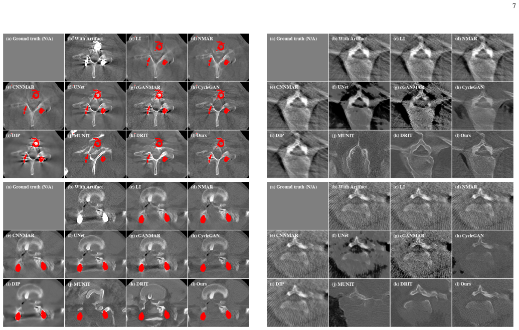
- [Sec. IV-E and abstract] The abstract and Sec. IV-E claim that ADN shows 'better generalization ability over the supervised models' on clinical datasets. This claim is supported only by qualitative visual comparisons; there is no quantitative metric, reader study, blinded evaluation, or statistical test on CL1 or CL2. For a clinical MAR method, the more important claim is that the method removes artifacts without erasing anatomical structures. Some form of quantitative content-preservation check (e.g., lesion/landmark preservation, or comparison of anatomical contours against a reference such as NMAR) is needed to support the generalization claim. Without it, the abstract's central claim is overstated.
- [Eq. (8), Sec. III-B] The artifact consistency loss Lart = E[||(xa - xhat) - (yhat_a - y)||_1] enforces that the residual removed from an artifact-affected image equals the residual added to an artifact-free image. This assumes that the artifact component is additive and independent of the underlying anatomy. The physical mechanisms of metal artifacts (beam hardening, scatter, partial volume) cited in Sec. I do not guarantee such additivity. The paper's own caveat in Sec. IV-G, that an anatomical structure or lesion that looks like metal artifacts 'might also be transferred,' indicates that the artifact code can contain content-dependent information. This undermines the claim in Sec. IV-E that ADN removes dark shadings and streaks 'without introducing secondary artifacts' while preserving anatomy. The authors should either provide evidence that the artifact code does not carry anatomy-specific content (e.g., an experiment on paired data with inserted lesions) or explicitly discuss this as a limitation and potential failure mode.
- [Sec. IV-E, Figs. 7-8] The quantitative evaluation on SYN (Table II) is sound, but the clinical evaluation is purely qualitative without criteria, statistical analysis, or error bars. To justify the paper's contribution, the authors should provide a more rigorous clinical evaluation, for example a reader study with multiple evaluators and inter-rater agreement, or at least a quantitative proxy such as noise/streak metrics and anatomical-structure preservation scores. Alternatively, the claims in the abstract and Sec. I should be tempered to state that ADN shows promising qualitative generalization.
minor comments (5)
- [Sec. IV-E] Please ensure that the abstract's claim of 'better generalization ability' is consistent with the evaluation section, which itself states that only qualitative comparisons are performed on clinical data.
- [Table II] The MUNIT result (SSIM 7.5) is far lower than the other baselines; a brief note explaining that MUNIT prioritizes diverse outputs over structural fidelity would help the reader interpret this outlying value.
- [Sec. III-A] The notation for the artifact-affected encoder E_Ia as a set of two encoders (Ec_Ia and Ea_Ia) could be visually clarified in the text; the current superscript/subscript placement in the PDF is easy to misread.
- [References] References [18] and [24] both refer to CycleGAN, and [19] and [25] both refer to MUNIT; these duplicates should be consolidated.
- [Sec. III-C] Minor grammatical issue: 'the padding of all the convolutional layers in the blocks ... are reflection padding' should be 'is reflection padding.'
Circularity Check
No significant circularity: ADN's self-supervision losses are consistency objectives trained on unpaired data, and the reported SYN metrics are measured on a held-out set against ground truth.
full rationale
The paper's central claim—unsupervised MAR via artifact disentanglement—does not reduce to its inputs by construction. The training objective (Eq. 5) combines adversarial losses, reconstruction (Eq. 7), artifact consistency (Eq. 8), and self-reduction (Eq. 9). Eq. 8 enforces that the residual (xa−x̂) matches the residual (ŷa−y); this is a self-supervised cycle assumption about artifact additivity, not a definitional identity that makes the artifact-removed output equal to an input or a fitted label. Eq. 9 likewise is a cycle-consistency regularizer, and the reported PSNR/SSIM on SYN are computed against ground-truth artifact-free test images withheld from training, so the numerical result is not forced by a fitted parameter. The preliminary-version self-citation [22] is disclosed rather than used as load-bearing justification, and no uniqueness theorem from the authors is invoked to rule out alternatives. The paper's own caveat in Sec. IV.G—that a lesion resembling metal artifacts might be transferred—is a limitation of the artifact-additivity assumption, not a circular step; it affects clinical reliability but does not make the derivation equivalent to its input. The clinical comparison is qualitative, which limits the strength of the generalization claim, but that is an evidence-quality concern outside the circularity definition.
Assumptions & free parameters
free parameters (2)
- Loss weights λadv, λrec, λself, λart =
λadv=1.0; λrec=λself=λart=20.0 for SYN/CL1, 5.0 for CL2
- CL1 group selection thresholds =
HU > 2,500 and metal region > 400 pixels for artifact-affected; max HU < 2,000 for artifact-free
assumptions (3)
- domain assumption Any artifact-affected CT image can be decomposed into independent content and artifact components in latent space.
- domain assumption The artifact code a satisfies (xa - x̂) ≈ (ŷa - y), i.e., the artifact is additive and independent of content.
- domain assumption Images can be reliably grouped into artifact-affected and artifact-free domains.
Cite this review
Pith. "Pith review of ADN: Artifact Disentanglement Network for Unsupervised Metal Artifact Reduction." pith.science (2026). https://pith.science/paper/BI3GYISD
@misc{pith2026190801104,
  author       = {Pith},
  title        = {Pith review of: ADN: Artifact Disentanglement Network for Unsupervised Metal Artifact Reduction},
  year         = {2026},
  howpublished = {\url{https://pith.science/paper/BI3GYISD}},
  note         = {Machine review of arXiv:1908.01104}
}
read the original abstract
Current deep neural network based approaches to computed tomography (CT) metal artifact reduction (MAR) are supervised methods that rely on synthesized metal artifacts for training. However, as synthesized data may not accurately simulate the underlying physical mechanisms of CT imaging, the supervised methods often generalize poorly to clinical applications. To address this problem, we propose, to the best of our knowledge, the first unsupervised learning approach to MAR. Specifically, we introduce a novel artifact disentanglement network that disentangles the metal artifacts from CT images in the latent space. It supports different forms of generations (artifact reduction, artifact transfer, and self-reconstruction, etc.) with specialized loss functions to obviate the need for supervision with synthesized data. Extensive experiments show that when applied to a synthesized dataset, our method addresses metal artifacts significantly better than the existing unsupervised models designed for natural image-to-image translation problems, and achieves comparable performance to existing supervised models for MAR. When applied to clinical datasets, our method demonstrates better generalization ability over the supervised models. The source code of this paper is publicly available at https://github.com/liaohaofu/adn.
Figures


Figures from the paper (5 more)




Reference graph
Works this paper leans on
-
[22]
Artifact disentan- glement network for unsupervised metal artifact reduction,
H. Liao, W.-A. Lin, J. Yuan, S. K. Zhou, and J. Luo, “Artifact disentan- glement network for unsupervised metal artifact reduction,” in Medical Image Computing and Computer Assisted Intervention – MICCAI 2019 , MICCAI. Cham: Springer International Publishing, 2019
work page 2019
-
[1]
Metal artifact reduction in CT: where are we after four decades?
L. Gjesteby, B. D. Man, Y . Jin, H. Paganetti, J. Verburg, D. Giantsoudi, and G. Wang, “Metal artifact reduction in CT: where are we after four decades?” IEEE Access , vol. 4, pp. 5826–5849, 2016
work page 2016
-
[2]
Reduction of ct artifacts caused by metallic implants
W. A. Kalender, R. Hebel, and J. Ebersberger, “Reduction of ct artifacts caused by metallic implants.” Radiology, vol. 164, no. 2, pp. 576–577, 1987
work page 1987
-
[3]
Normalized metal artifact reduction (nmar) in computed tomography,
E. Meyer, R. Raupach, M. Lell, B. Schmidt, and M. Kachelrieß, “Normalized metal artifact reduction (nmar) in computed tomography,” Medical physics , 2010
work page 2010
-
[4]
Convolutional neural network based metal artifact reduction in x-ray computed tomography,
Y . Zhang and H. Yu, “Convolutional neural network based metal artifact reduction in x-ray computed tomography,” IEEE Trans. Med. Imaging , vol. 37, no. 6, pp. 1370–1381, 2018
work page 2018
-
[5]
Conditional genera- tive adversarial networks for metal artifact reduction in ct images of the ear,
J. Wang, Y . Zhao, J. H. Noble, and B. M. Dawant, “Conditional genera- tive adversarial networks for metal artifact reduction in ct images of the ear,” in Medical Image Computing and Computer Assisted Intervention – MICCAI 2018 , 2018
work page 2018
-
[6]
Metal artifact reduction on cervical ct images by deep residual learning,
X. Huang, J. Wang, F. Tang, T. Zhong, and Y . Zhang, “Metal artifact reduction on cervical ct images by deep residual learning,” Biomedical engineering online , vol. 17, no. 1, p. 175, 2018
work page 2018
-
[7]
Deep neural network for ct metal artifact reduction with a perceptual loss function,
L. Gjesteby, H. Shan, Q. Yang, Y . Xi, B. Claus, Y . Jin, B. De Man, and G. Wang, “Deep neural network for ct metal artifact reduction with a perceptual loss function,” in In Proceedings of The Fifth International Conference on Image F ormation in X-ray Computed Tomography , 2018
work page 2018
Show all 33 references
-
[8]
Challenging common assumptions in the unsupervised learning of disentangled representations,
F. Locatello, S. Bauer, M. Lucic, S. Gelly, B. Sch ¨olkopf, and O. Bachem, “Challenging common assumptions in the unsupervised learning of disentangled representations,” arXiv preprint arXiv:1811.12359 , 2018
2018 arXiv
-
[9]
Metal artifact reduction for polychromatic x-ray ct based on a beam-hardening corrector,
H. S. Park, D. Hwang, and J. K. Seo, “Metal artifact reduction for polychromatic x-ray ct based on a beam-hardening corrector,” IEEE transactions on medical imaging , vol. 35, no. 2, pp. 480–487, 2015
2015
-
[10]
An iterative approach to the beam hardening correction in cone beam ct,
J. Hsieh, R. C. Molthen, C. A. Dawson, and R. H. Johnson, “An iterative approach to the beam hardening correction in cone beam ct,” Medical physics, vol. 27, no. 1, pp. 23–29, 2000
2000
-
[11]
Empirical scatter correction (esc): A new ct scatter correction method and its application to metal artifact reduction,
E. Meyer, C. Maaß, M. Baer, R. Raupach, B. Schmidt, and M. Kachel- rieß, “Empirical scatter correction (esc): A new ct scatter correction method and its application to metal artifact reduction,” in IEEE Nuclear Science Symposuim & Medical Imaging Conference . IEEE, 2010, pp. 2036–2041
2010
-
[12]
An algorithm for the reduction of metal clip artifacts in ct reconstructions,
G. H. Glover and N. J. Pelc, “An algorithm for the reduction of metal clip artifacts in ct reconstructions,” Medical physics , vol. 8, no. 6, pp. 799–807, 1981. 10
1981
-
[13]
Image-to-image translation with conditional adversarial networks,
P. Isola, J.-Y . Zhu, T. Zhou, and A. A. Efros, “Image-to-image translation with conditional adversarial networks,” in Proceedings of the IEEE conference on computer vision and pattern recognition , 2017, pp. 1125– 1134
2017
-
[14]
Adver- sarial sparse-view cbct artifact reduction,
H. Liao, Z. Huo, W. J. Sehnert, S. K. Zhou, and J. Luo, “Adver- sarial sparse-view cbct artifact reduction,” in International Conference on Medical Image Computing and Computer-Assisted Intervention . Springer, 2018, pp. 154–162
2018
-
[15]
Perceptual losses for real-time style transfer and super-resolution,
J. Johnson, A. Alahi, and L. Fei-Fei, “Perceptual losses for real-time style transfer and super-resolution,” inEuropean conference on computer vision. Springer, 2016, pp. 694–711
2016
-
[16]
Generative mask pyramid network for ct/cbct metal artifact reduction with joint projection-sinogram correction,
H. Liao, W.-A. Lin, Z. Huo, L. V ogelsang, W. J. Sehnert, S. K. Zhou, and J. Luo, “Generative mask pyramid network for ct/cbct metal artifact reduction with joint projection-sinogram correction,” in International Conference on Medical Image Computing and Computer- Assisted Int...
2019
-
[17]
Dudonet: Dual domain network for ct metal artifact reduction,
W.-A. Lin, H. Liao, C. Peng, X. Sun, J. Zhang, J. Luo, R. Chellappa, and S. K. Zhou, “Dudonet: Dual domain network for ct metal artifact reduction,” in Proceedings of the IEEE Conference on Computer Vision and Pattern Recognition , 2019, pp. 10 512–10 521
2019
-
[18]
Unpaired image-to-image translation using cycle-consistent adversarial networks,
J.-Y . Zhu, T. Park, P. Isola, and A. A. Efros, “Unpaired image-to-image translation using cycle-consistent adversarial networks,” in The IEEE Conference on Computer Vision and Pattern Recognition (CVPR) , 2017
2017
-
[19]
Multimodal unsuper- vised image-to-image translation,
X. Huang, M. Liu, S. J. Belongie, and J. Kautz, “Multimodal unsuper- vised image-to-image translation,” in Computer Vision - ECCV 2018 , 2018
2018
-
[20]
Diverse image-to- image translation via disentangled representations,
H. Lee, H. Tseng, J. Huang, M. Singh, and M. Yang, “Diverse image-to- image translation via disentangled representations,” in Computer Vision - ECCV 2018 , 2018
2018
-
[21]
Deep image prior,
D. Ulyanov, A. Vedaldi, and V . S. Lempitsky, “Deep image prior,” inThe IEEE Conference on Computer Vision and Pattern Recognition (CVPR) , June 2018
2018
-
[23]
Generative adversarial nets,
I. Goodfellow, J. Pouget-Abadie, M. Mirza, B. Xu, D. Warde-Farley, S. Ozair, A. Courville, and Y . Bengio, “Generative adversarial nets,” in Advances in neural information processing systems , 2014
2014
-
[24]
Unpaired image-to-image translation using cycle-consistent adversarial networks,
J. Zhu, T. Park, P. Isola, and A. A. Efros, “Unpaired image-to-image translation using cycle-consistent adversarial networks,” CoRR, vol. abs/1703.10593, 2017
2017 arXiv
-
[25]
Multimodal unsuper- vised image-to-image translation,
X. Huang, M. Liu, S. J. Belongie, and J. Kautz, “Multimodal unsuper- vised image-to-image translation,” CoRR, vol. abs/1804.04732, 2018
2018 arXiv
-
[26]
Unsupervised representation learning with deep convolutional generative adversarial networks,
A. Radford, L. Metz, and S. Chintala, “Unsupervised representation learning with deep convolutional generative adversarial networks,”arXiv preprint arXiv:1511.06434, 2015
2015 arXiv
-
[27]
Deep residual learning for image recognition,
K. He, X. Zhang, S. Ren, and J. Sun, “Deep residual learning for image recognition,” in Proceedings of the IEEE conference on computer vision and pattern recognition , 2016, pp. 770–778
2016
-
[28]
Deconvolution and checkerboard artifacts,
A. Odena, V . Dumoulin, and C. Olah, “Deconvolution and checkerboard artifacts,” Distill, vol. 1, no. 10, p. e3, 2016
2016
-
[29]
Feature pyramid networks for object detection,
T.-Y . Lin, P. Doll´ar, R. Girshick, K. He, B. Hariharan, and S. Belongie, “Feature pyramid networks for object detection,” in Proceedings of the IEEE Conference on Computer Vision and Pattern Recognition , 2017, pp. 2117–2125
2017
-
[30]
U-net: Convolutional networks for biomedical image segmentation,
O. Ronneberger, P. Fischer, and T. Brox, “U-net: Convolutional networks for biomedical image segmentation,” in International Conference on Medical image computing and computer-assisted intervention , 2015
2015
-
[31]
Deeplesion: automated mining of large-scale lesion annotations and universal lesion detection with deep learning,
K. Yan, X. Wang, L. Lu, and R. M. Summers, “Deeplesion: automated mining of large-scale lesion annotations and universal lesion detection with deep learning,” Journal of Medical Imaging , 2018
2018
-
[32]
Deep image prior,
D. Ulyanov, A. Vedaldi, and V . Lempitsky, “Deep image prior,” arXiv:1711.10925, 2017
2017 arXiv
-
[33]
Diverse image-to-image translation via disentangled representations,
H. Lee, H. Tseng, J. Huang, M. K. Singh, and M. Yang, “Diverse image-to-image translation via disentangled representations,” CoRR, vol. abs/1808.00948, 2018
2018 arXiv
Reviewed August 14, 2026 · model on record in the stance chip above.
Discussion (0). Continue with ORCID to comment.