REVIEW 4 major objections 6 minor 38 references
Unsupervised Microvascular Image Segmentation Using an Active Contours Mimicking Neural Network
T0 review · 4 major / 6 minor · reviewed 2026-08-14 · deepseek-v4-flash
Pith's one-line read An unsupervised network using an active-contours loss outperforms supervised vessel segmenters on both benchmark datasets and across datasets.
desk verdict A genuinely new unsupervised vessel-segmentation loss plus morphological pooling layers, but the headline superiority claim rests on very small test sets and no error bars. read the letter →
The pith
A machine-rendered reading of the paper's core claim, the machinery that carries it, and where it could break.
The reading
What carries the argument
The load-bearing mechanism is the morphological ACWE pipeline split into two network ingredients. The image-attachment term $\Gamma$ becomes the active contour loss $L_{AC}$, computed on the intermediate segmentation $\bar S$; the ranking loss $L_{\text{rank}}=\exp(c_2-c_1)$ encodes the prior that vessels are bright; and the curvature operator $(SI\circ IS)^\mu$ is realized by morphological pooling layers that take masked max/min over nine $3\times3\times3$ structuring elements, applied $\mu=3$ times to smooth $\bar S$ into the final mask $S$. The ablation shows both $L_{AC}$ and $L_{\text{rank}}$ are necessary, and replacing $S$ with $\bar S$ in the loss causes tearing artifacts.
What would settle it
A direct test: apply the trained network to the same volumes with intensities inverted, so vessels become darker than the background. If the central claim is right, the segmentation should invert and F1 should collapse; if it does not, the stated ranking-loss mechanism is not the actual driver.
Extended reading notes
Core claim
The paper claims that an unsupervised network can outperform supervised approaches for microvascular segmentation. The segmentation network maps an input intensity volume $I$ to a soft mask $S$, and is trained by minimizing an ACWE-inspired loss in which each voxel is penalized when it disagrees with the sign of the image-attachment term $\Gamma = \|\nabla \bar S\|_1(\alpha(I-c_1)^2-\beta(I-c_2)^2)$, where $c_1,c_2$ are the intensity means inside and outside the current mask and, following the morphological ACWE literature, $\alpha=1,\beta=2$. A ranking loss $\exp(c_2-c_1)$ forces the interior mean above the exterior mean, and non-learned morphological pooling layers implement the $SI\circ IS$ curvature smoothing. On the DeepVess and VesselNN datasets the method reports the best F1, AP, JI, DICE, and mIoU among the compared methods, and in cross-dataset transfer the gap over supervised baselines grows; unsupervised fine-tuning on the test volume improves results further.
Load-bearing premise
The method assumes vessels appear brighter than their surroundings, because the ranking loss pushes the mean intensity inside the segmentation above the mean outside; with inverted contrast it would likely segment the background instead.
Editorial extensions
If this is right
- On both benchmark datasets, the method reports the best F1, AP, JI, DICE, and mIoU among the compared supervised and unsupervised baselines.
- When trained on one dataset and tested on another, the unsupervised method maintains a larger advantage over supervised baselines, and unsupervised fine-tuning on unlabeled target data (Ours-FT) yields the highest cross-dataset scores.
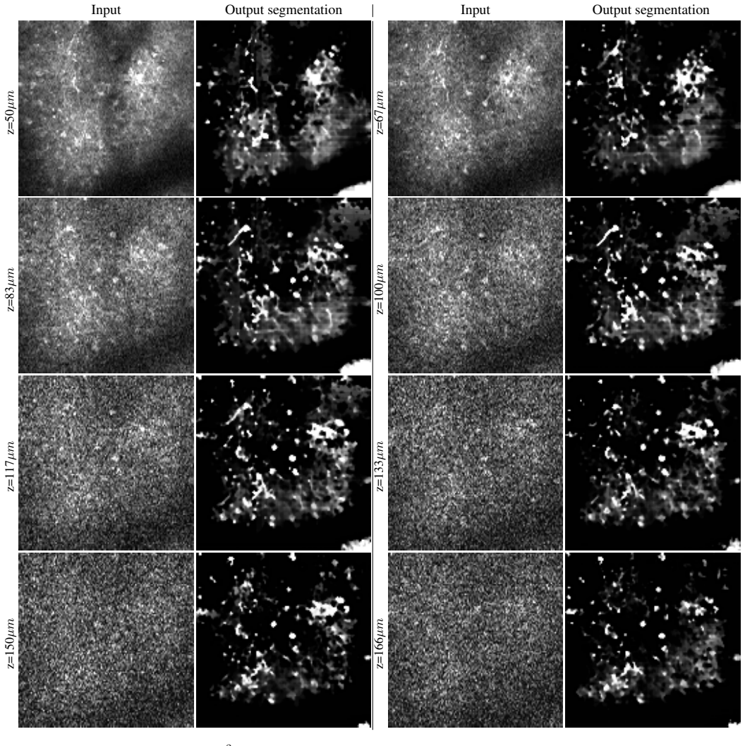
- Without any labels, the method can be applied directly to low-SNR 4D intravital movies; expert reviewers preferred its output over DeepVess in every presented case.
- The ablation indicates the ACWE loss alone already produces competitive segmentation, while adding the ranking loss and morphological smoothing gives the full gain.
Reading between the lines
- The contrast sign is hard-coded, so a natural next step would be to let the network learn whether vessels are bright or dark from the data rather than assuming brightness; the authors do not test inverted-contrast data.
- Because the loss only needs intensity statistics and a smoothness prior, the same architecture could be adapted to other bright tubular structures, such as plant vasculature or engineered microfluidics, though the paper does not claim this.
- The method's success in transductive fine-tuning suggests a broader recipe: unsupervised losses can serve as a domain-adaptation layer on top of any segmentation network, a use the paper mentions but does not develop.
Signed reviews
Editorial analysis
A structured set of objections, weighed in public.
Referee Report
Summary. This paper presents an unsupervised deep learning method for 3D blood vessel segmentation. The key idea is to replace the level-set evolution of morphological Active Contours Without Edges with a differentiable loss and with morphological pooling layers embedded in a 3D ResNet-based autoencoder. The training objective combines an ACWE-inspired image attachment loss, a ranking loss that enforces brighter vessels, a reconstruction loss, a minimal-segmentation loss, and two disjunctive losses. Experiments on the DeepVess and VesselNN datasets, including cross-dataset transfer, compare against supervised baselines (VesselNN, DeepVess) and classical methods (VIDA, Morph-ACWE); further qualitative results are presented on a 4D intravital dataset. The paper claims state-of-the-art unsupervised performance, and the ablation study attributes the gain to the proposed ACWE loss and the ranking loss.
Significance. The idea of designing a network architecture and loss that mimic morphological ACWE is novel and well motivated, and the ablation study (Table 4) demonstrates that the ACWE loss and the ranking loss are important for performance. The method addresses a real need: unsupervised segmentation across imaging-domain shifts, and the transductive fine-tuning protocol (Sec. 4.4) is an interesting way to exploit unlabeled test data. However, the quantitative evidence for the headline claim is not yet statistically convincing, and an internal inconsistency in the reported results must be resolved before the strengths can be fully credited.
major comments (4)
- [Sec. 5.2, Table 4 vs. Table 1] Table 4 reports JI=0.708 for the full method "OursL", while Table 1 reports JI=0.811 for the same method on the same dataset; the other overlapping metrics (AP, F1, DICE, mIoU) match exactly. This is an internal contradiction. Please correct the value or explain the discrepancy (e.g., a different evaluation protocol or train/test split). Without this, the ablation table cannot be considered reliable.
- [Sec. 5.2, Tables 1-3] The central claim that the method outperforms supervised baselines is based on small margins without any measure of uncertainty. On DeepVess, which has a single test volume, the gains over DeepVess are AP 0.909 vs. 0.889, F1 0.829 vs. 0.820, JI 0.811 vs. 0.807. No error bars, number of runs, or significance tests are provided anywhere in the paper. Please report mean ± std over multiple training runs (e.g., different random seeds) and, if possible, bootstrap confidence intervals or paired significance tests over the test volumes. With the current evidence, the observed differences are within plausible run-to-run and train/test-split variability, so the stated outperformance claim is not established.
- [Sec. 4.4, Table 3] The FT (unsupervised fine-tuning) protocol uses the test data, giving the method an information and compute advantage over the supervised baselines, which are not fine-tuned. While the paper marks FT explicitly, the narrative in the abstract and in Sec. 5.2 ("our unsupervised method is able to outperform such previous methods") relies on the FT results. Please either present the no-FT results as the primary cross-domain comparison or provide a fair baseline that also uses test data (e.g., test-time adaptation or a transductive variant). The no-FT rows in Table 3 are often favorable, but the current presentation makes the headline comparison difficult to interpret fairly.
- [Sec. 5 (baseline reimplementations)] The paper states that the reimplementations of DeepVess and VesselNN achieve the same level of results as the original implementations, but it does not report the original numbers or the details of the reimplementation procedure (e.g., hyperparameters, training duration, preprocessing beyond normalization). Without this information, the reader cannot verify that the baselines are not disadvantaged. Please include a side-by-side comparison with the original published metrics and provide the reimplementation settings in the supplementary material or appendix.
minor comments (6)
- [Author affiliations] The first affiliation contains a typo: "School of Computer Sceince" should be "School of Computer Science".
- [Eq. (12)] Equation (12) is garbled: the notation "SI(IS(... (SI(IS| {z } SI◦IS µtimes" is not readable and should be typeset properly, e.g., as an iteration of the composite operator (SI ◦ IS) applied µ times.
- [Sec. 5.2] The sentence "the F1 score and the Average Precision are the more informative metrics" should likely read "the most informative metrics".
- [Sec. 5.2, 4D-NVIV evaluation] The qualitative evaluation on the 4D-NVIV dataset is only descriptive; the expert-preference statement lacks a protocol, the number of experts, and any measure of agreement. A more formal evaluation would strengthen the subjective claim.
- [Sec. 4.3] For reproducibility, the paper should state the batch size, the number of training epochs or iterations, and the learning-rate schedule in addition to the fixed loss weights and optimizer.
- [Eq. (21)] It would be helpful to state explicitly that the default loss in Eq. (21) uses the smoothed output S and that gradients therefore flow through the morphological pooling layers; the ablation that replaces S with S̄ is clear, but the default choice is currently implicit.
Circularity Check
No significant circularity; the ACWE-inspired loss and morphological layers are derived from external classical methods, and the empirical claims are evaluated against external labels.
full rationale
The paper's central derivation takes the morphological ACWE functional from external prior work (Chan-Vese [4] and Marquez-Neila et al. [27]) and translates the PDE evolution into a loss (Eq. 18-21) and the morphological operators into fixed pooling layers (Eq. 14-15). No step defines a prediction in terms of the labels it is later compared against: the loss uses only the input volume I and the network's own segmentation S, while the evaluation metrics use expert annotations that are never inserted into the loss or the network's training procedure. Hyperparameters are stated as fixed across all experiments (Sec. 4.3), and no parameter is fitted to the test labels and then renamed as a prediction. The unsupervised fine-tuning protocol (Sec. 4.4) uses only unlabeled test inputs, which is transductive learning rather than circularity. The paper does cite prior work with overlapping authors, notably [14] for the 4D imaging setup and [34] for a VIDA baseline and dataset details, but these citations are not load-bearing for the central methodological claim; the ACWE foundation itself is external and well-established. Concerns about tiny margins, absence of error bars, and the bright-vessel assumption are statistical robustness or applicability issues, not circular reductions. Under the requirement to exhibit a specific reduction, no circular step can be identified.
Assumptions & free parameters
free parameters (4)
- Loss weights λ1..λ6 =
λ1=1, λ2=1e-2, λ3=λ4=1e-3, λ5=1e-3, λ6=1e-6
- Morphological smoothing iterations μ =
3
- ACWE coefficients α, β =
α=1, β=2
- Input patch size and stride =
32×128×128, stride 8×16×16
assumptions (4)
- domain assumption Vessels are brighter than their surroundings in the input images.
- domain assumption Vessels occupy a small fraction of the image volume.
- ad hoc to paper The gradient magnitude of the network output |∇S̄| can stand in for |∇u| in the ACWE level-set evolution.
- standard math Morphological operators SI and IS can be implemented as masked max/min pooling over the nine structuring elements of a 3×3×3 cube.
Cite this review
Pith. "Pith review of Unsupervised Microvascular Image Segmentation Using an Active Contours Mimicking Neural Network." pith.science (2026). https://pith.science/paper/KBCRE5KU
@misc{pith2026190801373,
  author       = {Pith},
  title        = {Pith review of: Unsupervised Microvascular Image Segmentation Using an Active Contours Mimicking Neural Network},
  year         = {2026},
  howpublished = {\url{https://pith.science/paper/KBCRE5KU}},
  note         = {Machine review of arXiv:1908.01373}
}
read the original abstract
The task of blood vessel segmentation in microscopy images is crucial for many diagnostic and research applications. However, vessels can look vastly different, depending on the transient imaging conditions, and collecting data for supervised training is laborious. We present a novel deep learning method for unsupervised segmentation of blood vessels. The method is inspired by the field of active contours and we introduce a new loss term, which is based on the morphological Active Contours Without Edges (ACWE) optimization method. The role of the morphological operators is played by novel pooling layers that are incorporated to the network's architecture. We demonstrate the challenges that are faced by previous supervised learning solutions, when the imaging conditions shift. Our unsupervised method is able to outperform such previous methods in both the labeled dataset, and when applied to similar but different datasets. Our code, as well as efficient PyTorch reimplementations of the baseline methods VesselNN and DeepVess is available on GitHub - https://github.com/shirgur/UMIS.
Figures


Figures from the paper (5 more)


Reference graph
Works this paper leans on
-
[1]
P. Bouillot, B. M. A. Delattre, O. Brina, R. Ouared, M. Farhat, C. Chnafa, D. A. Steinman, K. olof Lovblad, V . M. Pereira, and M. I. Vargas. 3d phase contrast mri: Par- tial volume correction for robust blood flow quantification in small intracranial vessels. Magnetic resonance in medicine, 79 1:129–140, 2018. 2
work page 2018
-
[2]
V . Caselles, F. Catt ´e, T. Coll, and F. Dibos. A geometric model for active contours in image processing. Numerische mathematik, 66(1):1–31, 1993. 2
work page 1993
-
[3]
V . Caselles, R. Kimmel, and G. Sapiro. Geodesic active con- tours. International journal of computer vision, 22(1):61–79,
-
[4]
T. F. Chan and L. A. Vese. Active contours without edges. IEEE Transactions on image processing , 10(2):266–277,
-
[5]
K. Chhabria, K. Plant, O. Bandmann, R. N. Wilkinson, C. Martin, E. Kugler, P. A. Armitage, P. L. Santoscoy, V . T. Cunliffe, J. Huisken, et al. The effect of hyperglycemia on neurovascular coupling and cerebrovascular patterning in ze- brafish. Journal of Cerebral Blood Flow & Metabolism, page 0271678X18810615, 2018. 1
work page 2018
-
[6]
¨O. C ¸ ic ¸ek, A. Abdulkadir, S. S. Lienkamp, T. Brox, and O. Ronneberger. 3d u-net: learning dense volumetric seg- mentation from sparse annotation. In International confer- ence on medical image computing and computer-assisted in- tervention, pages 424–432. Springer, 2016. 2, 7
work page 2016
- [7]
-
[8]
W. Denk, J. H. Strickler, and W. W. Webb. Two-photon laser scanning fluorescence microscopy. Science, 248 4951:73–6,
Show all 38 references
-
[9]
A. P. Di Giovanna, A. Tibo, L. Silvestri, M. C. M¨ullenbroich, I. Costantini, A. L. A. Mascaro, L. Sacconi, P. Frasconi, and F. S. Pavone. Whole-brain vasculature reconstruction at the single capillary level. Scientific reports, 8(1):12573, 2018. 2
2018
-
[10]
Fischl, D
B. Fischl, D. H. Salat, E. Busa, M. Albert, M. E. Dieterich, C. Haselgrove, A. J. W. van der Kouwe, R. J. Killiany, D. Kennedy, S. Klaveness, A. Montillo, N. Makris, B. Rosen, and A. M. Dale. Whole brain segmentation automated label- ing of neuroanatomical structures in the hu...
2002
-
[11]
H. Fu, Y . Xu, D. W. K. Wong, and J. Liu. Retinal vessel segmentation via deep learning network and fully-connected conditional random fields. 2016 IEEE 13th International Symposium on Biomedical Imaging (ISBI) , pages 698–701,
2016
-
[12]
Guichard, J.-M
F. Guichard, J.-M. Morel, and R. Ryan. Contrast invariant image analysis and pdes. preprint, 35, 2004. 2
2004
-
[13]
Haft-Javaherian, L
M. Haft-Javaherian, L. Fang, V . Muse, C. B. Schaffer, N. Nishimura, and M. R. Sabuncu. Deep convolutional neu- ral networks for segmenting 3d in vivo multiphoton images of vasculature in alzheimer disease mouse models. arXiv preprint arXiv:1801.00880, 2018. 2, 5, 6, 7
2018 arXiv
-
[14]
Har-Gil, L
H. Har-Gil, L. Golgher, S. Israel, D. Kain, O. Cheshnovsky, M. Parnas, and P. Blinder. Pysight: plug and play pho- ton counting for fast continuous volumetric intravital mi- croscopy. Optica, 5(9):1104–1112, 2018. 1, 10
2018
-
[15]
K. He, X. Zhang, S. Ren, and J. Sun. Deep residual learn- ing for image recognition. 2016 IEEE Conference on Com- puter Vision and Pattern Recognition (CVPR) , pages 770– 778, 2016. 3
2016
-
[16]
E. E. Hoover and J. A. Squier. Advances in multiphoton mi- croscopy technology. Nature photonics, 7 2:93–101, 2013. 1, 2
2013
-
[17]
P. Hu, B. Shuai, J. Liu, and G. Wang. Deep level sets for salient object detection. In Proceedings of the IEEE con- ference on computer vision and pattern recognition , pages 2300–2309, 2017. 2
2017
-
[18]
M. Kass, A. Witkin, and D. Terzopoulos. Snakes: Active contour models. International journal of computer vision , 1(4):321–331, 1988. 2
1988
-
[19]
Kichenassamy, A
S. Kichenassamy, A. Kumar, P. Olver, A. Tannenbaum, and A. Yezzi. Gradient flows and geometric active contour mod- els. In Proceedings of IEEE International Conference on Computer Vision, pages 810–815. IEEE, 1995. 2
1995
-
[20]
Y . Kim, S. Kim, T. Kim, and C. Kim. Cnn-based semantic segmentation using level set loss. In 2019 IEEE Winter Con- ference on Applications of Computer Vision (WACV), pages 1752–1760. IEEE, 2019. 2
2019
-
[21]
R. Kimmel. Numerical geometry of images: Theory, algo- rithms, and applications. Springer Science & Business Me- dia, 2003. 2
2003
-
[22]
L. Kong, J. Tang, J. P. Little, Y . Yu, T. L ¨ammermann, C. P. Lin, R. N. Germain, and M. Cui. Continuous volumetric imaging via an optical phase-locked ultrasound lens. Nature methods, 12(8):759, 2015. 1
2015
-
[23]
Lesage, E
D. Lesage, E. D. Angelini, I. Bloch, and G. Funka-Lea. A review of 3d vessel lumen segmentation techniques: Models, features and extraction schemes. Medical image analysis, 13 6:819–45, 2009. 2
2009
-
[24]
Liskowski and K
P. Liskowski and K. Krawiec. Segmenting retinal blood ves- sels with deep neural networks. IEEE Transactions on Med- ical Imaging, 35:2369–2380, 2016. 2
2016
-
[25]
Maninis, J
K.-K. Maninis, J. Pont-Tuset, P. A. Arbel´aez, and L. V . Gool. Deep retinal image understanding. In MICCAI, 2016. 2
2016
-
[26]
Marcos, D
D. Marcos, D. Tuia, B. Kellenberger, L. Zhang, M. Bai, R. Liao, and R. Urtasun. Learning deep structured active contours end-to-end. In Proceedings of the IEEE Conference on Computer Vision and Pattern Recognition , pages 8877– 8885, 2018. 2
2018
-
[27]
Marquez-Neila, L
P. Marquez-Neila, L. Baumela, and L. Alvarez. A morpho- logical approach to curvature-based evolution of curves and surfaces. IEEE Transactions on Pattern Analysis and Ma- chine Intelligence, 36(1):2–17, 2014. 2, 3, 4, 7
2014
-
[28]
Mille and L
J. Mille and L. D. Cohen. Deformable tree models for 2d and 3d branching structures extraction. 2009 IEEE Com- puter Society Conference on Computer Vision and Pattern Recognition Workshops, pages 149–156, 2009. 2
2009
-
[29]
Osher and J
S. Osher and J. A. Sethian. Fronts propagating with curvature-dependent speed: algorithms based on hamilton- jacobi formulations. Journal of computational physics , 79(1):12–49, 1988. 2
1988
-
[30]
Ricci and R
E. Ricci and R. Perfetti. Retinal blood vessel segmentation using line operators and support vector classification. IEEE Transactions on Medical Imaging, 26:1357–1365, 2007. 2
2007
-
[31]
Rupprecht, E
C. Rupprecht, E. Huaroc, M. Baust, and N. Navab. Deep active contours. arXiv preprint arXiv:1607.05074, 2016. 2
2016 arXiv
-
[32]
Teikari, M
P. Teikari, M. Santos, C. Poon, and K. Hynynen. Deep learning convolutional networks for multiphoton microscopy vasculature segmentation. arXiv preprint arXiv:1606.02382,
-
[33]
M. I. Todorov, J. C. Paetzold, O. Schoppe, G. Tetteh, V . Efre- mov, K. V oelgyi, M. Duering, M. Dichgans, M. Piraud, B. Menze, et al. Automated analysis of whole brain vascu- lature using machine learning. bioRxiv, page 613257, 2019. 2
2019
-
[34]
P. S. Tsai, J. P. Kaufhold, P. Blinder, B. Friedman, P. J. Drew, H. J. Karten, P. D. Lyden, and D. Kleinfeld. Correlations of neuronal and microvascular densities in murine cortex re- vealed by direct counting and colocalization of nuclei and vessels. The Journal of neuroscien...
2009
-
[35]
Urban, L
A. Urban, L. Golgher, C. Brunner, A. Gdalyahu, H. Har-Gil, D. Kain, G. Montaldo, L. Sironi, and P. Blinder. Understand- ing the neurovascular unit at multiple scales: advantages and limitations of multi-photon and functional ultrasound imag- ing. Advanced drug delivery reviews...
2017
-
[36]
A. Wu, Z. Xu, M. Gao, M. Buty, and D. J. Mollura. Deep vessel tracking: A generalized probabilistic approach via Layer In-Out Kernel Stride Padding ConvT3D-BR 512-64 (4,4,4) (2,2,2) (1,1,1) ConvT3D-BR 64-64 (1,3,3) (1,1,1) (0,1,1) Upsample - - - - ConvT3D-BR* 320-32 (4,4,4) (2...
2016
-
[37]
A. J. Yezzi, S. Kichenassamy, A. Kumar, P. J. Olver, and A. R. Tannenbaum. A geometric snake model for segmen- tation of medical imagery. IEEE Transactions on Medical Imaging, 16:199–209, 1997. 2
1997
-
[38]
W. R. Zipfel, R. M. E. Williams, and W. W. Webb. Nonlinear magic: multiphoton microscopy in the biosciences. Nature Biotechnology, 21:1369–1377, 2003. 1 A. The decoder architecture The Decoder architecture forDseg andDrec is described in the following table: We denote by ConvT...
2003
Reviewed August 14, 2026 · model on record in the stance chip above.
Discussion (0). Continue with ORCID to comment.