REVIEW 3 major objections 6 minor 9 references
Shadow Detection for Ultrasound Images Using Unlabeled Data and Synthetic Shadows
T0 review · 3 major / 6 minor · reviewed 2026-08-14 · deepseek-v4-flash
Pith's one-line read Ultrasound shadows can be detected without labeled training data by teaching an autoencoder to separate synthetic shadows.
desk verdict A genuinely label-free shadow detection idea with a small, noisy validation; worth engaging, but the evidence does not yet prove it beats supervised baselines. read the letter →
The pith
A machine-rendered reading of the paper's core claim, the machinery that carries it, and where it could break.
The reading
What carries the argument
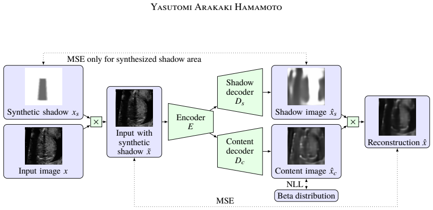
The mechanism is a restricted autoencoder with two output branches and a multiplicative synthetic-shadow prior. The encoder $E$ maps the augmented input $\tilde{x}=x\circ x_s$ to a latent code $z$; the shadow decoder $D_s$ and the content decoder $D_c$ produce $\hat{x}_s$ and $\hat{x}_c$, and the reconstruction is $\hat{x}=\hat{x}_s\circ\hat{x}_c$. Synthetic shadows are random annular sectors on a zero/one background, chosen to mimic convex-probe artifacts. The training loss combines reconstruction MSE, a shadow-prediction MSE evaluated only where the synthetic mask is not 1, a regularization pulling predicted shadows toward 1, and a negative log-likelihood that keeps the predicted content close to a $\beta$ distribution. The load-bearing design is that the network can satisfy the reconstruction loss by putting all shadow variation into $\hat{x}_s$ only if $D_s$ learns the statistics of the injected masks; the masked evaluation of the shadow loss prevents the decoder from needing to invent shadows outside the known injected area.
What would settle it
Take a set of fetal ultrasound frames with manually annotated shadows that clearly deviate from annular sectors (e.g., thin shadows cast by ribs or limbs); if the model's IoU on this set is no better than trivial thresholding, the synthetic-shadow prior is too narrow to support the general shadow-detection claim.
Extended reading notes
Core claim
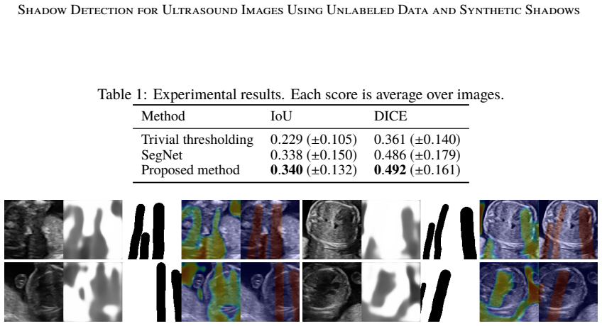
The paper's central claim is that shadows can be separated from ultrasound content by a two-decoder autoencoder trained with a known synthetic-shadow confusion signal, using no annotations in training. An input is decomposed as $\hat{x} = \hat{x}_s \circ \hat{x}_c$, and during training a synthetic mask $x_s$ is multiplied into the input to produce $\tilde{x} = x \circ x_s$; the shadow decoder is penalized for not reproducing $x_s$ exactly inside the synthesized region. This forces the shadow branch to learn the artifact subspace while the content branch models the underlying anatomy. On a test set of 52 clinician-annotated fetal heart frames, the paper reports IoU/DICE of 0.340/0.492 for the proposed method, 0.338/0.486 for supervised SegNet, and 0.229/0.361 for trivial thresholding. The paper reads this as evidence that label-free shadow detection is viable in small-annotation regimes.
Load-bearing premise
The load-bearing assumption is that real fetal ultrasound shadows are well approximated by the multiplicative annular-sector masks used in training; if real shadows differ in shape, opacity, or formation, the network will not have learned to detect them.
Editorial extensions
If this is right
- Hospitals and researchers can train shadow detectors from existing unlabeled ultrasound video archives, bypassing the cost of pixel-level annotation.
- In scenarios where only a small annotated set is available, the label-free method can serve as a viable alternative to supervised segmentation, matching or slightly exceeding its reported scores.
- The same training recipe could transfer to other ultrasound probes, machines, or organs by swapping in the appropriate synthetic-shadow generator, since no labels are required for the new domain.
- Output shadow maps can be used to screen low-quality frames, warn clinicians about shadowed regions, or exclude shadow-heavy images from downstream recognition pipelines.
Reading between the lines
- A fair reading of the reported numbers is that the method establishes parity with supervised training rather than superiority: the IoU gap is far smaller than the reported standard deviations, so a larger annotated test set would be needed to detect a real difference.
- Because the content decoder is regularized toward a beta distribution while the shadow decoder is only weakly constrained outside the synthetic area, the model's notion of "shadow" is likely to be sensitive to the chosen beta parameters; tuning them could act as a detection sensitivity dial in practice.
- The same autoencoding separation could be applied to other multiplicative image artifacts—glare, vignetting, attenuation—by replacing the annular-sector generator with masks that match those formation processes, giving a general template for label-free artifact detection.
Signed reviews
Editorial analysis
A structured set of objections, weighed in public.
Referee Report
Summary. The paper proposes an unsupervised/self-supervised shadow detection method for ultrasound images. A single encoder feeds two decoders that produce a shadow image x̂_s and a shadow-free content image x̂_c, and the input is reconstructed by their element-wise product. To prevent the auto-encoder from learning a trivial split, the authors inject synthetic multiplicative shadows (random annular sectors) into the input and train the shadow decoder to reproduce the known synthetic mask, using an additional beta-distribution prior on content and a regularizer that discourages overly dark shadow predictions. The method is trained on unlabeled fetal-heart ultrasound videos and evaluated on 52 held-out annotated images, where it reaches IoU 0.340 and DICE 0.492, slightly above a supervised SegNet baseline (IoU 0.338, DICE 0.486) and well above trivial thresholding (IoU 0.229). The authors conclude that the method works well in settings with little annotated data.
Significance. If validated, the core idea is practically significant: it replaces expensive pixel-level shadow annotations with automatically generated synthetic shadows while retaining a deep-network feature extractor, which could ease cross-domain and cross-organ transfer. The paper also provides a direct comparison to a supervised baseline, and the reported numbers are competitive. The strengths are the simplicity of the decomposition objective and the fact that the evaluation is performed on real, clinician-annotated shadows rather than only on the synthetic training signal, so the headline result is not a circular fit to the injected masks. The main limitations are the small test set (52 images), the absence of statistical tests, the ambiguous definition of the shadow loss, and the unverified assumption that real acoustic shadows are well approximated by multiplicative annular sectors. For these reasons the significance of the claimed advantage is currently not firmly established.
major comments (3)
- [§2.2, Eq. (1)] The indicator function in the shadow loss is written as 1[\hat x_{sij} \neq 1], but the accompanying sentence says the loss evaluates correctness only where synthetic shadows exist. These two conditions are not the same: the formula penalizes any predicted non-background value even in regions where x_s = 1, whereas the described intention would require an indicator on x_s, e.g., 1[x_{sij} < 1]. This changes the training objective materially. Please clarify the intended indicator and state explicitly which version was used in the reported experiments.
- [§3, Table 1] The proposed method is reported as IoU 0.340 (±0.132) versus SegNet IoU 0.338 (±0.150). The difference of 0.002 is an order of magnitude smaller than the standard deviations, and no paired significance test is provided. As written, the claim that the method is 'slightly better' than SegNet is not statistically supported, and the broader claim that it 'works well in situations with small annotated data' is not established by this single comparison. Please report per-image paired statistics (e.g., Wilcoxon signed-rank test or bootstrap confidence intervals) and, if possible, a supervised baseline trained with different label-budget sizes.
- [§2.2 and §3] The entire self-supervision signal relies on the assumption that real shadows in fetal-heart ultrasound are well approximated by the injected multiplicative annular-sector masks (\tilde x = x ∘ x_s, with x_s ∈ [0,1)). The paper gives no evidence for this assumption, such as a comparison of synthetic and real shadow intensity profiles, a sensitivity analysis over generation parameters, or an ablation that varies the synthetic shadow model. The admitted tendency to predict dark anatomical regions (amniotic fluid, cardiac cavity) as shadows further indicates that the learned decision is not specifically tied to the acoustic shadow formation process. Please provide direct validation of the synthetic shadow model, or at least a sensitivity analysis, to justify that the self-supervision transfers to real shadows.
minor comments (6)
- [§2.1] The text says the reconstruction is 'element-wise product of ˆxs and ˆxs'; presumably the second factor should be ˆxc. Please correct this typo.
- [§2.3] The loss weights are introduced as λ_AE, λ_s, λ_sreg, λ_c but then referred to as λ_recon, λ_shadow, λ_sreg, λ_content. Please unify the notation.
- [§2.2] The synthetic shadow generation process is described only as 'random annular sectors in a rule-based manner'. For reproducibility, please specify the sector radius range, angular width, opacity range, number of sectors per image, and any randomness in placement.
- [Figure 1] The figure labels the shadow loss as 'MSE only for synthesized shadow area', which appears to contradict Eq. (1) as written. This should be reconciled with the clarified indicator function.
- [§2.3] The beta-distribution NLL loss is introduced without reporting the values of α and β or the rationale for choosing them. Please provide the values used in the experiments and a short justification.
- [§3] The test set consists of 52 images from 7 videos, but no information is given about the distribution of shadow sizes or the number of shadow pixels per image. Reporting these statistics would help assess whether the IoU/DICE scores are dominated by easy or hard cases.
Circularity Check
No significant circularity: the synthetic-shadow training signal is an input to training, but the reported test numbers are measured against real clinician labels.
full rationale
The proposed method generates synthetic shadows xs, injects them as x~ = x ∘ xs, and trains the shadow decoder with loss ls only inside the synthetic mask. This is a self-supervised training signal, not the evaluation target. The paper's reported IoU/DICE (Table 1) are computed against pixel-level clinician annotations on held-out test images from separate videos, so the result is not forced by the training loss by construction. The synthetic sector model is an assumption that could limit generalization if real shadows are not multiplicative annular sectors, but that is an empirical correctness risk, not circularity: the paper does not define real-shadow correctness in terms of its synthetic mask. Hyperparameters are selected on a separate validation set, and the supervised baseline SegNet is trained on a subset of validation data; neither step makes the test comparison equivalent to the input. There are no self-citations nor any imported uniqueness theorem. The central derivation is self-contained, and the only strong modeling premise—that real shadows resemble the injected synthetic shadows—is tested indirectly through held-out real labels rather than assumed into the metric.
Assumptions & free parameters
free parameters (4)
- loss weights (λ_AE, λ_s, λ_sreg, λ_c) =
not reported
- Beta distribution parameters (α, β) =
not reported
- Synthetic shadow generation parameters =
not reported
- Network architecture hyperparameters =
not reported
assumptions (3)
- domain assumption Multiplicative shadow formation: x = xs ∘ xc
- domain assumption Real shadows resemble random annular sectors for convex probes
- ad hoc to paper Content pixels follow a Beta distribution
Cite this review
Pith. "Pith review of Shadow Detection for Ultrasound Images Using Unlabeled Data and Synthetic Shadows." pith.science (2026). https://pith.science/paper/L4LPUJYQ
@misc{pith2026190801439,
  author       = {Pith},
  title        = {Pith review of: Shadow Detection for Ultrasound Images Using Unlabeled Data and Synthetic Shadows},
  year         = {2026},
  howpublished = {\url{https://pith.science/paper/L4LPUJYQ}},
  note         = {Machine review of arXiv:1908.01439}
}
read the original abstract
Medical ultrasound is widely used technique for diagnosing internal organs. As common artifacts, shadows often appear in ultrasound images. Detecting such shadows is curious because they prevent accurate diagnosis. In this paper, we propose a novel shadow detection method based on auto-encoding structure. It once separates an input image into a shadow image and a content image using two decoders and combines them to reconstruct the input. To lead the network into separating the input, we inject synthetic shadows into the input and make the network to predict them as the shadow image. Since we know the rough shape of shadows as basic domain knowledge, we can generate plausible shadows. These processes are achieved by using only unlabeled data. Experiments on ultrasound images for fetal heart diagnosis shows the effectiveness of the method.
Figures
Reference graph
Works this paper leans on
-
[1]
SegNet : A deep convolutional encoder-decoder architecture for image segmentation
Vijay Badrinarayanan, Alex Kendall, and Roberto Cipolla. SegNet : A deep convolutional encoder-decoder architecture for image segmentation. IEEE transactions on pattern analysis and machine intelligence, 39 0 (12): 0 2481--2495, 2017
work page 2017
-
[2]
Christopher M. Bishop. Pattern Recognition and Machine Learning. Springer , 2006
work page 2006
-
[3]
Cheng, Juan Shan, Wen Ju, Yanhui Guo, and Ling Zhang
H.D. Cheng, Juan Shan, Wen Ju, Yanhui Guo, and Ling Zhang. Automated breast cancer detection and classification using ultrasound images: A survey. Pattern Recognition, 43 0 (1): 0 299--317, 2010
work page 2010
-
[4]
Pierre Hellier, Pierrick Coup\'e, Xavier Morandi, and D. Louis Collins. An automatic geometrical and statistical method to detect acoustic shadows in intraoperative ultrasound brain images. Medical Image Analysis, 14 0 (2): 0 195--204, 2010
work page 2010
-
[5]
Ultrasound confidence maps using random walks
Athanasios Karamalis, Wolfgang Wein, Tassilo Klein, and Nassir Navab. Ultrasound confidence maps using random walks. Medical Image Analysis, 16 0 (6): 0 1101--1112, 2012
work page 2012
-
[6]
Automatic shadow detection in 2d ultrasound images
Qingjie Meng, Christian Baumgartner, Matthew Sinclair, James Housden, Martin Rajchl, Alberto Gomez, Benjamin Hou, Nicolas Toussaint, Veronika Zimmer, Jeremy Tan, Jacqueline Matthew, Daniel Rueckert, Julia Schnabel, and Bernhard Kainz. Automatic shadow detection in 2d ultrasound images. In Data Driven Treatment Response Assessment and Preterm, Perinatal, a...
work page 2018
-
[7]
Weakly Supervised Estimation of Shadow Confidence Maps in Fetal Ultrasound Imaging
Qingjie Meng, Matthew Sinclair, Veronika Zimmer, Benjamin Hou, Martin Rajchl, Nicolas Toussaint, Alberto Gomez, James Housden, Jacqueline Matthew, Daniel Rueckert, Julia Schnabel, and Bernhard Kainz. Weakly supervised estimation of shadow confidence maps in ultrasound imaging. arXiv:1811.08164 [cs], 2018 b
work page Pith review arXiv 2018
-
[8]
Alison Noble and Djamal Boukerroui
J. Alison Noble and Djamal Boukerroui. Ultrasound image segmentation: A survey. IEEE Transactions on Medical Imaging, 25 0 (8): 0 987--1010, 2006
work page 2006
Show all 9 references
-
[9]
Stacked denoising autoencoders: Learning useful representations in a deep network with a local denoising criterion
Pascal Vincent, Hugo Larochelle, Isabelle Lajoie, Yoshua Bengio, and Pierre-Antoine Manzagol. Stacked denoising autoencoders: Learning useful representations in a deep network with a local denoising criterion. Journal of Machine Learning Research, 11 0 (Dec): 0 3371--3408, 2010
2010
Reviewed August 14, 2026 · model on record in the stance chip above.
Discussion (0). Continue with ORCID to comment.