REVIEW 4 major objections 7 minor 49 references
Classification of Hand Movements from EEG using a Deep Attention-based LSTM Network
T0 review · 4 major / 7 minor · reviewed 2026-08-14 · deepseek-v4-flash
Pith's one-line read This paper claims that an attention-based LSTM classifies left versus right hand movements from EEG with 83.2% accuracy across subjects and 98.3% within a subject.
desk verdict Solid application paper with one credible internal result (attention beats their own LSTM by ~6 points) and a state-of-the-art claim that rests on shaky quoted baselines. read the letter →
The pith
A machine-rendered reading of the paper's core claim, the machinery that carries it, and where it could break.
The reading
What carries the argument
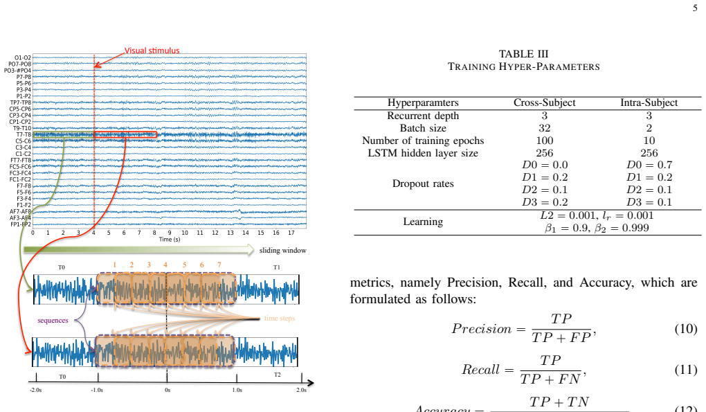
The load-bearing mechanism is the attention-weighted aggregation of LSTM hidden states. The network computes a trainable scalar score $u_i = \tanh(W_s h_i + b_s)$ for each time step's hidden state $h_i$, normalizes these scores with a softmax to get $\alpha_i$, and forms the sequence representation $v = \sum_i \alpha_i h_i$; the final sigmoid classifier reads $v$. This lets the model up-weight time steps that carry the most discriminative EEG information instead of relying only on the last hidden state. The input side is a fixed feature manifold: from each of 27 differential channels, seven time-domain features (mean, variance, skewness, kurtosis, zero crossings, absolute area, peak-to-peak) and four band-power features (delta, $\theta$, $\alpha$, $\beta$) are computed per time step, producing 297 features per step fed to seven LSTM cells.
What would settle it
Re-run the PLV and ANN methods of references [15] and [16] on the exact 103-subject set, 27 differential channels, 2-second segments, and 10-fold cross-subject splits used here; if either re-implementation matches or exceeds 83.2% accuracy, the state-of-the-art claim fails. A simpler proxy: report cross-subject accuracy of the proposed model on the original 109 subjects without excluding the six low-SNR subjects.
Extended reading notes
Core claim
On its own terms, the paper's central claim is that an LSTM with attention, trained on hand-crafted time and frequency features, establishes a new state of the art on the EEG Movement dataset for both validation schemes. Cross-subject accuracy is 83.2% ± 1.2% versus 78.9% for the best benchmark (PLV) and 77.2% for the same LSTM without attention; intra-subject accuracy is 98.3% ± 0.9% versus 88.6% for the previous best (QDA). The authors further claim that the attention layer, which scores each LSTM hidden state and aggregates them as a weighted sum, is the component that lets the network focus on the discriminative time steps, and that the sensors most used by the model sit over frontal-temporal and temporal regions, following a temporal flow from visual cortex to motor cortex that matches known neurophysiology.
Load-bearing premise
The central claim assumes the prior cross-subject accuracies of 78.9% (PLV) and 68.0% (ANN) came from the same preprocessing, channel selection, segment length, and evaluation splits; if those protocols differ, the reported improvement may be a comparison artifact.
Editorial extensions
If this is right
- Cross-subject BCI without per-user calibration moves from a near-chance exercise to an 83%-accurate task on this dataset, making off-the-shelf hand-movement decoding a realistic target.
- The 6-point gap between the LSTM+attention and the plain LSTM (83.2% vs 77.2%) indicates that time-step weighting, not recurrent depth alone, is what buys most of the improvement.
- A no-overlap 10-fold protocol yields accuracy estimates well below earlier overlap-based numbers, so the field's reported gains should be re-read with the evaluation protocol in mind.
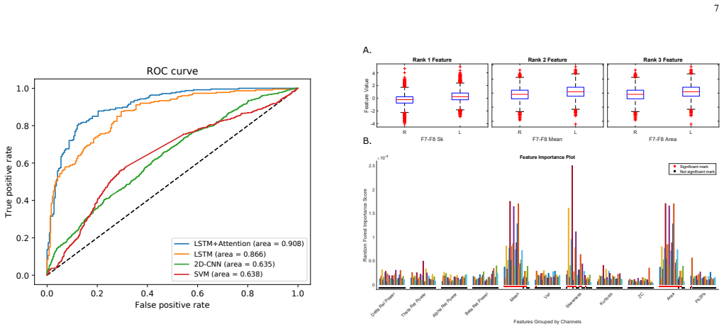
- The top-ranked sensor pairs (FT7-FT8, T9-T10, F7-F8, T7-T8) localize the discriminative signal to frontal-temporal and temporal regions, narrowing where future BCI hardware can place electrodes.
- Intra-subject accuracy of 98.3% suggests that, for a single user, EEG left/right movement discrimination is nearly saturated on this dataset, and remaining headroom is in generalization rather than basic decoding.
Reading between the lines
- If the protocol-match assumption holds, the method's margin over PLV and ANN is likely to shrink or grow depending on how those baselines are re-run; a clean head-to-head under one preprocessing pipeline would settle whether attention-LSTM is inherently better or the gains come from 2-second segments and channel selection.
- The same attention-over-time recipe could transfer to motor imagery EEG, where LSTM-based models already exist, since imagery shares the same time-evolving sensorimotor structure.
- Retraining the model on only the top few sensor pairs (FT7-FT8, T9-T10) is a cheap test of whether the high accuracy is achievable with a reduced montage, which would lower BCI hardware cost.
- The peak in feature importance at roughly 0.75 seconds after the visual stimulus suggests the model's decisions coincide with the completion of visual-to-motor information flow; this could be exploited for early-decision or reaction-time measures, though the paper does not make that claim.
Signed reviews
Editorial analysis
A structured set of objections, weighed in public.
Referee Report
Summary. The paper proposes an attention-based stacked LSTM pipeline for left/right hand movement classification from EEG. Raw signals are pre-processed (channel selection, filtering, normalization), hand-crafted time- and frequency-domain features are extracted over 2-second segments, and a 3-layer 7-cell LSTM with a soft attention layer followed by a sigmoid output is trained. Experiments on the PhysioNet EEG Movement dataset (103 subjects) report 83.2% accuracy in cross-subject 10-fold validation and 98.3% accuracy in intra-subject 10-fold validation, outperforming quoted prior results (PLV 78.9%, ANN 68.0%) and several implemented baselines. The paper also provides a feature-importance analysis and a spatio-temporal interpretation of the attention-informed sensor relevance, linking it to known neurophysiological stages of stimulus processing and movement execution.
Significance. If the reported results are reliable, the paper shows that an attention-based LSTM, combined with a wide feature set and careful sensor selection, can substantially improve cross-subject EEG hand-movement classification, a task known for low generalization. The strengths are the scale of the evaluation (103 subjects, both intra- and cross-subject schemes), the inclusion of multiple implemented baselines, and the attempt to connect model behavior to neuroscientific plausibility. However, the central state-of-the-art claim rests on externally quoted baselines whose protocols are not demonstrated to be matched, and the tuning/segmentation procedure may introduce optimism in the reported numbers. The architecture itself is standard, so the contribution is primarily empirical; the empirical claim therefore needs to be made robust.
major comments (4)
- [§IV-A, §IV-C, Table V] The claim that the proposed model 'significantly outperforms the best performing benchmark' is based on Table V, where the PLV (78.9%) and ANN (68.0%) results are adopted from [15] and [16] without reimplementation. The protocols are not shown to match: the paper excludes 6 of 109 subjects (§IV-A), while [16] is listed in Table I as using 109 subjects, so the ANN comparison may be against a different (larger) subject set. It is also not established that [15] and [16] used the same 27-channel differential montage, the same 2-second/7-step overlapping window construction, the same features, or the same fold definitions. The absence of standard deviations or confidence intervals for these baselines makes the word 'significantly' unsupported even under a matched protocol. The authors should either reimplement these baselines under their own protocol or clearly restrict the comparison to protocol-matched experiments and report uncertainty.
- [§IV-B, §IV-C, Tables III–IV] Hyperparameters (segment size, recurrent depth, batch size, epochs, hidden size, dropout rates, L2 coefficient, Adam parameters) were tuned to achieve the best results on the same 10-fold cross-validation setup used for the final evaluation. Table IV explicitly selects the segment size that maximizes cross-subject accuracy, and Table III lists separate optimal hyperparameter sets for the two validation schemes. Unless tuning was nested inside the training folds, the reported accuracies are optimistic estimates of generalization. The paper should clarify the tuning protocol or perform a nested cross-validation; as written, the cross-subject accuracy of 83.2% may partly reflect test-set selection.
- [§IV-C, Figure 3] The description of the data segmentation raises a potential leakage issue. Figure 3 indicates that the 2-second LSTM sequence consists of 7 time steps with 50% overlap between adjacent windows. If the 10-fold split is applied at the level of individual sequences rather than at the level of subjects or trials, overlapping windows from the same original trial could appear in both training and test sets, inflating the reported accuracy. The sentence 'no overlap existed in the training and testing segments at each fold' is not sufficient to rule out leakage; the authors should specify the split unit (subject, trial, or window) and confirm that no two overlapping windows straddle the train/test boundary.
- [§V, Table V] The paper states that the proposed method 'significantly outperforms' the best benchmark by 'a considerable 5%', but Table V shows a 4.3-percentage-point difference against PLV (83.2 vs. 78.9) and no statistical significance test is reported. For the intra-subject comparison the same issue appears: the 98.3% result is compared with QDA's 88.6% from [11] without confidence intervals for the prior work. The authors should either report paired significance tests (e.g., across folds or across subjects) or soften the 'significant' wording.
minor comments (7)
- [Abstract] The abstract contains a duplicated phrase: 'our proposed solution our method achieves improvements' — this should be corrected.
- [Introduction] There is a typo in Section I: 'cerebreal palsy' should be 'cerebral palsy'.
- [Table I] The table entry 'Rought set' should be 'Rough set'.
- [§IV-C] The text contains an orphaned LaTeX command 'vspace-2mm' that should be removed.
- [Figure 3] The caption contains the garbled text 'Visual s(mulus' which should be 'Visual stimulus'.
- [§V] The phrase 'non-parametric t-test' is an oxymoron; if the authors used a Wilcoxon or Mann-Whitney test, they should name it correctly, and if they used a t-test, they should remove 'non-parametric'.
- [§V] The paper reports 'p < 0.05/297' but does not state explicitly whether this is a Bonferroni correction across the 297 features; the text should say so.
Circularity Check
No circularity: the paper's accuracy claims are empirical results from held-out folds compared against external and implemented baselines; hyperparameter tuning and un-reimplemented literature baselines are correctness risks, not circular reasoning.
full rationale
The paper's central claim is an empirical accuracy comparison, not an analytical derivation. No equation in the paper defines a quantity in terms of the quantity it is claimed to predict. The LSTM+attention model is trained and evaluated on held-out folds (10-fold cross-validation with no overlap between training and testing segments), and its accuracy is compared against independently implemented benchmarks (SVM, 2D-CNN, VGG-16, plain LSTM) as well as literature-reported baselines (PLV [15], ANN [16]). The reported improvements are not forced by construction: the attention mechanism, feature extraction, and network weights are estimated from training folds, and accuracy is measured on test folds. The main adjacent issues—hyperparameters tuned on the same dataset and literature baselines not reimplemented—are overfitting and experimental-protocol concerns, not circular reasoning. There are no self-citations carrying load-bearing uniqueness claims, no fitted parameter renamed as a prediction, and no known result renamed as new. Accordingly, no circular step can be exhibited with a quote, so the score is 0.
Assumptions & free parameters
free parameters (10)
- Segment size =
2.0 s
- LSTM hidden size =
256
- Recurrent depth =
3
- Batch size =
32 (cross-subject), 2 (intra-subject)
- Number of training epochs =
100 (cross-subject), 10 (intra-subject)
- Dropout rates D0-D3 =
0.0/0.7, 0.2, 0.1, 0.2 (cross-subject); 0.7, 0.2, 0.1, 0.1 (intra-subject)
- L2 regularization coefficient =
0.001
- Adam learning rate / beta1 / beta2 =
0.001 / 0.9 / 0.999
- Number of time steps =
7
- Differential sensor pair selection =
27 pairs from 10-10 topology in [23]
assumptions (4)
- domain assumption The hand-crafted feature set (11 features per channel) preserves discriminative information for L/R movement classification.
- domain assumption The 27 differential sensor pairs formed from the non-central channels provide a symmetric and informative representation.
- domain assumption The PhysioNet dataset labels (T1 left, T2 right movements) and segmentation (2-second epochs aligned to stimulus) are correct.
- domain assumption Min-max normalization does not leak information between training and test subjects.
Cite this review
Pith. "Pith review of Classification of Hand Movements from EEG using a Deep Attention-based LSTM Network." pith.science (2026). https://pith.science/paper/ACNQFZKC
@misc{pith2026190802252,
  author       = {Pith},
  title        = {Pith review of: Classification of Hand Movements from EEG using a Deep Attention-based LSTM Network},
  year         = {2026},
  howpublished = {\url{https://pith.science/paper/ACNQFZKC}},
  note         = {Machine review of arXiv:1908.02252}
}
read the original abstract
Classifying limb movements using brain activity is an important task in Brain-computer Interfaces (BCI) that has been successfully used in multiple application domains, ranging from human-computer interaction to medical and biomedical applications. This paper proposes a novel solution for classification of left/right hand movement by exploiting a Long Short-Term Memory (LSTM) network with attention mechanism to learn the electroencephalogram (EEG) time-series information. To this end, a wide range of time and frequency domain features are extracted from the EEG signals and used to train an LSTM network to perform the classification task. We conduct extensive experiments with the EEG Movement dataset and show that our proposed solution our method achieves improvements over several benchmarks and state-of-the-art methods in both intra-subject and cross-subject validation schemes. Moreover, we utilize the proposed framework to analyze the information as received by the sensors and monitor the activated regions of the brain by tracking EEG topography throughout the experiments.
Figures


Figures from the paper (1 more)
Reference graph
Works this paper leans on
-
[15]
Discrimi- nation of EEG-based motor imagery tasks by means of a simple phase information method,
A. Loboda, A. Margineanu, G. Rotariu, and A. M. Lazar, “Discrimi- nation of EEG-based motor imagery tasks by means of a simple phase information method,” International Journal of Advanced Research in Artificial Intelligence, vol. 3, no. 10, 2014
work page 2014
-
[16]
N. T. M. Huong, H. Q. Linh, and L. Q. Khai, “Classification of left/right hand movement EEG signals using event related potentials and advanced features,” inInternational Conference on the Development of Biomedical Engineering in Vietnam . Springer, 2017, pp. 209–215
work page 2017
-
[11]
O. D. Eva and A. M. Lazar, “Comparison of classifiers and statistical analysis for EEG signals used in brain computer interface motor task paradigm,” International Journal of Advanced Research in Artificial Intelligence, vol. 4, no. 1, pp. 8–12, 2015
work page 2015
-
[1]
I. K ¨athner, S. C. Wriessnegger, G. R. M ¨uller-Putz, A. K ¨ubler, and S. Halder, “Effects of mental workload and fatigue on the p300, alpha and theta band power during operation of an erp (p300) brain–computer interface,” Biological Psychology, vol. 102, pp. 118–129, 2014
work page 2014
-
[2]
Brain computer interfacing: Applications and challenges,
S. N. Abdulkader, A. Atia, and M.-S. M. Mostafa, “Brain computer interfacing: Applications and challenges,” Egyptian Informatics Journal, vol. 16, no. 2, pp. 213–230, 2015
work page 2015
-
[3]
Brain–computer interfaces in neurological rehabilitation,
J. J. Daly and J. R. Wolpaw, “Brain–computer interfaces in neurological rehabilitation,” The Lancet Neurology , vol. 7, no. 11, pp. 1032–1043, 2008
work page 2008
-
[4]
Brain–computer interfaces for communication and control,
J. R. Wolpaw, N. Birbaumer, D. J. McFarland, G. Pfurtscheller, and T. M. Vaughan, “Brain–computer interfaces for communication and control,” Clinical Neurophysiology, vol. 113, no. 6, pp. 767–791, 2002. 9
work page 2002
-
[5]
M. Takahashi, K. Takeda, Y . Otaka, R. Osu, T. Hanakawa, M. Gouko, and K. Ito, “Event related desynchronization-modulated functional elec- trical stimulation system for stroke rehabilitation: a feasibility study,” Journal of neuroengineering and rehabilitation , vol. 9, no. 1, p. 56, 2012
work page 2012
Show all 49 references
-
[6]
Neuronal ensemble control of prosthetic devices by a human with tetraplegia,
L. R. Hochberg, M. D. Serruya, G. M. Friehs, J. A. Mukand, M. Saleh, A. H. Caplan, A. Branner, D. Chen, R. D. Penn, and J. P. Donoghue, “Neuronal ensemble control of prosthetic devices by a human with tetraplegia,” Nature, vol. 442, no. 7099, p. 164, 2006
2006
-
[7]
Current trends in graz brain-computer interface (bci) research,
G. Pfurtscheller, C. Neuper, C. Guger, W. Harkam, H. Ramoser, A. Schlogl, B. Obermaier, and M. Pregenzer, “Current trends in graz brain-computer interface (bci) research,” IEEE transactions on Rehabil- itation Engineering, vol. 8, no. 2, pp. 216–219, 2000
2000
-
[8]
An approach to improve the per- formance of subject-independent bcis-based on motor imagery allocating subjects by gender,
J. Cantillo-Negrete, J. Gutierrez-Martinez, R. I. Carino-Escobar, P. Carrillo-Mora, and D. Elias-Vinas, “An approach to improve the per- formance of subject-independent bcis-based on motor imagery allocating subjects by gender,” Biomedical Engineering Online , vol. 13, no. 1, ...
2014
-
[9]
Comparison of designs towards a subject-independent brain-computer interface based on motor imagery,
F. Lotte, C. Guan, and K. K. Ang, “Comparison of designs towards a subject-independent brain-computer interface based on motor imagery,” in 2009 Annual International Conference of the IEEE Engineering in Medicine and Biology Society , pp. 4543–4546
2009
-
[10]
Comprehensive common spatial patterns with temporal structure information of EEG data: minimizing nontask re- lated EEG component,
H. Wang and D. Xu, “Comprehensive common spatial patterns with temporal structure information of EEG data: minimizing nontask re- lated EEG component,” IEEE Transactions on Biomedical Engineering , vol. 59, no. 9, pp. 2496–2505, 2012
2012
-
[12]
Rough set-based classification of EEG signals related to real and imagery motion,
P. Szczuko, “Rough set-based classification of EEG signals related to real and imagery motion,” in IEEE Signal Processing: Algorithms, Architectures, Arrangements, and Applications (SPA) , 2016, pp. 34–39
2016
-
[13]
Comparison of classification ,ethods for EEG signals of real and imaginary motion,
P. Szczuko, M. Lech, and A. Czy ˙zewski, “Comparison of classification ,ethods for EEG signals of real and imaginary motion,” in Advances in Feature Selection for Data and Pattern Recognition . Springer, 2018, pp. 227–239
2018
-
[14]
Real and imaginary motion classification based on rough set analysis of EEG signals for multimedia applications,
P. Szczuko, “Real and imaginary motion classification based on rough set analysis of EEG signals for multimedia applications,” Multimedia Tools and Applications, vol. 76, no. 24, pp. 25 697–25 711, 2017
2017
-
[17]
Logistic regression for single trial EEG classification,
R. Tomioka, K. Aihara, and K.-R. M ¨uller, “Logistic regression for single trial EEG classification,” in Advances in Neural Information Processing Systems, 2007, pp. 1377–1384
2007
-
[18]
Performance analysis of left/right hand movement classification from EEG signal by intelligent algorithms,
S. Bhattacharyya, A. Khasnobish, A. Konar, D. Tibarewala, and A. K. Nagar, “Performance analysis of left/right hand movement classification from EEG signal by intelligent algorithms,” in IEEE Symposium on Computational Intelligence, Cognitive Algorithms, Mind, and Brain (CCMB),...
2011
-
[19]
Single-trial EEG classification of motor imagery using deep convolutional neural networks,
Z. Tang, C. Li, and S. Sun, “Single-trial EEG classification of motor imagery using deep convolutional neural networks,” Optik-International Journal for Light and Electron Optics , vol. 130, pp. 11–18, 2017
2017
-
[20]
A novel deep learning approach for classi- fication of eeg motor imagery signals,
Y . R. Tabar and U. Halici, “A novel deep learning approach for classi- fication of eeg motor imagery signals,” Journal of neural engineering , vol. 14, no. 1, p. 016003, 2016
2016
-
[21]
Lstm-based EEG classification in motor imagery tasks,
P. Wang, A. Jiang, X. Liu, J. Shang, and L. Zhang, “Lstm-based EEG classification in motor imagery tasks,” IEEE Transactions on Neural Systems and Rehabilitation Engineering, vol. 26, no. 11, pp. 2086–2095, 2018
2018
-
[22]
Eeg- based intention recognition from spatio-temporal representations via cascade and parallel convolutional recurrent neural networks,
D. Zhang, L. Yao, X. Zhang, S. Wang, W. Chen, and R. Boots, “Eeg- based intention recognition from spatio-temporal representations via cascade and parallel convolutional recurrent neural networks,” arXiv preprint arXiv:1708.06578, 2017
2017 arXiv
-
[23]
EEG-based emotion recognition in music listening,
Y .-P. Lin, C.-H. Wang, T.-P. Jung, T.-L. Wu, S.-K. Jeng, J.-R. Duann, and J.-H. Chen, “EEG-based emotion recognition in music listening,” IEEE Transactions on Biomedical Engineering, vol. 57, no. 7, pp. 1798–1806, 2010
2010
-
[24]
Automated classification of l/r hand movement EEG signals using advanced feature extraction and machine learning,
M. H. Alomari, A. Samaha, and K. AlKamha, “Automated classification of l/r hand movement EEG signals using advanced feature extraction and machine learning,” arXiv preprint arXiv:1312.2877 , 2013
2013 arXiv
-
[25]
A long short-term memory deep learning network for the prediction of epileptic seizures using EEG signals,
K. M. Tsiouris, V . C. Pezoulas, M. Zervakis, S. Konitsiotis, D. D. Koutsouris, and D. I. Fotiadis, “A long short-term memory deep learning network for the prediction of epileptic seizures using EEG signals,” Computers in Biology and Medicine , vol. 99, pp. 24–37, 2018
2018
-
[26]
Attention- based bidirectional long short-term memory networks for relation clas- sification,
P. Zhou, W. Shi, J. Tian, Z. Qi, B. Li, H. Hao, and B. Xu, “Attention- based bidirectional long short-term memory networks for relation clas- sification,” in Proceedings of Association for Computational Linguistics, vol. 2, 2016, pp. 207–212
2016
-
[27]
Attention-based lstm for aspect- level sentiment classification,
Y . Wang, M. Huang, L. Zhao et al. , “Attention-based lstm for aspect- level sentiment classification,” in Proceedings of the 2016 conference on empirical methods in natural language processing , 2016, pp. 606–615
2016
-
[28]
A critical review of recurrent neural networks for sequence learning,
Z. C. Lipton, J. Berkowitz, and C. Elkan, “A critical review of recurrent neural networks for sequence learning,” arXiv preprint arXiv:1506.00019, 2015
2015 arXiv
-
[29]
Lstm: A search space odyssey,
K. Greff, R. K. Srivastava, J. Koutn ´ık, B. R. Steunebrink, and J. Schmid- huber, “Lstm: A search space odyssey,” IEEE Transactions on Neural Networks and Learning Systems , vol. 28, no. 10, pp. 2222–2232, 2017
2017
-
[30]
Bci2000: a general-purpose brain-computer interface (BCI) system,
G. Schalk, D. J. McFarland, T. Hinterberger, N. Birbaumer, and J. R. Wolpaw, “Bci2000: a general-purpose brain-computer interface (BCI) system,” IEEE Transactions on Biomedical Engineering , vol. 51, no. 6, pp. 1034–1043, 2004
2004
-
[31]
Physionet: components of a new research resource for complex physiologic signals,
P. PhysioToolkit, “Physionet: components of a new research resource for complex physiologic signals,” Circulation. v101 i23. e215-e220
-
[32]
Adam: A method for stochastic optimization,
D. P. Kingma and J. Ba, “Adam: A method for stochastic optimization,” arXiv preprint arXiv:1412.6980 , 2014
2014 arXiv
-
[33]
Decision tree structure based classifi- cation of eeg signals recorded during two dimensional cursor movement imagery,
O. Aydemir and T. Kayikcioglu, “Decision tree structure based classifi- cation of eeg signals recorded during two dimensional cursor movement imagery,” Journal of neuroscience methods , vol. 229, pp. 68–75, 2014
2014
-
[34]
Random forest and filter bank common spatial patterns for eeg-based motor imagery classification,
M. Bentlemsan, E.-T. Zemouri, D. Bouchaffra, B. Yahya-Zoubir, and K. Ferroudji, “Random forest and filter bank common spatial patterns for eeg-based motor imagery classification,” in 2014 5th International Conference on Intelligent Systems, Modelling and Simulation . IEEE, 2014,...
2014
-
[35]
Imagenet: A large-scale hierarchical image database,
J. Deng, W. Dong, R. Socher, L.-J. Li, K. Li, and L. Fei-Fei, “Imagenet: A large-scale hierarchical image database,” in IEEE Conference on Computer Vision and Pattern Recognition (CVPR) , 2009, pp. 248–255
2009
-
[36]
Random forests,
L. Breiman, “Random forests,” Machine learning , vol. 45, no. 1, pp. 5–32, 2001
2001
-
[37]
An extensive empirical study of feature selection metrics for text classification,
G. Forman, “An extensive empirical study of feature selection metrics for text classification,” Journal of Machine Learning Research , vol. 3, no. Mar, pp. 1289–1305, 2003
2003
-
[38]
Exploring large virtual environments by thoughts using a brain–computer interface based on motor imagery and high-level commands,
F. Lotte, A. Van Langhenhove, F. Lamarche, T. Ernest, Y . Renard, B. Ar- naldi, and A. L´ecuyer, “Exploring large virtual environments by thoughts using a brain–computer interface based on motor imagery and high-level commands,” Presence: Teleoperators and Virtual Environments...
2010
-
[39]
A direct demonstration of functional specialization in human visual cortex,
S. Zeki, J. Watson, C. Lueck, K. J. Friston, C. Kennard, and R. Frack- owiak, “A direct demonstration of functional specialization in human visual cortex,” Journal of neuroscience , vol. 11, no. 3, pp. 641–649, 1991
1991
-
[40]
Two visual systems re-viewed,
A. D. Milner and M. A. Goodale, “Two visual systems re-viewed,” Neuropsychologia, vol. 46, no. 3, pp. 774–785, 2008
2008
-
[41]
Visual field maps in human cortex,
B. A. Wandell, S. O. Dumoulin, and A. A. Brewer, “Visual field maps in human cortex,” Neuron, vol. 56, no. 2, pp. 366–383, 2007
2007
-
[42]
The mental prosthesis: assessing the speed of a p300-based brain-computer interface,
E. Donchin, K. M. Spencer, and R. Wijesinghe, “The mental prosthesis: assessing the speed of a p300-based brain-computer interface,” IEEE Transactions on Neural Systems and Rehabilitation Engineering , vol. 8, no. 2, pp. 174–179, 2000
2000
-
[43]
On the usability of electroencephalographic signals for biometric recognition: A survey,
S. Yang and F. Deravi, “On the usability of electroencephalographic signals for biometric recognition: A survey,” IEEE Transactions on Human-Machine Systems (HMS) , vol. 47, no. 6, pp. 958–969, 2017
2017
-
[44]
Factors influencing the latency of simple reaction time,
D. L. Woods, J. M. Wyma, E. W. Yund, T. J. Herron, and B. Reed, “Factors influencing the latency of simple reaction time,” Human Neu- roscience, vol. 9, p. 131, 2015
2015
-
[45]
Separate visual pathways for perception and action,
M. A. Goodale and A. D. Milner, “Separate visual pathways for perception and action,” Trends in Neurosciences , vol. 15, no. 1, pp. 20–25, 1992
1992
-
[46]
Bottom-up visual integration in the medial parietal lobe,
T. Pflugshaupt, M. N ¨osberger, K. Gutbrod, K. P. Weber, M. Linnebank, and P. Brugger, “Bottom-up visual integration in the medial parietal lobe,” Cerebral Cortex, vol. 26, no. 3, pp. 943–949, 2014
2014
-
[47]
Motor functions of the parietal lobe,
L. Fogassi and G. Luppino, “Motor functions of the parietal lobe,” Current Opinion in Neurobiology , vol. 15, no. 6, pp. 626–631, 2005
2005
-
[48]
Motor areas in the frontal lobe of the primate,
R. P. Dum and P. L. Strick, “Motor areas in the frontal lobe of the primate,” Physiology & Behavior , vol. 77, no. 4-5, pp. 677–682, 2002
2002
-
[49]
Wgan domain adap- tation for eeg-based emotion recognition,
Y . Luo, S.-Y . Zhang, W.-L. Zheng, and B.-L. Lu, “Wgan domain adap- tation for eeg-based emotion recognition,” in International Conference on Neural Information Processing . Springer, 2018, pp. 275–286
2018
Reviewed August 14, 2026 · model on record in the stance chip above.
Discussion (0). Continue with ORCID to comment.