REVIEW 4 major objections 4 minor 43 references
Bias and variance reduction and denoising for CTF Estimation
T0 review · 4 major / 4 minor · reviewed 2026-08-14 · deepseek-v4-flash
Pith's one-line read Recovering several zero-crossing rings of the contrast transfer function from a cryo-EM micrograph — without additional assumptions — yields accurate defocus and astigmatism estimates.
desk verdict A solid CTF-estimation methods paper with one real but fixable flaw: the convex-background justification is mathematically wrong, yet the pipeline and experiments still deserve peer review. read the letter →
The pith
A machine-rendered reading of the paper's core claim, the machinery that carries it, and where it could break.
The reading
What carries the argument
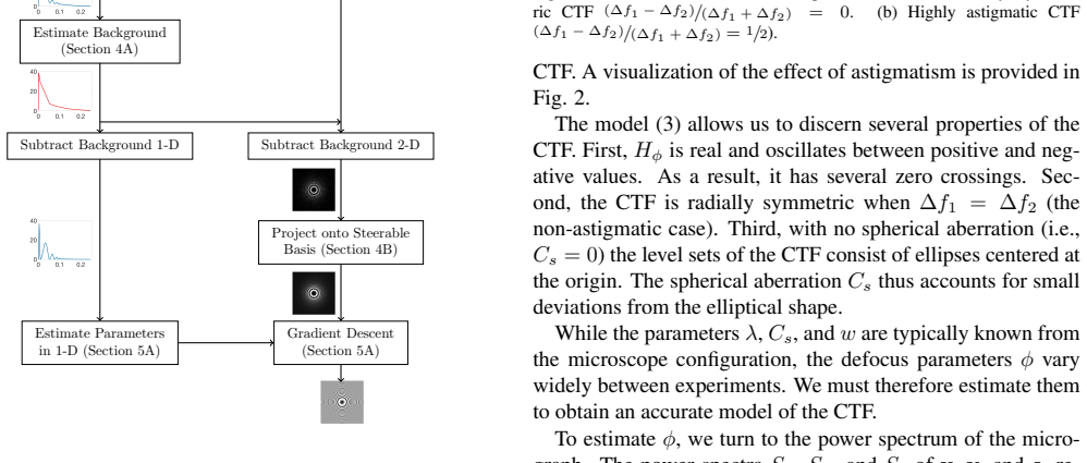
The identity carrying the method is the micrograph power spectrum model $S_y(g) = |H_\varphi(g)|^2 S_x(g) + S_e(g)$: the observed spectrum is the clean projection spectrum modulated by the squared CTF, plus a background. The machinery that runs on it has three parts. The multitaper estimator uses zeroth-order discrete prolate spheroidal sequences as tapers, averaged over half-overlapping blocks, to lower both bias and variance relative to the periodogram. The background model is the non-negative, convex, radially symmetric function, found by linear programming, that is closest to and nowhere larger than the multitaper estimate; the convexity constraint is justified by the claim that the counting-mode background decreases monotonically, or decreases and then increases. The variance-reduction step is the steerable-basis projection: because the CTF's Taylor expansion around zero astigmatism contains only angular frequencies $k = 0, \pm 2$, the cleaned spectrum is projected onto that span, smoothing away noise without removing CTF structure. The final mechanism is the zero-crossing rings: they appear as closed, approximately elliptical local minima of the cleaned spectrum, and each ring obeys $\chi_\varphi(g) = \pi \ell$, giving an overdetermined system of equations solved for the defocus parameters.
What would settle it
Simulate a micrograph from the paper's own model, $S_y = |H_\varphi|^2 S_x + S_e$, with known defocus and a deliberately non-convex background — for instance a radial profile that decreases, rises through a bump, and falls again — or a deliberately non-radial background; run ASPIRE-CTF on it and compare the recovered ring radii and defocus to the ground truth. If the rings shift or disappear whenever the convexity or radial-symmetry premise fails, the claim that several zero-crossing rings are recovered without additional assumptions is falsified. The simulation is fully determined by the paper's model and needs no experimental data.
Extended reading notes
Core claim
The paper's central claim is that the background-subtracted power spectrum $S_y - S_e = |H_\varphi|^2 S_x$ can be estimated cleanly enough that several zero-crossing rings of the CTF are recovered without additional assumptions. Three reductions carry the argument. First, a multitaper estimator built from discrete prolate spheroidal sequence tapers and averaged over half-overlapping blocks replaces the periodogram, cutting both the bias from frequency leakage and the variance that hides the Thon rings. Second, the radially symmetric background is estimated by a linear program that finds the non-negative, convex radial function closest to, and nowhere larger than, the multitaper estimate, leaving a residual that is non-negative and convex and in which the CTF oscillations survive. Third, because the astigmatic CTF's Taylor expansion around $(\Delta f_1 - \Delta f_2) = 0$ involves only the angular frequencies $k = 0, \pm 2$ of a steerable basis, projecting the square root of the residual onto that span suppresses noise without discarding CTF signal. From the cleaned spectrum, defocus and astigmatism are estimated either by maximizing the Pearson correlation of the square root with simulated CTF magnitudes, or by detecting the closed elliptical zero-crossing rings and solving the overdetermined system $\chi_\varphi(g) = \pi \ell$ for the defocus parameters $\varphi = (\Delta f_1, \Delta f_2, \alpha_f)$.
Load-bearing premise
The pipeline's load-bearing premise is that the background noise spectrum is radially symmetric and convex, justified by the claim that a background which decreases, or decreases and then increases, must be convex — a step that rules out wavy or sawtooth-shaped backgrounds, which would bias the subtraction and distort every recovered ring.
Editorial extensions
If this is right
- The power spectrum can be estimated directly from raw movie frames, so CTF estimation can run in parallel with motion correction and is immune to errors introduced by motion-correction software.
- A visibly clean spectrum benefits every downstream cryo-EM stage that assumes a known CTF, including particle picking, denoising, class averaging, ab initio reconstruction, and refinement.
- The zero-crossing-based solver is fast: ASPIRE-CTF takes about 22.5 seconds per micrograph versus about 541 seconds for CTFFIND4 in the paper's comparison, so the cleaner estimate does not cost throughput.
- Defocus and astigmatism estimates stay consistent as the number of summed frames drops from 43 to 5, a regime where the paper reports that Gctf begins to drift in astigmatism.
- The two parameter estimators agree on clean data, and the paper recommends the zero-crossings method for clean micrographs and the correlation method for very low-SNR micrographs.
Reading between the lines
- The convexity constraint is the component most likely to fail in practice; a natural extension is to test the radial profile for convexity before running the linear program, or to replace the fixed constraint with a monotone-plus-bump model — especially since the paper itself notes that zero-crossing positions are extremely sensitive to the background subtraction.
- The steerable-basis fact, that the squared CTF lives almost entirely in the $k = 0, \pm 2$ angular frequencies, is a transferable denoising result: the same projection could clean noise power spectra for Wiener filtering or per-particle defocus refinement, not just the CTF estimation step.
- A testable prediction of the ring-based mechanism is that defocus accuracy degrades once fewer than three closed rings survive, so the method's low-SNR floor could be characterized by counting rings rather than by defocus error alone.
- The 'first without additional assumptions' claim is comparative; an ablation on simulated micrographs with known ground truth — multitaper alone, multitaper plus background subtraction, and the full pipeline — would isolate how much of the ring recovery comes from the spectral estimator versus the background model, a separation the paper does not perform.
Signed reviews
Editorial analysis
A structured set of objections, weighed in public.
Referee Report
Summary. The paper presents ASPIRE-CTF, a pipeline for estimating the contrast transfer function in single-particle cryo-EM from micrograph power spectra. The pipeline combines multitaper spectral estimation to reduce bias and variance, a linear-programming background subtraction that models the background as a non-negative convex radial function (Section 2.3.1), projection of the background-subtracted spectrum onto a low-dimensional steerable basis containing squared CTFs (Section 2.3.2), and two CTF parameter estimation schemes, one based on correlation with simulated CTFs and one based on fitting zero-crossing rings (Section 2.4). The paper claims that this is the first method to produce power spectrum estimates in which several zero-crossing rings of the CTF are easily recovered without additional assumptions. The method is validated on EMPIAR datasets and on the CTF challenge datasets by comparing estimated defocus parameters with CTFFIND4 and Gctf, and a runtime comparison shows substantial speedup over CTFFIND4.
Significance. If the central claim holds, the method would be a useful contribution to cryo-EM CTF estimation. The multitaper-based variance reduction is well motivated, the steerable-basis truncation derivation in Eqs. (19)-(21) is sound under a small-astigmatism assumption, and the LP formulation is clearly stated. The open-source implementation, the reproducibility of the experiments on public data, and the systematic comparison with two widely used tools are notable strengths. However, the background model's convexity assumption is mathematically unjustified and, as the paper itself acknowledges in Section 2.4.2, the zero-crossing output is extremely sensitive to the background subtraction. Because background subtraction is a load-bearing component for the headline claim of accurate zero-crossing recovery, this issue must be addressed before the contribution can be fully credited.
major comments (4)
- [Section 2.3.1, LP (17)]
- [Section 2.3.1, radial symmetry assumption]
- [Section 3.2, Tables 2-3]
- [Section 1 and Section 2.3.1]
minor comments (4)
- [Appendix A, Eq. (32)]
- [Section 2.4.1, Eq. (24)]
- [Section 2.4.1]
- [Section 3.2, Tables 2-3]
Circularity Check
No significant circularity: the LP background fit, steerable-basis projection, and parameter estimations are data-driven and externally validated; the false convexity justification is a correctness issue, not circularity.
full rationale
The central derivation chain is not circular. The multitaper estimate (15) is a standard spectral estimator; the LP background estimate (17) is fit to the multitaper data under non-negativity, upper-bound, and convexity constraints, and the background-subtracted spectrum is then defined as the difference. Zero-crossings of this residual are, by construction, contact points where the fitted convex minorant equals the multitaper estimate, but the paper does not claim these are predicted from the CTF model independently; rather it uses the model (6) to interpret them as CTF zeros, which is a modeling assumption rather than a logical circle. The steerable-basis projection is derived in the paper by Taylor expansion of the CTF (19)-(21) and the expansion coefficients are computed from the data (22); using the same CTF family for subsequent correlation or zero-crossing fitting is model-based inference, not a reduction of the output to the input. The defocus parameters are fitted outputs that are then compared against CTFFIND4 and Gctf on the external CTF challenge datasets, providing independent support. The paper's assertion that a monotonically decreasing (or decrease-then-increase) background 'must be convex' is mathematically false, and Section 2.4.2 itself notes that estimated zero-crossings are 'extremely sensitive to the method of background subtraction'; however, an incorrect or restrictive modeling assumption is a correctness/robustness risk, not circularity. The self-citations to the authors' prior multitaper and steerable-basis work are not load-bearing: the necessary properties are either standard or re-derived here, and the empirical validation is external.
Assumptions & free parameters
free parameters (4)
- Number of DPSS tapers L =
4 (blocks 512x512), 16 (blocks 1024x1024)
- Radial cutoff m/K =
3/8
- Block size K and overlap =
512 or 1024, half-overlapping
- Zero-crossing detection rule =
pixel smaller than at least 6 of 8 neighbors; closed-ring and ellipse checks
assumptions (7)
- domain assumption The micrograph is y = h_phi * x + e with x and e stationary random fields (weak-phase object approximation, Eq. (2)).
- domain assumption The power spectrum satisfies S_y = |H_phi|^2 S_x + S_e (Eq. (6)).
- domain assumption The CTF is H_phi = -sin(chi_phi) with the phase model (4)-(5) and known lambda, C_s, w, p.
- domain assumption S_x and S_e are slowly varying, and S_e is radially symmetric.
- domain assumption Astigmatism is small, so the first-order Taylor expansion (19)-(21) with remainder bounded by ((Delta f1 - Delta f2)/(Delta f1 + Delta f2))^(P+1) is accurate.
- ad hoc to paper The background is non-negative and convex, and the LP constraints (17) are feasible.
- domain assumption Blocks of the micrograph are independent enough that averaging reduces variance by a factor of roughly 1/B (Section 2.2.2).
Cite this review
Pith. "Pith review of Bias and variance reduction and denoising for CTF Estimation." pith.science (2026). https://pith.science/paper/GWJXEBDT
@misc{pith2026190803454,
  author       = {Pith},
  title        = {Pith review of: Bias and variance reduction and denoising for CTF Estimation},
  year         = {2026},
  howpublished = {\url{https://pith.science/paper/GWJXEBDT}},
  note         = {Machine review of arXiv:1908.03454}
}
read the original abstract
When using an electron microscope for imaging of particles embedded in vitreous ice, the objective lens will inevitably corrupt the projection images. This corruption manifests as a band-pass filter on the micrograph. In addition, it causes the phase of several frequency bands to be flipped and distorts frequency bands. As a precursor to compensating for this distortion, the corrupting point spread function, which is termed the contrast transfer function (CTF) in reciprocal space, must be estimated. In this paper, we will present a novel method for CTF estimation. Our method is based on the multi-taper method for power spectral density estimation, which aims to reduce the bias and variance of the estimator. Furthermore, we use known properties of the CTF and of the background of the power spectrum to increase the accuracy of our estimation. We will show that the resulting estimates capture the zero-crossings of the CTF in the low-mid frequency range.
Figures


Figures from the paper (6 more)


Reference graph
Works this paper leans on
-
[1]
Multitaper estimation on arbitrary domains
author And\'en, J. , & author Romero, J. L. ( year 2019 ). title Multitaper estimation on arbitrary domains . note Submitted to SIAM J. Imag. Sci., arXiv:1812.03225 https://arxiv.org/abs/1812.03225
work page Pith review arXiv 2019
-
[2]
author And\'en, J. , & author Singer, A. ( year 2017 ). title Factor analysis for spectral estimation . In booktitle Proc. SampTA \/ (pp. pages 169--173 ). :10.1109/SAMPTA.2017.8024447
arXiv 2017
-
[3]
author Babadi , B. , & author Brown , E. N. ( year 2014 ). title A review of multitaper spectral analysis . journal IEEE Trans. Biomed. Eng. \/ , volume 61 \/ , pages 1555--1564
work page 2014
-
[4]
author Bai, X.-C. , author Fernandez, I. S. , author McMullan, G. , & author Scheres, S. H. W. ( year 2013 ). title Ribosome structures to near-atomic resolution from thirty thousand cryo- EM particles . journal eLife \/ , volume 2 \/ , pages e00461
work page 2013
-
[5]
author Bartesaghi, A. , author Matthies, D. , author Banerjee, S. , author Merk, A. , & author Subramaniam, S. ( year 2014 ). title Structure of -galactosidase at 3.2- A resolution obtained by cryo-electron microscopy . journal Proceedings of the National Academy of Sciences \/ , volume 111 \/ , pages 11709--11714 . :10.1073/pnas.1402809111
-
[6]
author Bhamre, T. , author Zhang, T. , & author Singer, A. ( year 2016 ). title Denoising and covariance estimation of single particle cryo-EM images . journal J. Struct. Biol. \/ , volume 195 \/ , pages 72--81
work page 2016
-
[7]
author Cheng, Y. , author Glaeser, R. M. , & author Nogales, E. ( year 2017 ). title How cryo- EM became so hot . journal Cell \/ , volume 171 \/ , pages 1229--1231
work page 2017
-
[8]
author Erickson, H. P. , & author Klug, A. ( year 1971 ). title Measurement and compensation of defocusing and aberrations by F ourier processing of electron micrographs . journal Phil. Trans. R. Soc. Lond. B \/ , volume 261 \/ , pages 105--118
work page 1971
Show all 43 references
-
[9]
author Fern\' a ndez, J. J. , author Sanjurjo, J. R. , & author Carazo, J.-M. ( year 1997 ). title A spectral estimation approach to contrast transfer function detection in electron microscopy . journal Ultramicroscopy \/ , volume 68 \/ , pages 267--295
1997
-
[10]
( year 1996 )
author Frank, J. ( year 1996 ). title Three-Dimensional Electron Microscopy of Macromolecular Assemblies \/ . publisher Academic Press
1996
-
[11]
, author Rohou, A
author Grant, T. , author Rohou, A. , & author Grigorieff, N. ( year 2018 ). title cisTEM , user-friendly software for single-particle image processing . journal eLife \/ , volume 7 \/ , pages e35383
2018
-
[12]
, author And \'e n, J
author Heimowitz, A. , author And \'e n, J. , & author Singer, A. ( year 2018 ). title APPLE picker: A utomatic particle picking, a low-effort cryo- EM framework . journal J. Struct. Biol. \/ , volume 204 \/ , pages 215--227
2018
-
[13]
author Herzik, M. A. , author Wu, M. , & author Lander, G. C. ( year 2019 ). title High-resolution structure determination of sub-100 kDa complexes using conventional cryo- EM . journal Nat. Commun. \/ , volume 10 \/ , pages 1032 . :10.1038/s41467-019-08991-8
2019 doi
-
[14]
, author Baldwin, P
author Huang, Z. , author Baldwin, P. R. , author Mullapudi, S. , & author Penczek, P. A. ( year 2003 ). title Automated determination of parameters describing power spectra of micrograph images in electron microscopy . journal J. Struct. Biol. \/ , volume 144 \/ , pages 79--94
2003
-
[15]
, author Korir, A
author Iudin, A. , author Korir, A. K. , author Salavert-Torres, J. , author Kleywegt, G. J. , & author Patwardhan, A. ( year 2016 ). title EMPIAR : A public archive for raw electron microscopy image data . journal Nat. Methods \/ , volume 13 \/
2016
-
[16]
, & author Shkolnisky, Y
author Landa, B. , & author Shkolnisky, Y. ( year 2017 ). title Approximation scheme for essentially bandlimited and space-concentrated functions on a disk. journal Appl. Comput. Harmon. Anal. \/ , volume 43 \/ , pages 381--403
2017
-
[17]
, & author Shkolnisky, Y
author Landa, B. , & author Shkolnisky, Y. ( year 2018 ). title Steerable principal components for space-frequency localized images . journal SIAM J. Imaging Sci. \/ , volume 10 \/ , pages 508--534
2018
-
[18]
, author Zheng, S
author Li, X. , author Zheng, S. Q. , author Egami, K. , author Agard, D. A. , & author Cheng, Y. ( year 2013 ). title Influence of electron dose rate on electron counting images recorded with the K2 camera . journal J. Struct. Biol. \/ , volume 184 \/ , pages 251--260
2013
-
[19]
, author Carragher, B
author Marabini, R. , author Carragher, B. , author Cheni, S. , author Chen, J. , author Cheng, A. , author Downing, K. H. et al. ( year 2015 ). title CTF C hallenge: R esult summary . journal J. Struct. Biol. \/ , volume 190 \/ , pages 348--359
2015
-
[20]
author Mindell, J. A. , & author Grigorieff, N. ( year 2003 ). title Accurate determination of local defocus and specimen tilt in electron microscopy . journal J. Struct. Biol. \/ , volume 142 \/ , pages 334--347
2003
-
[21]
author Oppenheim, A. V. , & author Schafer, R. W. ( year 1989 ). title Discrete-Time Signal Processing \/ . ( edition 1st ed.). publisher Prentice Hall
1989
-
[22]
author Percival, D. B. , & author Walden, A. T. ( year 1993 ). title Spectral Analysis for Physical Applications \/ . publisher Cambridge University Press
1993
-
[23]
author Powell, M. J. D. ( year 1970 ). title Numerical Methods for Nonlinear Algebraic Equations \/
1970
-
[24]
, author Rubinstein, J
author Punjani, A. , author Rubinstein, J. L. , author Fleet, D. J. , & author Brubaker, M. A. ( year 2017 ). title cryoSPARC : algorithms for rapid unsupervised cryo-EM structure determination . journal Nat. Methods \/ , volume 14 \/ , pages 290
2017
-
[25]
, & author Grigorieff, N
author Rohou, A. , & author Grigorieff, N. ( year 2015 ). title CTFFIND4 : Fast and accurate defocus estimation from electron micrographs . journal J. Struct. Biol. \/ , volume 192 \/ , pages 216--221
2015
-
[26]
author Scheres, S. H. ( year 2015 ). title Semi-automated selection of cryo-EM particles in RELION-1.3 . journal J. Struct. Biol. \/ , volume 189 \/ , pages 114--122 . :https://doi.org/10.1016/j.jsb.2014.11.010
2015 doi
-
[27]
author Scheres, S. H. W. ( year 2012 ). title RELION : Implementation of a Bayesian approach to cryo-EM structure determination . journal J. Struct. Biol. \/ , volume 180 \/ , pages 519--530
2012
-
[28]
( year 1978 )
author Slepian, D. ( year 1978 ). title Prolate spheroidal wave functions, Fourier analysis, and uncertainty--- V : The discrete case . journal Bell Syst. Tech. J. \/ , volume 57 \/ , pages 1371--1430
1978
-
[29]
, author Jonic, S
author Sorzano, C. , author Jonic, S. , author N\' u \ n ez-Ram\' i rez , R. , author Boisset, N. , & author Carazo, J. M. ( year 2007 ). title Fast, robust, and accurate determination of transmission electron microscopy contrast transfer function . journal J. Struct. Biol. \/...
2007
-
[30]
, author Peng, L
author Tang, G. , author Peng, L. , author Baldwin, P. R. , author Mann, D. S. , author Jiang, W. , author Rees, I. et al. ( year 2007 ). title EMAN2 : An extensible image processing suite for electron microscopy . journal J. Struct. Biol. \/ , volume 157 \/ , pages 38--46
2007
-
[31]
, author Sasab, H
author Tani, K. , author Sasab, H. , & author Toyoshima, C. ( year 1996 ). title A set of computer programs for determining defocus and astigmatism in electron images . journal Ultramicroscopy \/ , volume 65 \/ , pages 31--44
1996
-
[32]
author Thomson , D. J. ( year 1982 ). title Spectrum estimation and harmonic analysis . journal Proc. IEEE \/ , volume 70 \/ , pages 1055--1096
1982
-
[33]
( year 1971 )
author Thon, F. ( year 1971 ). title Phase contrast electron microscopy . In editor U. Valdr\' e (Ed.), booktitle Electron Microscopy in Material Science \/ . publisher Academic Press
1971
-
[34]
, author Franken, E
author Vulovic, M. , author Franken, E. M. , author Ravelli, R. B. G. , author van Vliet, L. J. , & author Rieger, B. ( year 2012 ). title Precise and unbiased estimation of astigmatism and defocus in transmission electron microscopy . journal Ultramicroscopy \/ , volume 116 \...
2012
-
[35]
author Welch, P. D. ( year 1967 ). title The use of fast Fourier transform for the estimation of power spectra: A method based on time averaging over short, modified periodograms . journal IEEE Trans. Audio Electroacoust. \/ , volume 15 \/ , pages 70--73
1967
-
[36]
, author Bai, X.-C
author Wong, W. , author Bai, X.-C. , author Brown, A. , author Fernandez, I. S. , author Hanssen, E. , author Condron, M. et al. ( year 2014 ). title Cryo- EM structure of the Plasmodium falciparum 80S ribosome bound to the anti-protozoan drug emetine . journal eLife \/ , volume 3 \/
2014
-
[37]
, author Li, K
author Yan, R. , author Li, K. , & author Jiang, W. ( year 2017 ). title Real-time detection and single-pass minimization of TEM objective lens astigmatism . journal J. Struct. Biol. \/ , volume 197 \/ , pages 210--219
2017
-
[38]
( year 2016 )
author Zhang, K. ( year 2016 ). title G ctf: R eal-time CTF determination and correction . journal J. Struct. Biol. \/ , volume 193 \/ , pages 1--12
2016
-
[39]
, author Falcon, B
author Zhang, W. , author Falcon, B. , author Murzin, A. G. , author Fan, J. , author Crowther, R. A. , author Goedert, M. et al. ( year 2019 ). title Heparin-induced tau filaments are polymorphic and differ from those in A lzheimer's and P ick's diseases . journal eLife \/ , ...
2019 doi
-
[40]
, author Shkolnisky, Y
author Zhao, Z. , author Shkolnisky, Y. , & author Singer, A. ( year 2016 ). title Fast steerable principal component analysis . journal IEEE Trans. Comput. Imaging \/ , volume 2 \/ , pages 1--12
2016
-
[41]
, & author Singer, A
author Zhao, Z. , & author Singer, A. ( year 2013 ). title F ourier-- B essel rotational invariant eigenimages . journal J. Opt. Soc. Am. A \/ , volume 30 \/ , pages 871--877 . :10.1364/JOSAA.30.000871
2013 doi
-
[42]
author Zheng, S. Q. , author Palovcak, E. , author Armache, J.-P. , author Verba, K. A. , author Cheng, Y. , & author Agard, D. A. ( year 2017 ). title MotionCor2 : anisotropic correction of beam-induced motion for improved cryo-electron microscopy . journal Nat. Methods \/ , ...
2017
-
[43]
, author Penczek, P
author Zhu, J. , author Penczek, P. A. , author Schr\" o der, R. , & author Frank, J. ( year 1997 ). title Three-dimensional reconstruction with contrast transfer function correction from energy-filtered cryoelectron micrographs: Procedure and application to the 70S Escherichi...
1997
Reviewed August 14, 2026 · model on record in the stance chip above.
Discussion (0). Continue with ORCID to comment.