REVIEW 2 major objections 6 minor 27 references
Star-convex Polyhedra for 3D Object Detection and Segmentation in Microscopy
T0 review · 2 major / 6 minor · reviewed 2026-08-14 · deepseek-v4-flash
Pith's one-line read This paper claims that predicting star-convex polyhedra directly from 3D microscopy volumes yields more accurate nuclear instance segmentation than watershed or U-Net baselines, especially in dense, low-signal, anisotropic data.
desk verdict A solid, well-engineered 3D extension of StarDist with a genuinely useful anisotropy fix; the main caveat is the tiny test set and missing per-volume variance. read the letter →
The pith
A machine-rendered reading of the paper's core claim, the machinery that carries it, and where it could break.
The reading
What carries the argument
The load-bearing object is the parameter-efficient star-convex polyhedron. A shape is star-convex if there exists an interior point from which every boundary point is visible along a straight segment. The paper represents the boundary by $n$ distances $d_k$ along unit rays $\vec{r}_k$, where the rays come from a spherical Fibonacci lattice, $z_k=-1+2k/(n-1)$, $x_k=\sqrt{1-z_k^2}\cos(2\pi(1-\phi^{-1})k)$, $y_k=\sqrt{1-z_k^2}\sin(2\pi(1-\phi^{-1})k)$ with $\phi=(1+\sqrt5)/2$, then scaled by an anisotropy factor $\vec{s}$ so that squeezed nuclei are represented accurately. The network predicts, for every grid pixel, the object probability $p$ and the radial distances $\{d_k\}$; non-maximum suppression then removes duplicate candidates using successively tighter bounds: bounding spheres (upper), inscribed spheres (lower), convex hulls (upper), kernels (lower), and exact rasterization only when needed. This combination makes the representation compact, anisotropy-aware, and fast enough to process volumes with over ten thousand candidates in seconds.
What would settle it
Run the trained model on a 3D dataset whose ground-truth nuclei include pronounced concave invaginations, such as lobed or folded nuclei; if those instances cannot be written as a star-convex polyhedron from any interior pixel, the radial-distance representation will miss boundary points, and the method's accuracy at high IoU thresholds should drop below the U-Net baseline.
Extended reading notes
Core claim
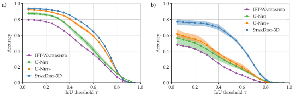
The central discovery is that star-convex polyhedra are a practical and accurate shape space for 3D instance segmentation of cell nuclei. The paper shows that, with only 64 to 96 radial directions arranged on a Fibonacci-lattice sphere and scaled by an anisotropy factor computed from ground-truth bounding boxes, a convolutional network can reconstruct nuclear shapes faithfully. Because each interior pixel predicts the full boundary distances, dense clusters of touching nuclei are disambiguated by non-maximum suppression over candidate polyhedra. On the WORM dataset the method reaches accuracy 0.765 at IoU threshold 0.5, versus 0.700 for the best U-Net variant, and on the harder PARHYALE dataset it reaches 0.593 versus 0.280. The authors conclude that the star-convex shape model, not just the network, is what carries the improvement, particularly in low-signal anisotropic volumes.
Load-bearing premise
The load-bearing premise is that every nucleus in the target data can be approximated by a star-convex polyhedron: from some interior point, the straight segment to every boundary point stays inside the nucleus.
Editorial extensions
If this is right
- For anisotropic fluorescence volumes, adapting the radial directions via the automatically computed factor $\vec{s}=(1,1,7.1)$ is critical: ignoring it drops accuracy from 0.593 to 0.291 at $\tau=0.5$ on PARHYALE.
- The method needs far less annotated data: with only 4.15% of the training and validation volumes (472 instances) it matches U-Net+'s accuracy of 0.7 at $\tau=0.5$ on WORM.
- The same pipeline transfers to any object whose shape is well represented by star-convex polyhedra, since bounding boxes and spheres are special cases.
- The NMS filtering scheme makes instance segmentation feasible on large stacks: a $1141\times140\times140$ volume with 12,000 candidates is processed in about 9 seconds.
Reading between the lines
- Beyond the paper's experiments, the same model and NMS machinery should transfer to any roughly star-shaped object in volumetric images, such as cell bodies, organelle clusters, or tumor spheroids, as long as the radial-visibility condition holds.
- A testable extension would be to estimate the anisotropy factor $\vec{s}$ from raw data statistics rather than from ground-truth bounding boxes; if that suffices, the method would need no annotated volume to set its shape prior.
- The successive-bounds NMS scheme is a general geometric pruning idea: any shape family with cheap outer and inner convex approximations could reuse it to accelerate overlap suppression.
- One could measure how accuracy varies with signal-to-noise ratio and packing density; the paper's PARHYALE results suggest the star-convex prior helps most precisely when neighboring nuclei fuse easily.
Signed reviews
Editorial analysis
A structured set of objections, weighed in public.
Referee Report
Summary. This paper extends the 2D StarDist approach to 3D fluorescence microscopy by representing cell nuclei as star-convex polyhedra parameterized by radial distances along rays sampled from a spherical Fibonacci lattice, adapted to anisotropic voxel sizes via a median-bounding-box anisotropy factor. A ResNet-style 3D CNN densely predicts object probability and radial distances, followed by non-maximum suppression that uses successively tighter bounds (bounding spheres, inscribed spheres, convex hulls, kernels) before exact rasterization. The method is evaluated on two datasets, WORM and PARHYALE, against IFT-Watershed, 3D U-Net, and U-Net with watershed postprocessing, and the authors claim that StarDist-3D outperforms these baselines, especially on low-signal, anisotropic data. The paper also includes an ablation over the amount of training data and a reconstruction-fidelity study of the ray representation.
Significance. If the empirical claim is supported, the paper is a useful and practical contribution: it makes star-convex shape prediction feasible in 3D with only 96 radial distances instead of a naive 1024, handles anisotropic volumes without manual tuning, and provides an efficient NMS procedure for star-convex polyhedra. The method builds transparently on prior work [21], uses a standard evaluation protocol (Hungarian matching, validation-tuned thresholds), averages results over five training trials, and releases code. The main limitation is statistical: the comparative claim rests on very small test sets, and the paper does not report per-volume variability or significance tests, which is load-bearing for the stated conclusion of superior performance.
major comments (2)
- [Section 3.2, Table 1 and Fig. 4] The central comparative claim is supported by averages over 5 training trials, but the test data consist of only 2 volumes for PARHYALE and 7 for WORM, and no per-volume accuracy, standard deviation, or significance test is reported. Because the accuracy gap at tau=0.5 on PARHYALE (0.593 vs. 0.280 for U-Net+) is computed from two test volumes with thresholds tuned on a single validation volume, one anomalous volume could drive the reported difference. Please report per-volume accuracies for at least tau in {0.3, 0.5, 0.7}, provide standard deviations or confidence intervals, and run a paired comparison across test volumes (or state explicitly that the sample size precludes significance testing).
- [Section 3.2, paragraph after Table 1] The anisotropy ablation is reported only as "not shown" in the text, with the claim that accuracy drops from 0.593 to 0.291 for tau=0.5 if the radial directions are not adapted to anisotropy. Since adapting the representation to anisotropy is one of the three stated contributions, this result should be shown in a table or figure with the same trial and variance reporting as the main experiments, including the exact configuration compared (presumably s=(1,1,1) versus s=(1,1,7.1) on PARHYALE) and the number of trials.
minor comments (6)
- [Section 3.1, hyperparameter list] The NMS overlap threshold used to suppress candidates is not stated, even though it directly controls the final instance count and is a free parameter of the method; please specify its value and whether it was fixed or tuned on validation data.
- [Table 1] The table reports averages over 5 independent trials without any measure of variance; adding standard deviations or ranges would make the comparison much more informative, especially since Fig. 4 only shows best and worst results as shading.
- [Section 2.1] The description of rays as "approximately evenly distributed on an ellipsoid representative of the objects" is slightly confusing because the rays are normalized to unit length after anisotropic scaling; please clarify that the final unit rays are on the sphere but with a density adapted to the anisotropy of the objects.
- [Section 3.1, baselines] The baseline set is limited to IFT-Watershed and 3D U-Net variants; a brief justification of why these are representative baselines for 3D fluorescence microscopy instance segmentation, or a comparison with at least one more recent deep instance-segmentation method, would strengthen the comparative claim.
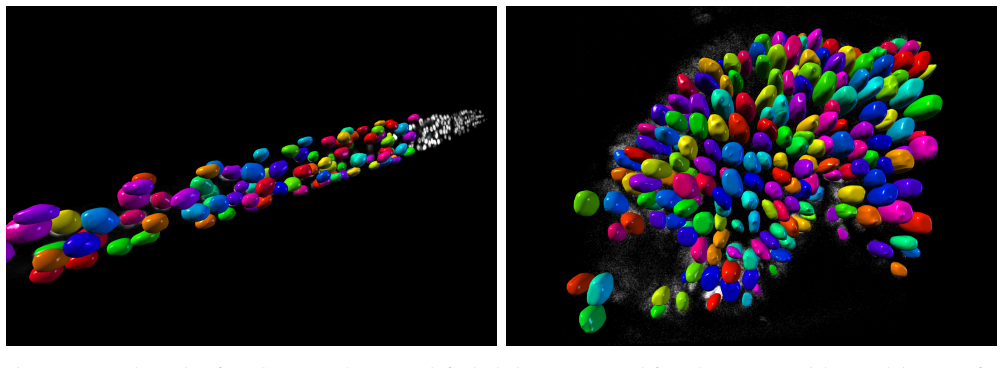
- [Figure 6] The example results in Fig. 6 would benefit from scale bars, as the lateral and axial views are otherwise difficult to compare quantitatively.
- [Discussion] The paper correctly notes that the method applies to objects well approximated by star-convex polyhedra, but this limitation is not tested on objects with significant concave indentations; a quantitative experiment on such data would make the scope claim more robust.
Circularity Check
No circularity: StarDist-3D's claims rest on held-out evaluation with independently defined baselines.
full rationale
The paper's derivation chain is self-contained. StarDist-3D trains a CNN to predict star-convex polyhedron parameters and object probabilities from input volumes, with the loss defined in Eqs. (1)-(3); all quantities entering the method, including the number of rays, the Fibonacci-lattice ray directions, and the anisotropy vector s, are set from training/validation data before evaluation on held-out test volumes. The anisotropy factor is computed as the median bounding box size of objects in the training images and is used only to define the ray set; Fig. 2 measures reconstruction fidelity of ground-truth instances and is not presented as a test-set prediction, so no fitted value is renamed as a prediction. The only self-citation is the authors' prior 2D StarDist paper [21], which is cited transparently as the methodological starting point and is not used as evidence for the empirical comparison; the central claim of superior results is supported by Table 1 and Fig. 4 against external methods (IFT-Watershed, U-Net, U-Net+). No uniqueness theorem is imported, no ansatz is smuggled in via citation, and no known result is merely renamed. The small size of the test sets and absence of per-volume confidence intervals are legitimate statistical-evidence concerns, but they concern correctness and robustness, not circularity.
Assumptions & free parameters
free parameters (6)
- Distance loss weight lambda_d =
0.1
- Regularization weight lambda_reg =
1e-4
- Number of radial directions n =
96
- Number of residual blocks m =
3
- Anisotropy vector s =
(1,1,1) for WORM, (1,1,7.1) for PARHYALE
- NMS overlap threshold =
not reported
assumptions (4)
- domain assumption Cell nuclei are approximately star-convex from an interior point
- standard math The spherical Fibonacci lattice gives sufficiently uniform ray directions
- domain assumption A single anisotropy factor derived from training bounding boxes generalizes to test volumes
- domain assumption The CNN can learn the mapping from image intensities to radial distances and object probability
Cite this review
Pith. "Pith review of Star-convex Polyhedra for 3D Object Detection and Segmentation in Microscopy." pith.science (2026). https://pith.science/paper/RZ6XDH6C
@misc{pith2026190803636,
  author       = {Pith},
  title        = {Pith review of: Star-convex Polyhedra for 3D Object Detection and Segmentation in Microscopy},
  year         = {2026},
  howpublished = {\url{https://pith.science/paper/RZ6XDH6C}},
  note         = {Machine review of arXiv:1908.03636}
}
read the original abstract
Accurate detection and segmentation of cell nuclei in volumetric (3D) fluorescence microscopy datasets is an important step in many biomedical research projects. Although many automated methods for these tasks exist, they often struggle for images with low signal-to-noise ratios and/or dense packing of nuclei. It was recently shown for 2D microscopy images that these issues can be alleviated by training a neural network to directly predict a suitable shape representation (star-convex polygon) for cell nuclei. In this paper, we adopt and extend this approach to 3D volumes by using star-convex polyhedra to represent cell nuclei and similar shapes. To that end, we overcome the challenges of 1) finding parameter-efficient star-convex polyhedra representations that can faithfully describe cell nuclei shapes, 2) adapting to anisotropic voxel sizes often found in fluorescence microscopy datasets, and 3) efficiently computing intersections between pairs of star-convex polyhedra (required for non-maximum suppression). Although our approach is quite general, since star-convex polyhedra include common shapes like bounding boxes and spheres as special cases, our focus is on accurate detection and segmentation of cell nuclei. Finally, we demonstrate on two challenging datasets that our approach (StarDist-3D) leads to superior results when compared to classical and deep learning based methods.
Figures


Figures from the paper (4 more)


Reference graph
Works this paper leans on
-
[21]
U. Schmidt, M. Weigert, C. Broaddus, and G. Myers. Cell detection with star-convex polygons. In MICCAI, 2018
work page 2018
- [1]
- [2]
-
[3]
C. B. Barber, D. P. Dobkin, D. P. Dobkin, and H. Huhdanpaa. The quickhull algorithm for convex hulls.ACM Transactions on Mathematical Software (TOMS), 22(4):469–483, 1996
work page 1996
-
[4]
S. Beucher and F. Meyer. The morphological approach to segmentation : The watershed transformation. Mathematics of Morphology in Image Processing, pages 433–482, 1993
work page 1993
-
[5]
Y . Boykov and G. Funka-Lea. Graph cuts and efficient ND image segmentation. International Journal of Computer Vi- sion, 70(2), 2006. 6 a) WORM Input GT IFT-W ATERSHED U-N ET U-N ET+ S TARDIST -3D b) PARHYALE Input GT IFT-W ATERSHED U-N ET U-N ET+ S TARDIST -3D Figure 6: Example results (τ = 0.5) of all methods for both datasets. Colors denote nucleus iden...
work page 2006
-
[6]
J. C. Caicedo, J. Roth, A. Goodman, T. Becker, K. W. Karhohs, M. Broisin, C. Molnar, C. McQuin, S. Singh, F. J. Theis, and A. E. Carpenter. Evaluation of deep learning strategies for nucleus segmentation in fluorescence images. Cytometry Part A, 95(9):952–965, 2019
work page 2019
- [7]
Show all 27 references
-
[8]
Chollet et al
F. Chollet et al. Keras. https://keras.io, 2015
2015
-
[9]
Çiçek, A
Ö. Çiçek, A. Abdulkadir, S. S. Lienkamp, T. Brox, and O. Ronneberger. 3D U-Net: learning dense volumetric seg- mentation from sparse annotation. In MICCAI, 2016
2016
-
[10]
González
Á. González. Measurement of areas on a sphere using Fi- bonacci and latitude–longitude lattices. Mathematical Geo- sciences, 42(1), 2010
2010
-
[11]
K. He, G. Gkioxari, P. Dollár, and R. Girshick. Mask R- CNN. In ICCV, 2017
2017
-
[12]
K. He, X. Zhang, S. Ren, and J. Sun. Deep residual learning for image recognition. In CVPR, 2016
2016
-
[13]
Jones, T
E. Jones, T. Oliphant, P. Peterson, et al. SciPy: Open source scientific tools for Python. http://www.scipy.org, 2001
2001
-
[14]
H. W. Kuhn. The Hungarian method for the assignment prob- lem. Naval Research Logistics, 2(1-2):83–97, 1955
1955
-
[15]
F. Long, H. Peng, X. Liu, S. K. Kim, and E. Myers. A 3D digital atlas of C. elegans and its application to single-cell analyses. Nature Methods, 6(9), 2009
2009
-
[16]
R. A. Lotufo, A. X. Falcao, and F. A. Zampirolli. IFT- Watershed from gray-scale marker. In XV Brazilian Symp. on Comp. Graphics and Image Processing, Oct 2002
2002
-
[17]
Meijering
E. Meijering. Cell segmentation: 50 years down the road. IEEE Signal Processing Magazine, 29(5), 2012
2012
-
[18]
Redmon, S
J. Redmon, S. Divvala, R. Girshick, and A. Farhadi. You only look once: Unified, real-time object detection. In CVPR, 2016
2016
-
[19]
S. Ren, K. He, R. Girshick, and J. Sun. Faster R-CNN: To- wards real-time object detection with region proposal net- works. In NIPS, 2015
2015
-
[20]
Schindelin, I
J. Schindelin, I. Arganda-Carreras, E. Frise, V . Kaynig, M. Longair, T. Pietzsch, S. Preibisch, C. Rueden, S. Saalfeld, B. Schmid, J.-Y . Tinevez, D. J. White, V . Hartenstein, K. Eli- ceiri, P. Tomancak, and A. Cardona. Fiji: an open-source platform for biological-image anal...
2012
-
[22]
Ulman, M
V . Ulman, M. Maška, K. E. Magnusson, O. Ronneberger, C. Haubold, N. Harder, P. Matula, P. Matula, D. Svoboda, M. Radojevic, et al. An objective comparison of cell-tracking algorithms. Nature Methods, 14(12), 2017
2017
-
[23]
van der Walt, J
S. van der Walt, J. L. Schönberger, J. Nunez-Iglesias, F. Boulogne, J. D. Warner, N. Yager, E. Gouillart, T. Yu, and the scikit-image contributors. scikit-image: image pro- cessing in Python. PeerJ, 2:e453, 6 2014
2014
-
[24]
D. A. Van Valen, T. Kudo, K. M. Lane, D. N. Macklin, N. T. Quach, M. M. DeFelice, I. Maayan, Y . Tanouchi, E. A. Ash- ley, and M. W. Covert. Deep learning automates the quanti- tative analysis of individual cells in live-cell imaging experi- ments. PLoS Computational Biology, ...
2016
-
[25]
W. Xie, J. A. Noble, and A. Zisserman. Microscopy cell counting and detection with fully convolutional regression networks. Computer methods in biomechanics and biomedi- cal engineering: Imaging & Visualization, 6(3), 2018
2018
-
[26]
Z. Xu, Z. Wu, and J. Feng. CFUN: Combining Faster R-CNN and U-net network for efficient whole heart segmentation. arXiv:1812.04914, 2018
2018 arXiv
-
[27]
Z. Zhao, L. Yang, H. Zheng, I. H. Guldner, S. Zhang, and D. Z. Chen. Deep learning based instance segmentation in 3D biomedical images using weak annotation. In MICCAI, 2018. 8
2018
Reviewed August 14, 2026 · model on record in the stance chip above.
Discussion (0). Continue with ORCID to comment.