REVIEW 5 major objections 5 minor 19 references
SkrGAN: Sketching-rendering Unconditional Generative Adversarial Networks for Medical Image Synthesis
T0 review · 5 major / 5 minor · reviewed 2026-08-14 · deepseek-v4-flash
Pith's one-line read Decomposing medical image generation into a sketching step and a color-rendering step preserves fine anatomical structures, and this paper reports that the resulting unconditional GAN outperforms four comparison GANs across four…
desk verdict SkrGAN is a plausible two-stage sketch-then-render generator for medical images, with a useful data-augmentation experiment, but the equations as printed don't train the two stages together; that has to be fixed before the central claim is trustworthy. read the letter →
The pith
A machine-rendered reading of the paper's core claim, the machinery that carries it, and where it could break.
The reading
What carries the argument
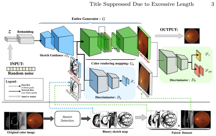
The load-bearing mechanism is a sketch prior enforced by two coupled modules. The sketch guidance module $G_S$ maps random noise to a structural sketch and answers to a sketch discriminator $D_S$ that compares against Sobel-and-morphology sketch drafts of real images; the color render mapping $G_P$, built on a U-net, embeds the sketch representation and produces the final color image under an image discriminator $D_P$ plus an $\ell_1$ reconstruction term. The two objectives are optimized jointly, and the sketch stage grows progressively in resolution. This isolates the anatomical geometry in a low-dimensional sketch space before the model attempts to match texture and background appearance.
What would settle it
Generate one fixed noise vector through SkrGAN, then compare the generated sketch with the Sobel/morphological sketch of the rendered color image; if thin vessels or bone edges diverge, the sketch prior is not preserving the structures claimed. A second check is to train with random line drawings of similar density in place of the real sketch drafts: if FID and segmentation scores remain unchanged, the specific sketch representation is doing none of the work.
Extended reading notes
Core claim
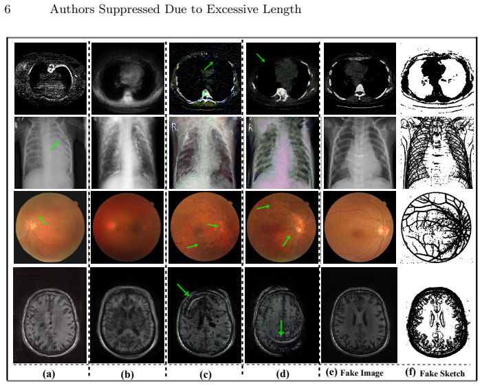
SkrGAN's central claim is that an unconditional GAN can generate medical images with anatomically faithful foreground structures if the generator first commits to a structural sketch and only then renders color. The sketch guidance module $G_S$ is trained against a sketch discriminator $D_S$ on sketch drafts extracted from real images by Sobel edge detection, Gaussian low-pass filtering, and morphological opening/closing; the color render mapping $G_P$, a U-net, is trained against an image discriminator $D_P$ and an $\ell_1$ reconstruction loss. The full objective joins the sketch-guidance loss with the color-rendering loss, with progressive growing used in the sketch stage. On the paper's experiments, this architecture performs better than DCGAN, ACGAN, WGAN, and PGGAN on FID, SWD, and MS-SSIM across four modalities, and its synthetic images raise U-net vessel-segmentation sensitivity on DRIVE from 0.778 to 0.846. The intended contribution is a general-purpose structure-preserving generator and data-augmentation tool that needs neither paired data nor manual labels.
Load-bearing premise
The paper's results depend on the hand-made sketch representation (Sobel edges, smoothing, and morphological cleaning) capturing exactly the anatomical structures that matter; if that sketch drops thin vessels or blurs bone boundaries, both the image-quality gains and the segmentation improvement would shrink.
Editorial extensions
If this is right
- On four modalities (retinal color fundus, chest X-ray, lung CT, brain MRI), SkrGAN reports lower FID and lower Sliced Wasserstein Distance, and higher MS-SSIM, than DCGAN, ACGAN, WGAN, and PGGAN.
- The generator requires no paired data, labels, or depth maps, because the sketch prior is derived automatically from training images.
- The synthetic images can be used as data augmentation: on DRIVE, pretraining a U-net with SkrGAN pairs raises sensitivity from 0.778 to 0.846, with accuracy and AUC also increasing.
- The same architecture transfers across modalities without per-modality redesign, which makes it a candidate general-purpose augmentation tool when training sets are small.
Reading between the lines
- Editorial inference: the sketch representation is probably interchangeable; if so, SkrGAN should transfer to ultrasound, OCT, or dermoscopy by swapping the sketch-preparation recipe, a test the paper does not run.
- Editorial inference: the gains could come mainly from constraining the image discriminator's input space rather than from the specific Sobel representation; this could be tested by replacing real sketch drafts with random line drawings of matched density.
- Editorial inference: because the generated sketches double as labels, SkrGAN is a source of weakly labeled training data; a natural extension is to measure how segmentation accuracy scales beyond 2,000 synthetic pairs.
- Editorial inference: the unconditional setting leaves room for a conditional extension that controls anatomy or pathology class, which would help class-imbalanced medical datasets; the paper does not explore this.
Signed reviews
Editorial analysis
A structured set of objections, weighed in public.
Referee Report
Summary. The paper proposes SkrGAN, an unconditional GAN for medical image synthesis that decomposes generation into a sketch guidance module (GS) and a color render mapping module (GP). Sketch drafts are extracted from real images using Sobel edge detection, Gaussian filtering, and morphological operations. GS is trained to generate sketches from noise under a sketch discriminator DS; GP is trained to translate sketches into color images under a color discriminator DP and an L1 loss. The paper evaluates the method on retinal fundus, chest X-ray, lung CT, and brain MRI datasets using MS-SSIM, SWD, and FID, and demonstrates a vessel-segmentation data-augmentation experiment on DRIVE. The abstract claims state-of-the-art synthesis and improved segmentation through SkrGAN-generated images.
Significance. If the claimed results hold, the paper would make a useful contribution by showing that an explicit structural sketch prior can improve unconditional medical image generation and provide a data-augmentation signal for segmentation. The application to four modalities and the DRIVE segmentation transfer experiment are appropriate tests, and the use of established metrics (FID, SWD, MS-SSIM) is commendable. However, the significance is currently undercut by formal problems in the stated objectives: the equations as printed do not implement the claimed sketch-guided generator, and the reported quantitative comparisons lack error bars or significance tests. The central mechanism must be correctly defined and verified before the empirical claims can be assessed.
major comments (5)
- [Sec. 2.2, Eq. (1)] The adversarial objective for the sketch guidance module is printed with inverted roles: Ls = Ez[log(DS(GS(z⊙l)))] + Ex[log(1−DS(y))]. Under the standard convention that DS outputs the probability of a real input, this trains DS to label generated sketches as real and real sketches as fake, and trains GS to move its output toward the fake label. If this is a typo, the correct form must be given, because the subsequent derivation and the interpretation of the training dynamics depend on it.
- [Sec. 2.3, Eq. (2)] The color render mapping objective has the same inversion: Ladv writes Ez,y[log(DP(GP(y),y))] for the generated pair and E(x,y)[log(1−DP(x,y))] for the real pair. As written, DP is rewarded for calling translated sketches real and original image-sketch pairs fake, which is the opposite of the standard conditional GAN loss. The authors should correct the sign convention or state explicitly if a nonstandard label convention is intended; as printed, the loss is not a valid adversarial objective.
- [Secs. 2.2–2.3, Eqs. (1)–(3)] The total objective in Eq. (3) does not contain any term that evaluates GP on the output of GS: no loss depends on GP(GS(z)). Equation (2) trains GP on pairs of real sketches y and real images x, while Eq. (1) trains GS alone. Therefore the gradients with respect to GS are independent of GP, and the color renderer is never trained on generated sketches, even though Fig. 2 and the inference procedure feed GS(z) into GP. This is not a cosmetic issue: the claimed sketch-prior constraint guiding final image generation is not realized by the equations as written. The reported FID/SWD improvements and the segmentation gain could, in principle, be produced by the supervised sketch-to-image translator alone, with the unconditional sketch generator playing no role in final image quality. The authors must report the actual objective used in training; if GP(GS(z)) is indeed trained, that term must be added to Eq. (3), and if it is not, the central claim of the paper needs to be substantially revised.
- [Sec. 3, Table 1] The quantitative comparison reports mean SWD, MS-SSIM, and FID computed from 100 generated images per method, with no variance, confidence intervals, or significance tests across independent training runs. FID and SWD are notoriously noisy at this sample size, and for some entries the gap to the runner-up is small (e.g., brain MRI FID of 27.51 versus 33.76 for PGGAN). The authors should report standard deviations over repeated generation/training runs or provide a statistical test to support the claim that SkrGAN is consistently better than the baselines.
- [Secs. 2.1 and 3, vessel segmentation experiment] The paper provides no quantitative validation that the handcrafted sketch draft representation preserves the anatomical structures that matter (small vessels, disc/cup boundaries, bone edges). This is load-bearing because the segmentation pretraining uses the generated sketches as labels: if the sketch extractor drops small vessels, the reported +8.78% sensitivity improvement may reflect a denoised or simplified label signal rather than a genuinely better structural prior. The authors should compare their sketch extraction against the full images at the relevant resolutions and report, for example, vessel detection recall on the DRIVE ground truth, or an ablation that varies the sketch-generation parameters.
minor comments (5)
- [Throughout] There are several typographical errors that should be corrected: 'Freshet Inception Distance' should be 'Fréchet Inception Distance', 'comparsions' should be 'comparisons', and Table 2 contains 'whithout' for 'without'. The PGGAN reference also contains 'et. al.o'.
- [Sec. 3, datasets and protocol] The description of the evaluation protocol is incomplete: it is not stated how the real-image set for FID/SWD is defined, whether all real images are used, and whether the 100 generated images are produced after a fixed number of training iterations or after convergence. These details affect reproducibility.
- [Eq. (3)] The notation 'arg[ min_GS max_DS Ls + min_GP max_DP (Ladv+L1) ]' is nonstandard and mixes the minimization and maximization arguments with the outer arg expression; this should be rewritten as separate min-max problems or as a single combined objective with clearly scoped optimization variables.
- [Sec. 2.2] The definition DS = D_S^(n)···D_S^(0) and GS = G_S^(n)···G_S^(0) is not explained with enough precision; it would be clearer to state that the generator and discriminator are progressive-growing networks following PGGAN, rather than introducing notation that suggests a composition of separate networks.
- [Sec. 3, hyperparameters] The statement that 'a small change of λ does not affect much the performance' is not supported by an ablation or sensitivity analysis; adding a short sensitivity experiment would strengthen the claim and the reproducibility of the method.
Circularity Check
No significant circularity: SkrGAN's evaluation is benchmarked externally and self-citations are background only.
full rationale
The paper's central derivation is not circular. The sketch guidance module GS is trained with a sketch discriminator against sketch drafts extracted from real images by Sobel edge detection, Gaussian filtering, and morphological operations (Sec. 2.1-2.2). The color render mapping GP is trained on real sketch-image pairs using an adversarial loss and an L1 reconstruction loss (Eq. 2), and the final objective in Eq. 3 is the sum of these two independent training losses. The reported image-quality results are measured against external baselines (DCGAN, ACGAN, WGAN, PGGAN) using FID, SWD, and MS-SSIM on public datasets, so no fitted parameter is renamed as a prediction. The segmentation experiment pretrains U-net on synthetic images with generated sketches as labels and then fine-tunes on the external DRIVE dataset; this is an independent empirical test rather than a conclusion contained in the training objective. The only self-citations, references [3] and [5], appear as generic background for deep learning in medical imaging and are not load-bearing. The skeptic's observation that Eqs. (1)-(3) never train GP on GS(z) is a possible architectural coherence or correctness concern, not a circularity: it does not show that any output is defined in terms of an input or that a result is forced by construction. The paper therefore receives a circularity score of 0.
Assumptions & free parameters
free parameters (6)
- lambda (L1 loss weight) =
100
- learning rate for GS and DS =
0.001
- learning rate for GP and DP =
0.0002
- batch size =
16
- number of generated images for evaluation =
100
- number of synthetic images for pretraining =
2000
assumptions (3)
- domain assumption The extracted sketch drafts preserve the structural information needed for generation and downstream segmentation.
- standard math The PGGAN backbone and U-net backbone are suitable for the two modules.
- ad hoc to paper The adversarial objectives in Eqs. (1) and (2) are standard GAN losses up to label convention.
Cite this review
Pith. "Pith review of SkrGAN: Sketching-rendering Unconditional Generative Adversarial Networks for Medical Image Synthesis." pith.science (2026). https://pith.science/paper/A66JEZTD
@misc{pith2026190804346,
  author       = {Pith},
  title        = {Pith review of: SkrGAN: Sketching-rendering Unconditional Generative Adversarial Networks for Medical Image Synthesis},
  year         = {2026},
  howpublished = {\url{https://pith.science/paper/A66JEZTD}},
  note         = {Machine review of arXiv:1908.04346}
}
read the original abstract
Generative Adversarial Networks (GANs) have the capability of synthesizing images, which have been successfully applied to medical image synthesis tasks. However, most of existing methods merely consider the global contextual information and ignore the fine foreground structures, e.g., vessel, skeleton, which may contain diagnostic indicators for medical image analysis. Inspired by human painting procedure, which is composed of stroking and color rendering steps, we propose a Sketching-rendering Unconditional Generative Adversarial Network (SkrGAN) to introduce a sketch prior constraint to guide the medical image generation. In our SkrGAN, a sketch guidance module is utilized to generate a high quality structural sketch from random noise, then a color render mapping is used to embed the sketch-based representations and resemble the background appearances. Experimental results show that the proposed SkrGAN achieves the state-of-the-art results in synthesizing images for various image modalities, including retinal color fundus, X-Ray, Computed Tomography (CT) and Magnetic Resonance Imaging (MRI). In addition, we also show that the performances of medical image segmentation method have been improved by using our synthesized images as data augmentation.
Figures

Reference graph
Works this paper leans on
- [1]
-
[2]
al.: End-to-end adversarial retinal image synthesis
Costa, P., Galdran, A., et. al.: End-to-end adversarial retinal image synthesis. IEEE TMI 37(99), 781–791 (2017)
work page 2017
-
[3]
IEEE transactions on medical imaging 37(7), 1597–1605 (2018)
Fu, H., Cheng, J., Xu, Y., Wong, D.W.K., Liu, J., Cao, X.: Joint optic disc and cup segmentation based on multi-label deep network and polar transformation. IEEE transactions on medical imaging 37(7), 1597–1605 (2018)
work page 2018
-
[4]
al.: Generative adversarial networks
Goodfellow, I.J., Pouget-Abadie, J., et. al.: Generative adversarial networks. NIPS (2014)
work page 2014
-
[5]
IEEE transactions on medical imaging (2019)
Gu, Z., Cheng, J., Fu, H., Zhou, K., Hao, H., Zhao, Y., Zhang, T., Gao, S., Liu, J.: Ce-net: Context encoder network for 2d medical image segmentation. IEEE transactions on medical imaging (2019)
work page 2019
-
[6]
al.: Image-to-image translation with conditional adversarial networks
Isola, P., Zhu, J.Y., et. al.: Image-to-image translation with conditional adversarial networks. In: CVPR. pp. 1125–1134 (2017)
work page 2017
-
[7]
al.o: Progressive growing of gans for improved quality, stability, and variation
Karras, T., Aila, T., et. al.o: Progressive growing of gans for improved quality, stability, and variation. arXiv (2017)
work page 2017
-
[8]
al.: Identifying medical diagnoses and treatable diseases by image-based deep learning
Kermany, D.S., Goldbaum, M., et. al.: Identifying medical diagnoses and treatable diseases by image-based deep learning. Cell 172(5), 1122–1131 (2018)
work page 2018
Show all 19 references
-
[9]
al.: Semi-supervised learning with generative adversar- ial networks for chest x-ray classification with ability of data domain adaptation
Madani, A., Moradi, M., et. al.: Semi-supervised learning with generative adversar- ial networks for chest x-ray classification with ability of data domain adaptation. In: ISBI. pp. 1038–1042 (2018)
2018
-
[10]
arXiv (2014) Title Suppressed Due to Excessive Length 9
Mirza, M., Osindero, S.: Conditional generative adversarial nets. arXiv (2014) Title Suppressed Due to Excessive Length 9
2014
-
[11]
In: ICML
Odena, A., Olah, C., Shlens, J.: Conditional image synthesis with auxiliary classifier GANs. In: ICML. pp. 2642–2651 (2017)
2017
-
[12]
Psychology of Aesthetics, Creativity, and the Arts 6(2), 124–136 (2012)
Ostrofsky, J., Kozbelt, A., Seidel, A.: Perceptual constancies and visual selection as predictors of realistic drawing skill. Psychology of Aesthetics, Creativity, and the Arts 6(2), 124–136 (2012)
2012
-
[13]
Foundations and Trends R© in Machine Learning 11(5-6), 355–607 (2019)
Peyr´ e, G., Cuturi, M., et al.: Computational optimal transport. Foundations and Trends R© in Machine Learning 11(5-6), 355–607 (2019)
2019
-
[14]
international conference on learning representations (2016)
Radford, A., Metz, L., Chintala, S.: Unsupervised representation learning with deep convolutional generative adversarial networks. international conference on learning representations (2016)
2016
-
[15]
In: MICCAI
Ronneberger, O., Fischer, P., Brox, T.: U-net: Convolutional networks for biomed- ical image segmentation. In: MICCAI. pp. 234–241 (2015)
2015
-
[16]
al.: Ridge-based vessel segmentation in color images of the retina
Staal, J., Abrmoff, M.D., et. al.: Ridge-based vessel segmentation in color images of the retina. IEEE TMI 23(4), 501–509 (2004)
2004
-
[17]
In: European Conference on Computer Vision
Wang, X., Gupta, A.: Generative image modeling using style and structure ad- versarial networks. In: European Conference on Computer Vision. pp. 318–335. Springer (2016)
2016
-
[18]
In: CVPR
Zhang, Z., Yang, L., Zheng, Y.: Translating and segmenting multimodal medi- cal volumes with cycle-and shape-consistency generative adversarial network. In: CVPR. pp. 9242–9251 (2018)
2018
-
[19]
al.: Synthesizing retinal and neuronal images with generative adversarial nets
Zhao, H., Li, H., et. al.: Synthesizing retinal and neuronal images with generative adversarial nets. Medical Image Analysis 49, 14–26 (2018)
2018
Reviewed August 14, 2026 · model on record in the stance chip above.
Discussion (0). Continue with ORCID to comment.