REVIEW 4 major objections 5 minor 13 references
The Channel Attention based Context Encoder Network for Inner Limiting Membrane Detection
T0 review · 4 major / 5 minor · reviewed 2026-08-14 · deepseek-v4-flash
Pith's one-line read Adding channel attention to a context encoder network lowers segmentation error for the inner limiting membrane in OCT images.
desk verdict Thin, honest, and statistically under-powered: a small SE-attention variant of CE-Net plus a 20-scan OCT dataset, where the claimed improvement over CE-Net is within noise. read the letter →
The pith
A machine-rendered reading of the paper's core claim, the machinery that carries it, and where it could break.
The reading
What carries the argument
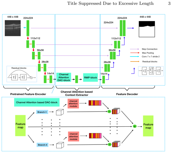
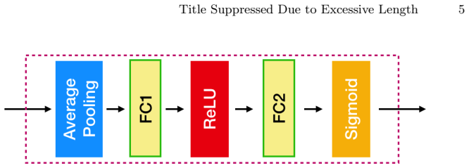
The load-bearing object is the channel attention based context encoder module (CACE module), inserted between the encoder and decoder. In each branch of the multi-scale atrous-convolution context extractor, it pools each feature channel spatially to get a channel descriptor $z_c$, passes $z_c$ through two linear transformations with a ReLU and a sigmoid to get a per-channel weight $s_c$, and multiplies $s_c$ back into the feature map before the residual multi-kernel pooling block. The effect is feature recalibration: channels that matter globally are amplified, channels that do not are suppressed, so the decoder receives context features weighted by their global importance. This module is the only component that distinguishes CACE-Net from CE-Net.
What would settle it
Compute per-volume error differences on repeated random 10/10 splits of the same 20 volumes. If CACE-Net is not lower than CE-Net in a majority of splits, or the typical split-to-split variation in the difference exceeds 0.268, the claimed 10.8% improvement is not established.
Extended reading notes
Core claim
On the authors' own terms, the central discovery is that reweighting each channel of the aggregated context features, via a squeeze-and-excitation-style attention module, makes the CE-Net architecture noticeably better at segmenting the ILM boundary. The proposed CACE-Net keeps CE-Net's encoder-decoder structure and its dense atrous convolution plus residual multi-kernel pooling, but inserts a channel attention module into the context extractor so that the network can emphasize informative feature channels before decoding. Evaluated on their 20-volume dataset, CACE-Net achieves a mean absolute error of 2.199±1.471, a 10.8% reduction from CE-Net's 2.467±1.989, with U-Net far behind at 6.404±16.407. The authors interpret this as evidence that the channel attention modification improves high-level semantic feature extraction for this boundary-detection task.
Load-bearing premise
The entire comparison rests on one random split of 20 right-eye OCT volumes into 10 training and 10 test scans, so the 0.268-point error gap over CE-Net could be a property of that split rather than of the network.
Editorial extensions
If this is right
- A channel-attention layer inside the context extractor is enough to reduce ILM segmentation error by about 10.8% over the base CE-Net architecture on this dataset.
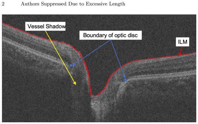
- The predicted ILM mask can be post-processed into a continuous boundary line, which is the input the authors use for locating optic-disc boundary points.
- The newly built 20-volume dataset with manual ILM annotations gives researchers a fixed ten-train/ten-test testbed for comparing ILM and optic-disc boundary detectors.
- The channel-attention context module is designed as a modification to CE-Net's context extractor, so other CE-Net-based segmentation models could adopt it directly.
Reading between the lines
- Beyond the paper: applying the same channel recalibration at multiple decoder stages, not only in the context encoder, could further sharpen the boundary localization.
- Beyond the paper: because the dataset is limited to 20 right-eye scans, validation on left-eye and diseased eyes is needed before the reported gain can be used for glaucoma screening.
- Beyond the paper: replacing the post-hoc morphological boundary extraction with a boundary-aware loss could make the predicted ILM line smoother and less dependent on denoising heuristics.
Signed reviews
Editorial analysis
A structured set of objections, weighed in public.
Referee Report
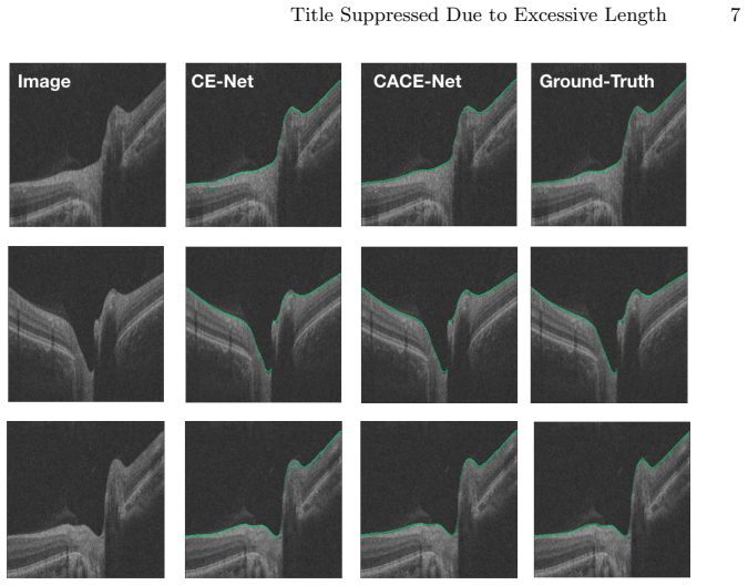
Summary. The paper introduces a new dataset of 20 optic-disc-centered 3D OCT volumes with manual annotations of the inner limiting membrane (ILM) boundary, and proposes CACE-Net, a modification of the authors' earlier CE-Net architecture in which a squeeze-and-excitation style channel attention module is inserted into the context extractor. The method is evaluated on a single random 10/10 train/test split of the dataset, reporting a mean absolute error (MAE) of 2.199, which the authors claim is a 10.8% improvement over CE-Net (2.467) and substantially better than U-Net (6.404). The paper includes visual sample results and a brief description of the boundary extraction post-processing.
Significance. The dataset contribution—20 manually labeled ILM boundaries in 3D OCT volumes—is potentially useful for future retinal layer segmentation research, and the proposed channel attention extension to CE-Net is a plausible, clearly described architectural variant. If the reported improvement over CE-Net were statistically robust, the work would be a modest but valid incremental contribution. However, the central claim of state-of-the-art performance currently rests on a single small test split with no significance testing, no cross-validation, and no ablation isolating the proposed module. The paper also provides no external validation on other OCT datasets. These shortcomings substantially limit the current significance of the results, though they are addressable with additional experiments.
major comments (4)
- [§3.3, Table 1] The claimed advantage of CACE-Net over CE-Net (2.199 vs. 2.467 MAE) is not statistically supported. With 10 test volumes and reported standard deviations of 1.471 and 1.989, the standard error of the difference is approximately 0.782, giving an unpaired t-statistic of roughly 0.34 (p > 0.7); the observed difference is thus well within plausible random variation. The paper reports no paired significance test, confidence interval, or per-volume errors. To support the claim of superiority, the authors should report per-volume errors and a paired test (e.g., Wilcoxon signed-rank or paired t-test), or provide bootstrap confidence intervals.
- [§3.1] The evaluation uses a single random 10/10 split of 20 OCT volumes, with no cross-validation or repeated splitting. The test set is small, and the extreme standard deviation for U-Net (16.407) indicates that individual outlier volumes can dominate the reported means. A single split cannot demonstrate that the 0.268 MAE difference between CACE-Net and CE-Net is reproducible. The authors should report results over multiple random splits (e.g., mean and standard deviation of the MAE across splits) or use k-fold cross-validation, and should identify whether any single test volume drives the difference.
- [§2.1 and §3.3] No ablation study is provided to isolate the proposed channel attention module. Since CACE-Net is exactly CE-Net plus a channel attention mechanism inside the context extractor, the architecture change is not isolated as the cause of the reported improvement. The authors should compare CE-Net with and without the channel attention module under identical training conditions, and ideally also ablate the placement of the attention module, to attribute the observed difference to the proposed component rather than to training details or randomness.
- [§3.2 and §3.3] The experimental protocol for the baselines is not described with sufficient control. The paper does not state whether U-Net and CE-Net were trained with the same hyperparameters, data augmentation, input size, pre-processing, and post-processing as CACE-Net. Because CE-Net is the authors' own prior publication, a home-team advantage cannot be ruled out without a controlled comparison. The authors should explicitly describe the identical training protocol for all methods and, if possible, use the original CE-Net implementation with the same settings as CACE-Net.
minor comments (5)
- [§1] The citations in the introduction do not match the described methods: reference [5] (Cheng et al., sparse dissimilarity-constrained coding) is described as an optic disc segmentation method, and reference [7] (Lee et al., macular edema segmentation) is described as applying a K-NN classifier to segment the optic disc cup and neuroretinal. Please verify and correct these references.
- [§2.1] The text says that 'a matrix multiplication ... is added' to obtain the final output, but the operation is a channel-wise scaling of the feature map by the excitation vector, not an addition. Please reword for accuracy.
- [§3.3] The paragraph states that CACE-Net 'outperforms the other algorithms based on deep learning image processing method,' but only two baseline algorithms are compared. The claim of state-of-the-art performance should be restricted to the compared methods or supported by additional comparisons with published ILM segmentation approaches.
- [Table 1] The table caption should specify the number of test volumes (10) and the units of the MAE (e.g., pixels per column or absolute pixel distance), since the error metric in Eq. (4) is otherwise ambiguous.
- [§4] The statement 'we will release our code and dataset on Github later' is a promise of future availability, not a current artifact. Either provide the repository or dataset link, or remove the claim, since reviewers and readers cannot verify an unreleased artifact.
Circularity Check
No circular derivation; the claimed improvement is an empirical, held-out test result rather than a construction-level tautology.
full rationale
The paper contains no derivation step that reduces to its own inputs. The proposed CACE-Net is admittedly an architectural modification of the authors' prior CE-Net ('The proposed CACE-Net is modified from the CE-Net [1]'), but the central claim is an empirical mean absolute error on a disjoint test set: 'we also randomly take 10 people's images for training, and others for testing.' The error is computed from network predictions against manual ground truth using the standard formula 'error = 1/n sum_i |yi - Yi|', so the reported 2.199 is a measured outcome, not an identity forced by the definition of the method. The channel attention module is taken from an external source (SE-Net [11]) and inserted into the DAC block, so CACE-Net is not a renaming of the prior work. The comparison with CE-Net is a self-citation, but the paper re-trains or re-evaluates CE-Net on its own data split ('we compare our method with two algorithms for the ILM segmentation') rather than importing the cited paper's reported numbers, so the baseline performance is independent evidence under the same protocol. The absence of significance testing and the small 10/10 split are correctness and statistical-evidence concerns, not circularity: no fitted parameter is relabeled as a prediction, and no equation equates the proposed result with the input by construction. Therefore no circular step is present.
Assumptions & free parameters
free parameters (2)
- Training hyperparameters (learning rate, weight decay, poly power, input size) =
lr=0.001, weight_decay=0.0001, power=0.9, input=448x448
- Network weights =
millions (not reported)
assumptions (5)
- domain assumption The 20 manually annotated OCT scans provide accurate ground-truth ILM boundaries.
- domain assumption The 10/10 train/test split is representative of the target population.
- domain assumption Mean absolute error (MAE) is a valid metric for ILM boundary segmentation quality.
- domain assumption Binary cross-entropy loss is appropriate for training this segmentation network.
- domain assumption The CE-Net architecture (reference [1]) is a valid state-of-the-art baseline.
Cite this review
Pith. "Pith review of The Channel Attention based Context Encoder Network for Inner Limiting Membrane Detection." pith.science (2026). https://pith.science/paper/33DJW7U2
@misc{pith2026190804413,
  author       = {Pith},
  title        = {Pith review of: The Channel Attention based Context Encoder Network for Inner Limiting Membrane Detection},
  year         = {2026},
  howpublished = {\url{https://pith.science/paper/33DJW7U2}},
  note         = {Machine review of arXiv:1908.04413}
}
read the original abstract
The optic disc segmentation is an important step for retinal image-based disease diagnosis such as glaucoma. The inner limiting membrane (ILM) is the first boundary in the OCT, which can help to extract the retinal pigment epithelium (RPE) through gradient edge information to locate the boundary of the optic disc. Thus, the ILM layer segmentation is of great importance for optic disc localization. In this paper, we build a new optic disc centered dataset from 20 volunteers and manually annotated the ILM boundary in each OCT scan as ground-truth. We also propose a channel attention based context encoder network modified from the CE-Net to segment the optic disc. It mainly contains three phases: the encoder module, the channel attention based context encoder module, and the decoder module. Finally, we demonstrate that our proposed method achieves state-of-the-art disc segmentation performance on our dataset mentioned above.
Figures
Figures from the paper (1 more)
Reference graph
Works this paper leans on
-
[1]
Gu, Z., et al.: Ce-net: Context encoder network for 2d medical image segmentation. IEEE Trans. Med. Imag. (2019)
work page 2019
-
[2]
Ophthalmology 121 (2014) 2081–2090
Tham, Y.C., Li, X., Wong, T.Y., Quigley, H.A., Aung, T., Cheng, C.Y.: Global prevalence of glaucoma and projections of glaucoma burden through 2040: a sys- tematic review and meta-analysis. Ophthalmology 121 (2014) 2081–2090
work page 2014
-
[3]
Journal of the American Medical Informatics Association 20 (2013) 1021–1027
Liu, J., Zhang, Z., Wong, D.W.K., Xu, Y., Yin, F., Cheng, J., Tan, N.M., Kwoh, C.K., Xu, D., Tham, Y.C., et al.: Automatic glaucoma diagnosis through medical imaging informatics. Journal of the American Medical Informatics Association 20 (2013) 1021–1027
work page 2013
-
[4]
Pattern recognition 40 (2007) 2063–2076
Xu, J., Chutatape, O., Sung, E., Zheng, C., Kuan, P.C.T.: Optic disk feature extraction via modified deformable model technique for glaucoma analysis. Pattern recognition 40 (2007) 2063–2076
work page 2007
-
[5]
IEEE Transactions on Biomedical Engineering 62 (2015) 1395–1403
Cheng, J., Yin, F., Wong, D.W.K., Tao, D., Liu, J.: Sparse dissimilarity-constrained coding for glaucoma screening. IEEE Transactions on Biomedical Engineering 62 (2015) 1395–1403
work page 2015
-
[6]
IEEE Journal of selected topics in quantum electronics 5 (1999) 1205–1215
Schmitt, J.M.: Optical coherence tomography (oct): a review. IEEE Journal of selected topics in quantum electronics 5 (1999) 1205–1215
work page 1999
-
[7]
Biomedical optics express 8 (2017) 3440–3448
Lee, C.S., Tyring, A.J., Deruyter, N.P., Wu, Y., Rokem, A., Lee, A.Y.: Deep- learning based, automated segmentation of macular edema in optical coherence tomography. Biomedical optics express 8 (2017) 3440–3448
work page 2017
-
[8]
Fu, H., Xu, D., Lin, S., Wong, D.W.K., Liu, J.: Automatic optic disc detection in oct slices via low-rank reconstruction. IEEE Transactions on Biomedical Engi- neering 62 (2014) 1151–1158 Title Suppressed Due to Excessive Length 9
work page 2014
Show all 13 references
-
[9]
In: Advances in neural information processing systems
Krizhevsky, A., Sutskever, I., Hinton, G.E.: Imagenet classification with deep con- volutional neural networks. In: Advances in neural information processing systems. (2012) 1097–1105
2012
-
[10]
In: Computational Pathology and Ophthalmic Medical Image Analysis
Gu, Z., Liu, P., Zhou, K., Jiang, Y., Mao, H., Cheng, J., Liu, J.: Deepdisc: Op- tic disc segmentation based on atrous convolution and spatial pyramid pooling. In: Computational Pathology and Ophthalmic Medical Image Analysis. Springer (2018) 253–260
2018
-
[11]
In: Proceedings of the IEEE conference on computer vision and pattern recognition
Hu, J., Shen, L., Sun, G.: Squeeze-and-excitation networks. In: Proceedings of the IEEE conference on computer vision and pattern recognition. (2018) 7132–7141
2018
-
[12]
In: International Conference on Medical image computing and computer-assisted intervention, Springer (2015) 234–241
Ronneberger, O., Fischer, P., Brox, T.: U-net: Convolutional networks for biomedi- cal image segmentation. In: International Conference on Medical image computing and computer-assisted intervention, Springer (2015) 234–241
2015
-
[13]
arXiv preprint arXiv:1712.07628 (2017)
Keskar, N.S., Socher, R.: Improving generalization performance by switching from adam to sgd. arXiv preprint arXiv:1712.07628 (2017)
2017 arXiv
Reviewed August 14, 2026 · model on record in the stance chip above.
Discussion (0). Continue with ORCID to comment.