REVIEW 3 major objections 5 minor 22 references
Generalizing Deep Whole Brain Segmentation for Pediatric and Post-Contrast MRI with Augmented Transfer Learning
T0 review · 3 major / 5 minor · reviewed 2026-08-14 · deepseek-v4-flash
Pith's one-line read Mixing original adult training data with pediatric or contrast-enhanced scans during transfer learning improves both new domains while limiting the loss in adult accuracy.
desk verdict A useful transfer-learning application for pediatric and post-contrast MRI; the pediatric result is solid, the contrast result is weakened by self-referential labels, and the adult 'drop' comparison needs a fold-matched baseline. read the letter →
The pith
A machine-rendered reading of the paper's core claim, the machinery that carries it, and where it could break.
The reading
What carries the argument
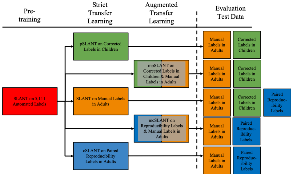
The load-bearing machinery is the augmented transfer-learning step, applied to the SLANT framework: SLANT divides the brain into 27 overlapping tiles, trains independent segmentation networks on each tile, and fuses their outputs by majority voting to label 132 volumetric regions. The pretrained model comes from 5,111 automatically labeled adult scans; SLANT's own transfer step then refines it on 45 manually traced adult atlases. The paper's modification is to compose that refinement set as a union of the original adult atlases and the new-domain images: 30 manually corrected pediatric scans, or 36 pre-contrast scans whose SLANT segmentations are transferred to co-registered post-contrast scans as training labels. All variants are fine-tuned with the Adam optimizer and Dice loss for 30 epochs and selected by validation Dice in five-fold cross-validation, so the only systematic difference between comparison arms is the training-set composition. The mechanism works, in the paper's account, because including adult examples anchors the weights to the original task while the new examples push the weights toward the new domain, controlling the accuracy-forgetting trade-off.
What would settle it
Have expert raters manually trace the 36 post-contrast images and score the mixed contrast-trained model against those manual labels rather than against the pre-contrast SLANT segmentation; if the reproducibility gain disappears or manual agreement does not improve, the result would be shown to reflect consistency with auto-generated labels, not anatomical truth.
Extended reading notes
Core claim
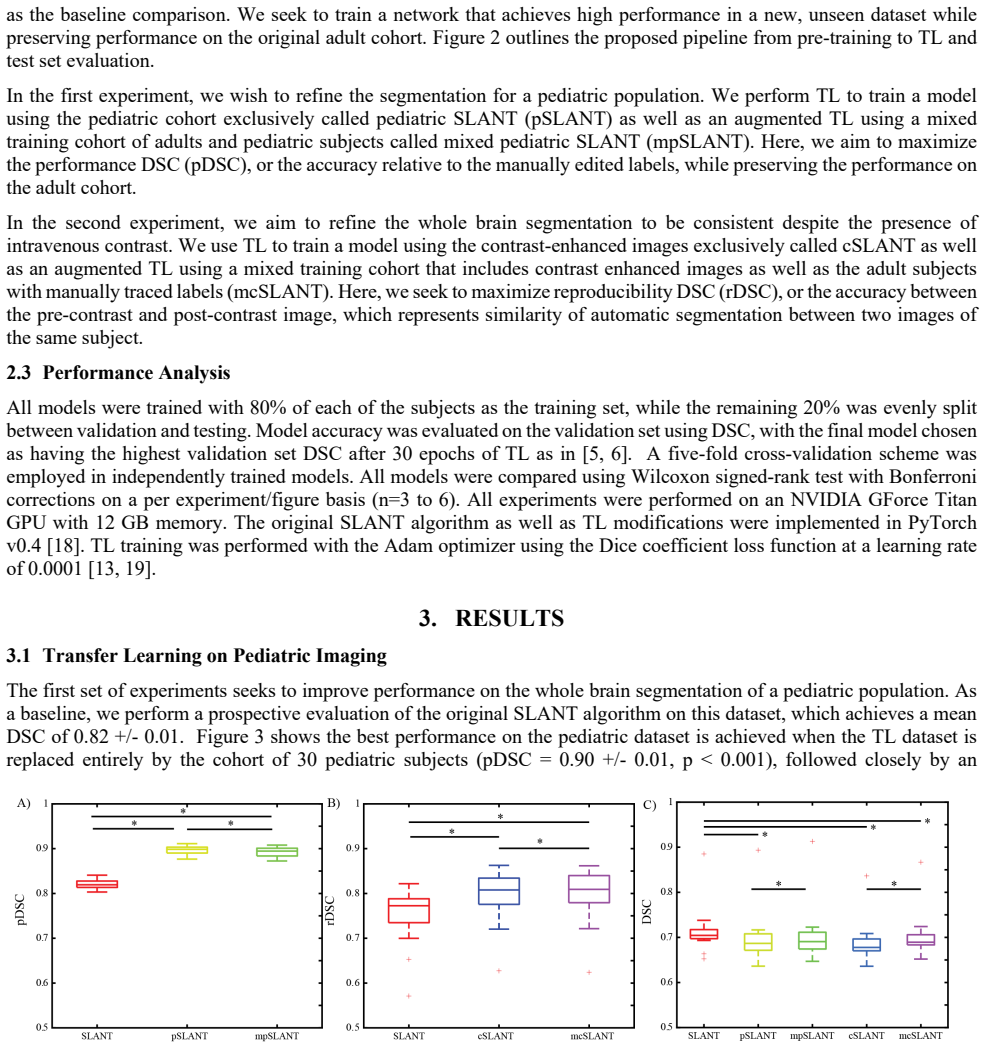
The central claim is that the composition of the fine-tuning set, not the network architecture or training schedule, is what determines whether SLANT generalizes to a new distribution without catastrophic forgetting. For pediatrics, the paper compares baseline SLANT, a model fine-tuned only on 30 manually corrected pediatric scans (pSLANT), and a model fine-tuned on those 30 plus the original 45 adult atlases (mpSLANT). mpSLANT reaches a pediatric Dice of 0.89 against manual truth, nearly matching pSLANT's 0.90 while losing less on the adult validation set (0.704 versus 0.698 adult DSC). For contrast, the paper compares baseline SLANT against cSLANT (only 36 paired pre- and post-contrast scans) and mcSLANT (paired scans plus adult atlases); mcSLANT reaches 0.80 rDSC between pre- and post-contrast segmentations, versus 0.76 baseline, and reduces the mean within-subject hippocampus volume change from 9.91% to 0.89%. The authors conclude that augmenting the original transfer-learning step with both old and new data is superior to strict transfer learning for adapting to anatomical variation and scanning protocol.
Load-bearing premise
In the contrast experiment, the paper uses the original SLANT segmentation of the pre-contrast image as the ground-truth labels for both training the post-contrast model and measuring its reproducibility, so if those labels carry systematic errors, the reported gains partly measure how well the model reproduces SLANT's own pre-contrast outputs rather than true anatomical accuracy.
Editorial extensions
If this is right
- A single mixed-cohort fine-tuning step can adapt a research-grade segmentation network to pediatric anatomy with 30 manually corrected examples, netting a 0.07 Dice gain over the unmodified model.
- The same recipe suppresses contrast-induced bias: within-subject hippocampus volume differences across pre- and post-contrast scans drop from about 10% to under 1%.
- Adult-cohort accuracy is not free: every adaptation in this paper lowers adult DSC slightly, but mixing new data with the original atlases reduces that drop compared with strict transfer learning.
- Because the method changes only the composition of the fine-tuning set, it can be dropped into existing SLANT pipelines without architectural changes or added inference cost.
Reading between the lines
- If the same mixing recipe works for other distribution shifts, such as infant brains, pathology, lower field strength, or different coil setups, it offers a generic way to extend any pretrained segmentation network to local clinical data, not just SLANT.
- The contrast experiment's training labels are themselves produced by the model being adapted, so the measured 0.80 rDSC may encode a preference for consistency with pre-contrast SLANT output rather than improved biological accuracy; a manual-label evaluation would separate the two.
- The pediatric labels came from one expert rater's corrections, and with only 30 subjects and no inter-rater variability reported, the true label noise could be larger than the numbers suggest; a multi-rater study would test how much of the 0.07 Dice gain is adaptation to that rater's style rather than to pediatric anatomy.
Signed reviews
Editorial analysis
A structured set of objections, weighed in public.
Referee Report
Summary. The paper addresses generalizability of the SLANT whole-brain segmentation method to two new domains: pediatric T1-weighted MRI and clinically acquired post-contrast T1-weighted MRI. The authors propose "augmented transfer learning," in which the original adult transfer-learning data are mixed with new-domain data during fine-tuning, and compare this with strict transfer learning using only the new-domain data and with the original SLANT baseline. In the pediatric experiment (n=30, manual labels), both pSLANT and mpSLANT improve DSC over baseline (0.89-0.90 vs. 0.82). In the contrast experiment (n=36, pre-/post-contrast pairs with labels derived from SLANT on the pre-contrast image), mcSLANT improves reproducibility DSC (0.803 vs. 0.757). The paper further reports that new-data-only transfer learning reduces adult DSC more than augmented transfer learning, and concludes that mixing original data offers a favorable tradeoff between new-domain performance and preservation of original-domain performance.
Significance. If the central comparison were sound, the paper would offer a practical and inexpensive strategy for adapting a large pretrained medical segmentation model to new clinical populations and acquisition protocols while mitigating catastrophic forgetting. The pediatric experiment has real strengths: manual labels, five-fold cross-validation, and explicitly stated statistical tests. The proposed augmented TL idea is simple and likely useful. However, the load-bearing adult-cohort comparison is confounded by a train-versus-test discrepancy between the baseline and the new methods, and the contrast reproducibility metric is partly self-referential because the same SLANT-derived labels are used for training and evaluation. As a result, the main claim that augmented TL is superior to strict TL for preserving original-domain performance is not established by the reported experiments. The significance is therefore contingent on a fair re-analysis.
major comments (3)
- [§2.2, §2.3, Results 3.1 and 3.2] The adult-cohort comparison is confounded. The baseline is defined as Huo et al.'s SLANT model after transfer learning on the full 45-subject OASIS adult cohort, while the adult performance of pSLANT/mpSLANT and cSLANT/mcSLANT is measured on withheld adult subjects under five-fold cross-validation. Consequently, the reported drops from 0.715 to 0.698/0.704 and from 0.715 to 0.688/0.701 can be explained entirely by the difference between evaluating on training subjects (baseline) and held-out subjects (new models). A fold-matched baseline—for example, retraining the original SLANT transfer-learning step on the same 80% adult folds and evaluating on the same withheld adults—is required. Without this, the central claim that augmented TL reduces the drop in original-domain performance relative to strict TL is not supported.
- [§2.1 and §3.2] The contrast reproducibility metric is partly self-referential. Labels for the post-contrast images are generated by applying the original SLANT algorithm to the co-registered pre-contrast image, and the rDSC between pre- and post-contrast SLANT outputs is used both as the training objective and as the evaluation metric. The reported improvement from 0.757 to 0.803 therefore demonstrates that the fine-tuned model reproduces SLANT's own pre-contrast labels more consistently, not that the post-contrast segmentations are more anatomically accurate. The manuscript should explicitly acknowledge this limitation, and, if feasible, validate on a subset with manual labels or with an independent volumetric measurement.
- [§3.1 and §3.2] The comparison between strict and augmented transfer learning on the new-domain metrics is under-specified. In the pediatric experiment, pSLANT reaches pDSC 0.90 while mpSLANT reaches 0.89, but no significance test is reported for this pairwise difference; the same is true for cSLANT (0.799) versus mcSLANT (0.803) on rDSC. Since the paper's tradeoff conclusion rests on the adult drop comparison, which is affected by the first major comment, the authors should report fold-matched adult DSC for all methods and per-pair significance tests or effect sizes for the new-domain metric comparisons.
minor comments (5)
- [Abstract and §1] The phrase "are to examples" should be corrected to "are two examples."
- [§3.2 and Figure 5] The units for hippocampus volume error are inconsistent: the text reports "RMSE = 0.512 cm3" for SLANT and "RMSE = 0.250 mm3" for mcSLANT; the latter should presumably be 0.250 cm3.
- [§3.2 and Figure 3 caption] "occipital loves" should be "occipital lobes" in the text and in the Figure 3 caption.
- [Abstract and §2.1] The abstract states 132 volumetric labels while the methods section says 133 manual labels; the relationship between these counts (e.g., background label) should be clarified.
- [§3.1] The phrase "withheld sample from the adult dataset" is ambiguous given that the baseline adult DSC is computed on the full adult cohort; please state explicitly which adult subjects are used for each reported number.
Circularity Check
Contrast-enhancement rDSC 'improvement' is the training objective with labels generated by the baseline algorithm; adult drop comparison is additionally confounded.
-
self definitional
[Section 2.1 / Abstract / Section 3.2 (contrast experiment)]
"accuracy of the post-contrast segmentations assessed relative to the pre-contrast automated assessment // Labels for the same 133 regions were generated for the pre-contrast image and used as labels for the post-contrast image using the original SLANT algorithm. // We use labels generated on the pre-contrast image as truth to train a segmentation on the contrast-enhanced image."
The post-contrast model is trained with Dice loss against pre-contrast SLANT segmentations, and the evaluation metric rDSC is defined as Dice similarity between pre- and post-contrast automatic segmentations. The 'truth' labels are produced by the same baseline SLANT algorithm to which the result is compared, so the reported gain (0.803 vs 0.757 rDSC) measures how well the model learned to reproduce the baseline's own pre-contrast output on post-contrast input. This is a self-generated target renamed as a generalizability improvement; the comparison is favorable by construction because the original SLANT was never optimized for this objective. No independent manual labels are involved in the contrast experiment.
full rationale
The pediatric experiment is not circular: accuracy is measured against independent manually corrected labels, and both pSLANT and mpSLANT improve on those labels. The adult-cohort comparison used to claim 'data augmentation was superior to strict transfer learning' is not circular but is confounded: the baseline adult DSC (0.715) is inherited from Huo et al.'s SLANT model after TL on the full 45-subject adult dataset, whereas pSLANT/mpSLANT/cSLANT/mcSLANT adult numbers are measured on adult subjects withheld from the new TL under five-fold cross-validation. Comparing a trained-on-all-adults baseline to models evaluated on held-out adults can explain part or all of the reported drop; I flag this as a correctness/validity concern rather than a circularity step. The circularity score is driven by the contrast experiment, where the evaluation metric is exactly the training objective and the labels are generated by the very algorithm used as baseline. The paper is transparent about this design, but the abstract's wording 'Over baseline SLANT, both approaches yielded significantly improved performance' presents the contrast result as independent evidence of generalizability, which it is not.
Assumptions & free parameters
free parameters (3)
- learning rate =
0.0001
- number of transfer learning epochs =
30
- train/validation/test split =
80/10/10
assumptions (3)
- domain assumption SLANT pretrained weights from 5,111 subjects are a valid starting point
- domain assumption Manual pediatric labels and adult OASIS labels are accurate ground truth
- domain assumption Affine registration accurately aligns pre- and post-contrast images
Cite this review
Pith. "Pith review of Generalizing Deep Whole Brain Segmentation for Pediatric and Post-Contrast MRI with Augmented Transfer Learning." pith.science (2026). https://pith.science/paper/5UT7Y5CK
@misc{pith2026190804702,
  author       = {Pith},
  title        = {Pith review of: Generalizing Deep Whole Brain Segmentation for Pediatric and Post-Contrast MRI with Augmented Transfer Learning},
  year         = {2026},
  howpublished = {\url{https://pith.science/paper/5UT7Y5CK}},
  note         = {Machine review of arXiv:1908.04702}
}
read the original abstract
Generalizability is an important problem in deep neural networks, especially in the context of the variability of data acquisition in clinical magnetic resonance imaging (MRI). Recently, the Spatially Localized Atlas Network Tiles (SLANT) approach has been shown to effectively segment whole brain non-contrast T1w MRI with 132 volumetric labels. Enhancing generalizability of SLANT would enable broader application of volumetric assessment in multi-site studies. Transfer learning (TL) is commonly used to update the neural network weights for local factors; yet, it is commonly recognized to risk degradation of performance on the original validation/test cohorts. Here, we explore TL by data augmentation to address these concerns in the context of adapting SLANT to anatomical variation and scanning protocol. We consider two datasets: First, we optimize for age with 30 T1w MRI of young children with manually corrected volumetric labels, and accuracy of automated segmentation defined relative to the manually provided truth. Second, we optimize for acquisition with 36 paired datasets of pre- and post-contrast clinically acquired T1w MRI, and accuracy of the post-contrast segmentations assessed relative to the pre-contrast automated assessment. For both studies, we augment the original TL step of SLANT with either only the new data or with both original and new data. Over baseline SLANT, both approaches yielded significantly improved performance (signed rank tests; pediatric: 0.89 vs. 0.82 DSC, p<0.001; contrast: 0.80 vs 0.76, p<0.001). The performance on the original test set decreased with the new-data only transfer learning approach, so data augmentation was superior to strict transfer learning.
Figures

Reference graph
Works this paper leans on
-
[1]
Medical image analysis 24, 205-219 (2015)
Iglesias, J.E., Sabuncu, M.R.: Multi-atlas segmentation of biomedical images: a survey. Medical image analysis 24, 205-219 (2015)
work page 2015
-
[2]
Heckemann, R.A., Hajnal, J.V., Aljabar, P., Rueckert, D., Hammers, A.: Automatic anatomical brain MRI segmentation combining label propagation and decision fusion. NeuroImage 33, 115-126 (2006)
work page 2006
-
[3]
In: 2004 2nd IEEE International Symposium on Biomedical Imaging: Nano to Macro (IEEE Cat No
Rohlfing, T., Russakoff, D.B., Brandt, R., Menzel, R., Maurer, C.J.: Performance-based multi-classifier decision fusion for atlas-based segmentation of biomedical images. In: 2004 2nd IEEE International Symposium on Biomedical Imaging: Nano to Macro (IEEE Cat No. 04EX821), pp. 404-407. IEEE, (Year)
work page 2004
-
[4]
In: Medical Imaging 2010: Image Processing, pp
Doan, N.T., de Xivry, J.O., Macq, B.: Effect of inter-subject variation on the accuracy of atlas-based segmentation applied to human brain structures. In: Medical Imaging 2010: Image Processing, pp. 76231S. International Society for Optics and Photonics, (Year)
work page 2010
-
[5]
Huo, Y., Xu, Z., Xiong, Y., Aboud, K., Parvathaneni, P., Bao, S., Bermudez, C., Resnick, S.M., Cutting, L.E., Landman, B.A.: 3D whole brain segmentation using spatially localized atlas network tiles. NeuroImage (2019)
work page 2019
-
[6]
Spatially Localized Atlas Network Tiles Enables 3D Whole Brain Segmentation from Limited Data
Huo, Y., Xu, Z., Aboud, K., Parvathaneni, P., Bao, S., Bermudez, C., Resnick, S.M., Cutting, L.E., Landman, B.A.: Spatially Localized Atlas Network Tiles Enables 3D Whole Brain Segmentation from Limited Data. arXiv preprint arXiv:1806.00546 (2018)
work page Pith review arXiv 2018
-
[7]
In: Medical Imaging 2019: Image Processing, pp
Xiong, Y., Huo, Y., Wang, J., Davis, L.T., McHugo, M., Landman, B.A.: Reproducibility evaluation of SLANT whole brain segmentation across clinical magnetic resonance imaging protocols. In: Medical Imaging 2019: Image Processing, pp. 109492V. International Society for Optics and Photonics, (Year)
work page 2019
-
[9]
Torrey, L., Shavlik, J.: Transfer learning. Handbook of research on machine learning applications and trends: algorithms, methods, and techniques, pp. 242-264. IGI Global (2010)
work page 2010
Show all 22 references
-
[10]
Magnetic resonance imaging (2019)
Mostapha, M., Styner, M.: Role of deep learning in infant brain MRI analysis. Magnetic resonance imaging (2019)
2019
-
[11]
Journal of cognitive neuroscience 19, 1498-1507 (2007)
Marcus, D.S., Wang, T.H., Parker, J., Csernansky, J.G., Morris, J.C., Buckner, R.L.: Open Access Series of Imaging Studies (OASIS): cross-sectional MRI data in young, middle aged, nondemented, and demented older adults. Journal of cognitive neuroscience 19, 1498-1507 (2007)
2007
-
[13]
Medical image analysis 42, 60-88 (2017)
Litjens, G., Kooi, T., Bejnordi, B.E., Setio, A.A.A., Ciompi, F., Ghafoorian, M., van der Laak, J.A., van Ginneken, B., Sánchez, C.I.: A survey on deep learning in medical image analysis. Medical image analysis 42, 60-88 (2017)
2017
-
[14]
Medical image analysis 26, 82-91 (2015)
Asman, A.J., Huo, Y., Plassard, A.J., Landman, B.A.: Multi-atlas learner fusion: An efficient segmentation approach for large-scale data. Medical image analysis 26, 82-91 (2015)
2015
-
[16]
Human brain mapping 38, 599-616 (2017)
Huo, Y., Asman, A.J., Plassard, A.J., Landman, B.A.: Simultaneous total intracranial volume and posterior fossa volume estimation using multi‐atlas label fusion. Human brain mapping 38, 599-616 (2017)
2017
-
[17]
Image and vision computing 19, 25-31 (2001)
Ourselin, S., Roche, A., Subsol, G., Pennec, X., Ayache, N.: Reconstructing a 3D structure from serial histological sections. Image and vision computing 19, 25-31 (2001)
2001
-
[18]
Paszke, A., Gross, S., Chintala, S., Chanan, G., Yang, E., DeVito, Z., Lin, Z., Desmaison, A., Antiga, L., Lerer, A.: Automatic differentiation in pytorch. (2017)
2017
-
[19]
arXiv preprint arXiv:1412.6980 (2014)
Kingma, D.P., Ba, J.: Adam: A method for stochastic optimization. arXiv preprint arXiv:1412.6980 (2014)
2014 arXiv
-
[20]
Neuroscience & biobehavioral reviews 28, 273-283 (2004)
Bannerman, D., Rawlins, J., McHugh, S., Deacon, R., Yee, B., Bast, T., Zhang, W.-N., Pothuizen, H., Feldon, J.: Regional dissociations within the hippocampus—memory and anxiety. Neuroscience & biobehavioral reviews 28, 273-283 (2004)
2004
-
[21]
Science 225, 1168-1170 (1984)
Hyman, B.T., Van Hoesen, G.W., Damasio, A.R., Barnes, C.L.: Alzheimer's disease: cell-specific pathology isolates the hippocampal formation. Science 225, 1168-1170 (1984)
1984
-
[22]
Archives of general psychiatry 56, 133-141 (1999)
Velakoulis, D., Pantelis, C., McGorry, P.D., Dudgeon, P., Brewer, W., Cook, M., Desmond, P., Bridle, N., Tierney, P., Murrie, V.: Hippocampal volume in first-episode psychoses and chronic schizophrenia: a high-resolution magnetic resonance imaging study. Archives of general ps...
1999
-
[23]
Epilepsy research 20, 137-150 (1995)
Yonekawa, W.D., Kapetanovic, I.M., Kupferberg, H.J.: The effects of anticonvulsant agents on 4-aminopyridine induced epileptiform activity in rat hippocampus in vitro. Epilepsy research 20, 137-150 (1995)
1995
-
[24]
In: Medical Imaging 2017: Image Processing, pp
Plassard, A.J., McHugo, M., Heckers, S., Landman, B.A.: Multi-scale hippocampal parcellation improves atlas-based segmentation accuracy. In: Medical Imaging 2017: Image Processing, pp. 101332D. International Society for Optics and Photonics, (Year)
2017
-
[25]
Human brain mapping 35, 2674-2697 (2014)
Hao, Y., Wang, T., Zhang, X., Duan, Y., Yu, C., Jiang, T., Fan, Y., Initiative, A.s.D.N.: Local label learning (LLL) for subcortical structure segmentation: application to hippocampus segmentation. Human brain mapping 35, 2674-2697 (2014)
2014
Reviewed August 14, 2026 · model on record in the stance chip above.
Discussion (0). Continue with ORCID to comment.