REVIEW 4 major objections 5 minor 10 references
Segmentation of Multimodal Myocardial Images Using Shape-Transfer GAN
T0 review · 4 major / 5 minor · reviewed 2026-08-14 · deepseek-v4-flash
Pith's one-line read By turning bSSFP cine images into synthetic LGE images with a shape-preserving GAN, this paper trains an LGE segmentor without using any LGE labels and reports Dice of 0.847, 0.776, and 0.686 on a 40-patient test set.
desk verdict Plausible cross-modality segmentation pipeline with a useful ablation, but the headline test numbers rest on untested synthetic-image quality and post-hoc exclusion of failures. read the letter →
The pith
A machine-rendered reading of the paper's core claim, the machinery that carries it, and where it could break.
The reading
What carries the argument
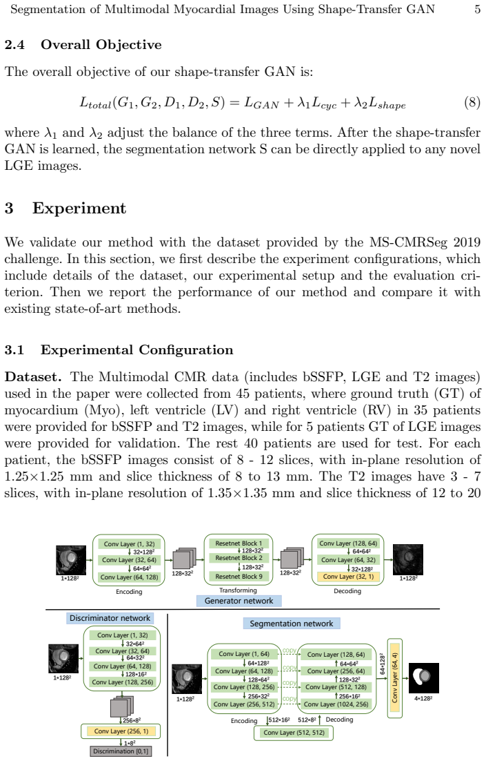
The load-bearing object is the Shape-Transfer GAN objective \(L_{\mathrm{total}} = L_{\mathrm{GAN}} + \lambda_1 L_{\mathrm{cyc}} + \lambda_2 L_{\mathrm{shape}}\), with \(L_{\mathrm{shape}}(S,G_1) = \mathbb{E}_x[\mathrm{CE}(m_x, S(G_1(x)))]\). This shape-preservation term forces the segmentor's prediction on a fake LGE image to match the known bSSFP mask, anchoring anatomy during unpaired style transfer. Cycle consistency stabilizes the translation, the discriminators enforce LGE-like intensity and texture, and the same \(S\) that serves as the shape regularizer becomes the deployed LGE segmentor.
What would settle it
Give the trained segmentor held-out real LGE slices with known masks and stratify Dice by whether a slice contains a bright infarct; if accuracy stays high on healthy slices but collapses on infarct slices while the shape-preservation loss is satisfied, the transfer of pathological appearance, not overall anatomy, is what failed.
Extended reading notes
Core claim
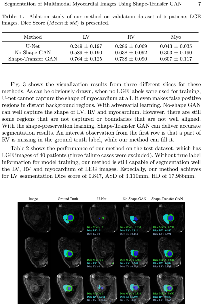
The central discovery is that unpaired style transfer, constrained by a segmentation network that must reproduce the source-domain mask, produces a usable target-domain segmentor. Concretely, the generator \(G_1\) maps bSSFP slices \(x\) to fake LGE slices, and the segmentor \(S\) is trained with cross-entropy against the bSSFP mask \(m_x\) on those fake slices; after training, \(S\) is deployed unchanged on real LGE slices. The paper's evidence includes reported Dice scores on the test set and an ablation in which the shape-preservation term lifts validation myocardium Dice from 0.303 to 0.607.
Load-bearing premise
The method assumes that fake LGE images produced from bSSFP are faithful enough in the clinically relevant places, especially bright diseased myocardium, that a segmentor trained on them transfers to real LGE; the shape-preservation loss only checks that the segmentor's output matches the bSSFP mask, not that the fake image reproduces real LGE pathology.
Editorial extensions
If this is right
- A label-free LGE segmentor can be built from bSSFP labels alone, which addresses the shortage of manual LGE annotations in practice.
- At inference the method is a single forward pass on real LGE slices; there is no per-patient registration, iterative optimization, or LGE label fine-tuning.
- The ablation attributes most of the transfer gain to the shape-preservation term: on the five-patient validation set, myocardium Dice rises from 0.303 without it to 0.607 with it.
- The method does not outperform a deep network trained on real LGE labels (myocardium Dice 0.686 vs 0.812 in the paper's comparison), so supervised labels still add accuracy when they exist.
- The test-set averages cover 37 of 40 patients; the three excluded failure cases are not part of the reported Dice numbers.
Reading between the lines
- The same cycle-plus-shape recipe could be pointed at other unlabeled cardiac MRI targets, such as T2-weighted images, reusing the same bSSFP masks; the paper does not test this.
- A slice-level comparison of segmentor Dice on synthetic versus real LGE would separate style fidelity from anatomy transfer, something the aggregate Dice averages do not reveal.
- The three excluded failures argue that a deployable version would need an automatic check for when the shape prior is violated, since the network itself would not flag low confidence.
- If bright infarct regions are not faithfully generated, average Dice can stay moderate while diseased myocardium is systematically under-segmented; stratifying test Dice by infarct presence would settle this.
Signed reviews
Editorial analysis
A structured set of objections, weighed in public.
Referee Report
Summary. The paper proposes Shape-Transfer GAN for segmenting the left ventricle, right ventricle, and myocardium in late gadolinium enhancement (LGE) cardiac MRI without using any LGE segmentation labels. The method learns a bidirectional bSSFP-to-LGE image translation with cycle-consistency and an additional shape-preservation loss: a segmentation network S is trained on synthetic LGE images produced from bSSFP inputs, supervised by bSSFP ground-truth masks, so that after training S can be applied directly to real LGE images. The authors report an ablation study on 5 validation patients and final test results on 40 patients with Dice scores of 0.847 (LV), 0.776 (RV), and 0.686 (myocardium), after excluding three failure cases, and compare against GMM-based and deep-learning baselines.
Significance. If the central claim is validated, the work would be practically valuable: it offers a way to train an LGE segmenter using only bSSFP labels plus unpaired LGE images, which is relevant to clinical settings where LGE annotations are scarce. The paper's strengths include a clear problem formulation, an explicit objective combining adversarial, cycle-consistency, and shape-preservation terms, and an ablation study showing that each component (adversarial learning, then shape preservation) improves over a U-Net baseline on the small validation set. The method is falsifiable and the experimental design is straightforward to reproduce in principle. However, the published evidence does not yet substantiate the key assumption that the synthetic LGE images preserve the pathological hyperenhanced appearance that makes real LGE segmentation difficult, and the reporting of the test set is incomplete.
major comments (4)
- [Section 3.1] The dataset description is internally inconsistent. The text states that data were collected from 45 patients, that ground truth was provided for bSSFP and T2 images of 35 patients, that LGE ground truth was provided for 5 patients for validation, and then that 'the rest 40 patients are used for test.' Since 35 + 5 = 40, this leaves only 5 patients from the 45-patient collection, not 40. Please clarify the total number of patients, how the 40-patient test set is formed, whether the 35 bSSFP-labeled patients overlap with the test set, and how the 5 validation patients relate to the 40 test patients. This is necessary to interpret the headline Dice scores.
- [Section 3.2, Table 2] The test metrics are reported after excluding three failure cases, but no exclusion criterion is stated. Excluding failures without a transparent rule can arbitrarily inflate the reported Dice scores, and the reader cannot tell whether the excluded cases correspond to missing or atypical hyperenhancement, which is precisely the failure mode most relevant to LGE segmentation. Please describe the exclusion rule, report the metrics with and without the excluded cases, and state how many patients were used for each reported number.
- [Section 2.3, Eq. (7)] The shape-preservation loss only enforces that the synthetic LGE image G1(x) is segmentable into the bSSFP mask mx; it does not constrain the intensity distribution of pathological, hyperenhanced myocardium, which is the main source of difficulty in real LGE segmentation. The cycle-consistency and adversarial losses constrain global structure and image realism, but they do not guarantee that the synthetic images reproduce the bright, heterogeneous appearance of infarcted tissue. Without any visual examples of generated LGE images or a quantitative analysis of translation fidelity (e.g., intensity statistics in infarct versus remote myocardium, or a domain-distance measure), the central claim that the segmentor trained on synthetic LGE generalizes to real LGE is not established. Please add such an analysis.
- [Sections 2.3 and 2.4] The segmentation network S is co-trained with the generator G1 through the shape-preservation loss, and the same S is then applied to real LGE images at test time. This creates a potential co-adaptation: G1 may learn to produce synthetic images that are easy for S to segment using boundary or shape cues inherited from bSSFP, without S learning to handle the actual intensity/texture of real LGE. The ablation on 5 validation patients (Table 1) is too small to rule out this degenerate solution. I recommend reporting a direct comparison of synthetic and real LGE appearance (for example, a reader study or feature-space distances) and, if possible, an analysis of segmentation performance stratified by the presence of hyperenhancement.
minor comments (5)
- [Abstract and Introduction] There are several typos: 'Its worth' should be 'It's worth', 'to be segment' should be 'to segment', and 'LEG images' in the caption of Figure 3 should be 'LGE images'.
- [Section 2.1, Eq. (2)] In the second term of LGAN2, the argument of G2 should be y, not x, to be consistent with the text and with the expectation over y: the generated bSSFP image is G2(y), not G2(x).
- [Section 3.1, Evaluation Metrics] The formula for the Jaccard index in Eq. (9) incorrectly includes a factor of 2; the Jaccard index is |A ∩ B| / |A ∪ B|.
- [Figure 2] The label 'Resetnet Block' should read 'Resnet Block'.
- [References] The text cites Goodfellow et al. as 2016, but reference [4] is the 2014 GAN paper; please correct the year or the reference.
Circularity Check
No significant circularity: the segmentation network is supervised by external bSSFP masks, not by its own predictions or by LGE labels.
full rationale
The paper's derivation chain is self-contained. The segmentation network S is trained under the shape-preservation loss in Eq. (7), Lshape(S,G1) = E[CE(mx, S(G1(x)))], where mx is the externally provided bSSFP ground-truth mask. The supervision signal therefore comes from real anatomical labels in the bSSFP domain, not from the segmentor's own output or from fitted LGE measurements. The subsequent claim that S can be applied directly to real LGE images is a generalization claim supported by the adversarial realism of the synthetic LGE images, but the target Dice scores are measured against held-out LGE ground truth and are not used as training inputs. No load-bearing self-citation or uniqueness argument appears; the cited works are standard external references. One reporting concern is noted but is not circular: in Section 3.2, Table 2 reports results on 40 test patients 'with three failure cases excluded' without specifying exclusion criteria, and the paper does not display generated LGE images or quantify translation quality. These are evaluation and generalization risks, not instances where a prediction reduces to its input by construction.
Assumptions & free parameters
free parameters (2)
- lambda1 =
not reported
- lambda2 =
not reported
assumptions (3)
- domain assumption CycleGAN adversarial and cycle-consistency losses produce realistic LGE images in the unpaired setting.
- domain assumption The bSSFP ground-truth masks mx are valid supervision for synthetic LGE images G1(x), meaning the generator inherits the exact spatial geometry of the input bSSFP slice.
- domain assumption A segmentor trained on synthetic LGE images generalizes to real LGE images without fine-tuning on real LGE labels.
Cite this review
Pith. "Pith review of Segmentation of Multimodal Myocardial Images Using Shape-Transfer GAN." pith.science (2026). https://pith.science/paper/KF4ISZ75
@misc{pith2026190805094,
  author       = {Pith},
  title        = {Pith review of: Segmentation of Multimodal Myocardial Images Using Shape-Transfer GAN},
  year         = {2026},
  howpublished = {\url{https://pith.science/paper/KF4ISZ75}},
  note         = {Machine review of arXiv:1908.05094}
}
read the original abstract
Myocardium segmentation of late gadolinium enhancement (LGE) Cardiac MR images is important for evaluation of infarction regions in clinical practice. The pathological myocardium in LGE images presents distinctive brightness and textures compared with the healthy tissues, making it much more challenging to be segment. Instead, the balanced-Steady State Free Precession (bSSFP) cine images show clearly boundaries and can be easily segmented. Given this fact, we propose a novel shape-transfer GAN for LGE images, which can 1) learn to generate realistic LGE images from bSSFP with the anatomical shape preserved, and 2) learn to segment the myocardium of LGE images from these generated images. It's worth to note that no segmentation label of the LGE images is used during this procedure. We test our model on dataset from the Multi-sequence Cardiac MR Segmentation Challenge. The results show that the proposed Shape-Transfer GAN can achieve accurate myocardium masks of LGE images.
Figures

Reference graph
Works this paper leans on
-
[1]
Multivariate mixture model for myocardial segmentation combining multi-source images,
X. Zhuang, "Multivariate mixture model for myocardial segmentation combining multi-source images," in IEEE Transactions on Pattern Analysis and Machine Intelligence. doi: 10.1109/TPAMI.2018.2869576
arXiv 2018
-
[2]
Z. Xiong, V. V. Fedorov, X. Fu, E. Cheng, R. Macleod and J. Zhao, "Fully Automatic Left Atrium Segmentation From Late Gadolinium Enhanced Magnetic Resonance Imaging Using a Dual Fully Convolutional Neural Network," in IEEE Transactions on Medical Imaging, vol. 38, no. 2, pp. 515-524, Feb. 2019
work page 2019
-
[3]
Cardiac Segmentation from LGE MRI Using Deep Neural Network Incorporating Shape and Spatial Priors
Qian Yue, Xinzhe Luo, Qing Ye, Lingchao Xu, Xiahai Zhuang . "Cardiac Segmentation from LGE MRI Using Deep Neural Network Incorporating Shape and Spatial Priors." MICCAI 2019
work page 2019
-
[4]
Goodfellow, Ian, et al. "Generative adversarial nets." Advances in neural information processing systems. 2014
work page 2014
-
[5]
Unpaired image-to-image translation using cycle-consistent adversarial networks
Zhu, Jun-Yan, et al. "Unpaired image-to-image translation using cycle-consistent adversarial networks." Proceedings of the IEEE international conference on computer vision. 2017
work page 2017
-
[6]
Isola P, Zhu J Y, Zhou T, et al. Image-to-image translation with conditional adversarial networks[C]//Proceedings of the IEEE conference on computer vision and pattern recognition. 2017: 1125-1134
work page 2017
-
[7]
Unsupervised representation learning with deep convolutional generative adversarial networks[J]
Radford A, Metz L, Chintala S. Unsupervised representation learning with deep convolutional generative adversarial networks[J]. arXiv preprint arXiv:1511.06434, 2015
arXiv 2015
-
[8]
Mao X, Li Q, Xie H, et al. Least squares generative adversarial networks[C]//Proceedings of the IEEE International Conference on Computer Vision. 2017: 2794-2802
work page 2017
Show all 10 references
-
[9]
, " * write output.state after.block = add.period write
ENTRY address author booktitle chapter doi edition editor eid howpublished institution journal key month note number organization pages publisher school series title type url volume year label INTEGERS output.state before.all mid.sentence after.sentence after.block FUNCTION in...
-
[10]
write newline
" write newline "" before.all 'output.state := FUNCTION n.dashify 't := "" t empty not t #1 #1 substring "-" = t #1 #2 substring "--" = not "--" * t #2 global.max substring 't := t #1 #1 substring "-" = "-" * t #2 global.max substring 't := while if t #1 #1 substring * t #2 gl...
Reviewed August 14, 2026 · model on record in the stance chip above.
Discussion (0). Continue with ORCID to comment.