REVIEW 3 major objections 5 minor 33 references
Using Near Infrared Spectroscopy and Machine Learning to diagnose Systemic Sclerosis
T0 review · 3 major / 5 minor · reviewed 2026-08-14 · deepseek-v4-flash
Pith's one-line read The paper claims that near-infrared spectra of the hand, classified by a linear support-vector machine with recursive feature elimination, distinguish systemic sclerosis patients from controls, with the 1270 nm singlet-oxygen band as the…
desk verdict The 1270 nm singlet-oxygen claim is not supported by the analysis as written; the synthetic-sample step and the contradictory statistics make the central result unverifiable. read the letter →
The pith
A machine-rendered reading of the paper's core claim, the machinery that carries it, and where it could break.
The reading
What carries the argument
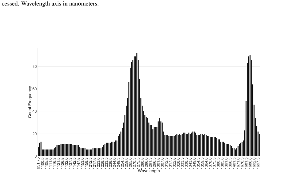
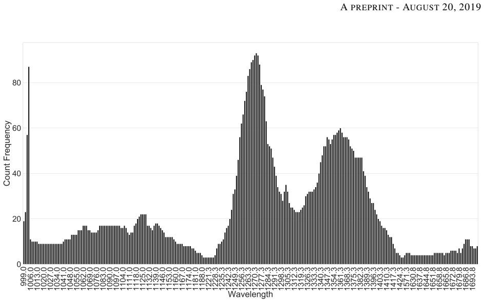
The central machinery is the pairing of near-infrared absorbance spectroscopy with Recursive Feature Elimination with Cross-Validation (RFECV) driving a linear Support Vector Classifier (SVC). RFECV repeatedly trains the classifier, removes the least informative wavelength, and uses cross-validation to decide how many wavelengths to keep, so that the output is a short list of diagnostic wavelengths rather than a full spectrum. Repeating the selection over 100 random train/test splits turns that list into a histogram, and the most frequent band, 1270 nm, is then interpreted biochemically as singlet-oxygen luminescence. This gives the argument a concrete optical marker instead of a purely statistical black box.
What would settle it
The decisive check is to rerun the same SVC+RFECV pipeline on real spectra only, using leave-one-out classification of the 17 SSc patients against the 25 controls, and see whether accuracy holds and 1270 nm remains the most-selected wavelength; if either fails, the reported diagnostic signal is an artifact of the synthetic samples.
Extended reading notes
Core claim
In its own terms, the paper finds that systemic sclerosis leaves a measurable optical signature in the near-infrared absorbance of hand skin. A linear support-vector classifier, guided by recursive feature elimination, separates SSc patients from controls across 100 training/test runs, and the wavelength selected most often is 1270 nm, attributed to singlet-oxygen luminescence, consistent with the oxidative-stress component of SSc pathogenesis. The accuracy distribution is best for spectra taken at the proximal interphalangeal joints, and statistical tests rank that region above the metacarpal and proximal phalanges sites. The authors therefore propose NIRS plus machine learning as a path toward faster, non-invasive SSc diagnosis.
Load-bearing premise
The entire accuracy result rests on the assumption that the eight synthetic spectra added to balance the SSc group behave exactly like real patient spectra; if they do not, the reported accuracies and the 1270 nm selection are artifacts of the balancing procedure.
Editorial extensions
If this is right
- A standard, inexpensive near-infrared spectrometer paired with SVC could serve as a screening aid in rheumatology clinics, shortening the time from symptom onset to diagnosis.
- The proximal interphalangeal joints should be the standard measurement region in any follow-up protocol, since they produced the most accurate classifications.
- The 1270 nm band gives a concrete optical target for future work: instrument designers and clinicians can focus on a narrow spectral window rather than whole-spectrum diagnostics.
- Because NIRS uses non-ionizing light and no biopsy, the approach could be repeated safely to track disease progression or response to treatment.
Reading between the lines
- A direct test the paper leaves implicit: retraining on the 17 real SSc spectra with leave-one-out validation would reveal whether the reported accuracy survives without the synthetic balancing samples; this is the single most informative follow-up.
- If the 1270 nm association is real, the same pipeline could be tested on patients with Raynaud's phenomenon alone to see whether the optical signal precedes clinical skin fibrosis, an early-diagnosis claim the paper does not make.
- The authors interpret 1270 nm as singlet-oxygen luminescence but do not establish a dose-response link; correlating the 1270 nm intensity with a clinical fibrosis score, such as the modified Rodnan skin score, would test that biochemical reading.
- More broadly, the method implies a cheap optical oxidative-stress readout for autoimmune disease, which could extend to other fibrotic conditions, but that extension is speculative until the SSc result is replicated independently.
Signed reviews
Editorial analysis
A structured set of objections, weighed in public.
Referee Report
Summary. The manuscript reports a pilot study using near-infrared spectroscopy (NIRS) of three hand regions to distinguish patients with systemic sclerosis (SSc) from healthy controls. Spectra were preprocessed by baseline removal and normalization; to balance group sizes, the authors created 'false samples' so that both groups had 25 spectra. A support vector classifier with recursive feature elimination and cross-validation (RFECV) was run 100 times with a 20% test split, and the most frequently selected wavelength bands were recorded. The paper claims that the proximal interphalangeal joints region yields the best accuracy and that the 1270 nm band, attributed to singlet oxygen luminescence, is the most diagnostically important.
Significance. If the central claim were established, a low-cost, non-invasive optical diagnostic for systemic sclerosis would be a valuable clinical tool, and the specific wavelength marker at 1270 nm would provide a testable biological hypothesis. The authors use standard open-source machine-learning software, report a reproducible procedure repeated 100 times, and identify a concrete wavelength band, which are commendable features. However, the significance of the result depends entirely on the validity of the synthetic data used to balance the groups and on the statistical interpretation of the accuracy comparisons; as presented, the evidence does not support the diagnostic claim.
major comments (3)
- [Section 2.1, 'Spectra acquiring and preprocessing'] The creation of 'false samples' is unexplained and load-bearing. The text states only that 'false samples were created, so the number of 25 samples was established for each of the two groups.' With 25 controls and 17 SSc patients, this implies that roughly eight fabricated spectra were added to the SSc group, but the generation procedure (e.g., resampling, interpolation, or noise injection) is not described. Every downstream result—the RFECV-selected wavelengths, the 1270 nm band, and the reported accuracy distribution—is computed on this mixed dataset. If the synthetic spectra are not statistically exchangeable with real SSc spectra, the classifier may learn to distinguish synthetic from real spectra rather than SSc from controls, and a 20% held-out split that includes synthetic samples does not measure real diagnostic performance. The authors must either describe the synthetic-data generation method and justify exchangeability, or validate the classifier on a real patient cohort.
- [Section 3, 'Results'] The statistical comparison between regions is contradictory and does not support the claim that the proximal interphalangeal joints region is best. The text states: 'U-tests were performed... all ρ values resulted were greater than 0.99.' If these are p-values, values greater than 0.99 indicate no statistically significant difference; if these are some other statistic, the test description is incomplete. Either way, the conclusion that one region is superior is not backed by the reported numbers. In addition, accuracy is reported as histograms without confidence intervals or standard errors, and the 100 random 80/20 splits on a dataset of at most 50 samples are not independent replications, so the displayed distributions do not provide a reliable estimate of generalization performance.
- [Sections 2.2 and 3, feature selection and evaluation] The identification of the 1270 nm band as the 'most important wavelength' is derived from the same cohort used to evaluate the classifier. The frequency of wavelength selection is obtained by running RFECV on random training subsets of the same 50 samples, and the accuracy is measured on the corresponding test subsets of the same cohort. This makes the wavelength importance a descriptive statistic of this dataset rather than an independent predictor. The singlet oxygen explanation in Section 4 is a post hoc attribution and does not establish a biological link. Supporting the claim would require an external validation cohort, or at least a nested cross-validation in which feature selection is performed without seeing the test data, and ideally an independent experiment designed to probe the 1270 nm feature.
minor comments (5)
- [Abstract and Introduction] The phrase 'hand's volunteers' should be 'volunteers' hands' or 'the hands of volunteers'; there are also minor grammatical errors elsewhere (e.g., 'The results suggests', 'better accuracy's scores').
- [Section 2.1] The preprocessing description is vague: 'the baseline of the raw data had to be removed' by subtracting the minimum value, but the exact order of operations (subtraction, division by source spectrum, filtering) is not fully specified, making the results difficult to reproduce.
- [Section 2.2] The grid search over SVC parameters is mentioned but not described: it is not reported which parameter ranges were searched, how performance was evaluated during the search, or whether the 'penalty parameter of 103 order of magnitude' means C = 10^3.
- [Section 3 and Figures] Figures 2-4 show histograms of selected wavelengths, but the x-axis is partially omitted 'for better visualization', and the y-axis is not labeled; the number of runs represented by each histogram is stated only in the captions. Table 1 would benefit from a definition of how the 'means' were computed from the counts.
- [General] The paper does not state where the data or analysis code can be obtained; given the small and synthetic-augmented dataset, public availability would be essential for auditing the results.
Circularity Check
No circularity in the derivation; the ML pipeline uses a held-out test split and external references, though the uncharacterized 'false samples' are a serious data-validity concern rather than a circular step.
full rationale
The paper's core derivation is not circular: spectra are split into training (80%) and test (20%) subsets, grid search selects SVC hyperparameters, RFECV selects wavelengths on the training portion, and accuracy is reported on the held-out portion. The 'most important wavelength' (1270 nm) is a feature-selection output, not a parameter fitted to the test labels, and the singlet-oxygen explanation is an external interpretation applied after the fact rather than an input to the model. No self-citation is load-bearing: the cited baseline-removal, SVM, scikit-learn, and singlet-oxygen references are all external works, and none supplies a uniqueness theorem or ansatz introduced by the present authors. The serious flaw in the manuscript is the uncharacterized creation of 'false samples' to balance the SSc group (25 controls vs. 17 patients). Because the paper does not describe how these synthetic spectra were generated, the reported accuracy and the selected 1270 nm band may be artifacts of the balancing procedure. However, this is a data-integrity and external-validity problem, not a derivation that reduces by construction to its own inputs: without a specified generative procedure, one cannot exhibit the kind of equation-level reduction that would constitute circularity. The paper's own equations and quoted references do not smuggle the conclusion into the premises, so the circularity score is 0.
Assumptions & free parameters
free parameters (3)
- SVC penalty parameter C =
1e3
- Number of synthetic samples =
8 (17 SSc supplemented to 25)
- Test split fraction =
20%
assumptions (3)
- domain assumption Support vector machines with linear kernels separate the two spectral groups because the absorbance differences are real and stable.
- ad hoc to paper The synthetic 'false samples' preserve the statistical properties of real SSc spectra.
- standard math The U-test p-values are computed and interpreted correctly.
Cite this review
Pith. "Pith review of Using Near Infrared Spectroscopy and Machine Learning to diagnose Systemic Sclerosis." pith.science (2026). https://pith.science/paper/KCNUVFDS
@misc{pith2026190806137,
  author       = {Pith},
  title        = {Pith review of: Using Near Infrared Spectroscopy and Machine Learning to diagnose Systemic Sclerosis},
  year         = {2026},
  howpublished = {\url{https://pith.science/paper/KCNUVFDS}},
  note         = {Machine review of arXiv:1908.06137}
}
read the original abstract
The motivation of this work is the use of non-invasive and low cost techniques to obtain a faster and more accurate diagnosis of systemic sclerosis (SSc), rheumatic, autoimmune, chronic and rare disease. The technique in question is Near Infrared Spectroscopy (NIRS). Spectra were acquired from three different regions of hand's volunteers. Machine learning algorithms are used to classify and search for the best optical wavelength. The results demonstrate that it is easy to obtain wavelength bands more important for the diagnosis. We use the algorithm RFECV and SVC. The results suggests that the most important wavelength band is at 1270 nm, referring to the luminescence of Singlet Oxygen. The results indicates that the Proximal Interphalangeal Joints region returns better accuracy's scores. Optical spectrometers can be found at low prices and can be easily used in clinical evaluations, while the algorithms used are completely diffused on open source platforms.
Figures


Figures from the paper (2 more)

Reference graph
Works this paper leans on
-
[1]
Epidemiology and natural history of systemic sclerosis
VD Steen and Jr TA Medsger. Epidemiology and natural history of systemic sclerosis. Rheumatic diseases clinics of North America, 16(1):1–10, 1990
work page 1990
-
[2]
Pulmonary arterial hypertension in systemic sclerosis: the need for early detection and treatment
SM Proudman, WM Stevens, J Sahhar, and D Celermajer. Pulmonary arterial hypertension in systemic sclerosis: the need for early detection and treatment. Internal medicine journal, 37(7):485–494, 2007
work page 2007
-
[3]
The clinical relevance of autoantibodies in scleroderma
Khanh T Ho and John D Reveille. The clinical relevance of autoantibodies in scleroderma. Arthritis Res Ther, 5(2):80, 2003
work page 2003
-
[4]
Incidence and preva- lence of systemic sclerosis: a systematic literature review
H ´el`ene Chifflot, Bruno Fautrel, Christelle Sordet, Emmanuel Chatelus, and Jean Sibilia. Incidence and preva- lence of systemic sclerosis: a systematic literature review. In Seminars in arthritis and rheumatism, volume 37, pages 223–235. Elsevier, 2008
work page 2008
-
[5]
Biomedical applications of collagen
Chi H Lee, Anuj Singla, and Yugyung Lee. Biomedical applications of collagen. International journal of pharmaceutics, 221(1-2):1–22, 2001
work page 2001
-
[6]
Collagen structure and stability
Matthew D Shoulders and Ronald T Raines. Collagen structure and stability. Annual review of biochemistry , 78:929–958, 2009
work page 2009
-
[7]
Preliminary criteria for the classification of systemic sclerosis (scleroderma)
Alfonse T Masi. Preliminary criteria for the classification of systemic sclerosis (scleroderma). Arthritis & Rheumatism, 23(5):581–590, 1980. 7 A PREPRINT - AUGUST 20, 2019
work page 1980
-
[8]
J ´erˆome Avouac, J Fransen, UA Walker, V Riccieri, Valdemar Smith, Christoph Muller, I Miniati, IH Tarner, S Bellando Randone, M Cutolo, et al. Preliminary criteria for the very early diagnosis of systemic sclerosis: results of a delphi consensus study from eular scleroderma trials and research group. Annals of the rheumatic diseases, 70(3):476–481, 2011
work page 2011
Show all 33 references
-
[9]
2013 classification criteria for systemic sclerosis: an american college of rheumatology/european league against rheumatism collaborative initiative
Frank Van Den Hoogen, Dinesh Khanna, Jaap Fransen, Sindhu R Johnson, Murray Baron, Alan Tyndall, Marco Matucci-Cerinic, Raymond P Naden, Thomas A Medsger Jr, Patricia E Carreira, et al. 2013 classification criteria for systemic sclerosis: an american college of rheumatology/eur...
2013
-
[10]
Gene expression of types i and iii collagen, decorin, matrix metalloproteinases and tissue inhibitors of metalloproteinases in skin fibroblasts from patients with systemic sclerosis
K Kuroda and H Shinkai. Gene expression of types i and iii collagen, decorin, matrix metalloproteinases and tissue inhibitors of metalloproteinases in skin fibroblasts from patients with systemic sclerosis. Archives of dermatological research, 289(10):567–572, 1997
1997
-
[11]
Patologia oral e maxilofacial
Brad Neville. Patologia oral e maxilofacial. Elsevier Brasil, 2016
2016
-
[12]
Small mouths
Jonathan B Albilia, David K Lam, Nick Blanas, Cameron ML Clokie, and George KB S ´andor. Small mouths. . . big problems? a review of scleroderma and its oral health implications. Journal of the Canadian Dental Associ- ation, 73(9), 2007
2007
-
[13]
Systemic sclerosis
Yannick Allanore, Robert Simms, Oliver Distler, Maria Trojanowska, Janet Pope, Christopher P Denton, and John Varga. Systemic sclerosis. Nature reviews Disease primers, 1:15002, 2015
2015
-
[14]
A study on oxygen saturation images constructed from the skin tissue of human hand
Hsin-Yi Tsai, Kuo-Cheng Huang, Han-Chao Chang, and Chung-Hsing Chang. A study on oxygen saturation images constructed from the skin tissue of human hand. In 2013 IEEE International Instrumentation and Mea- surement Technology Conference (I2MTC), pages 58–62. IEEE, 2013
2013
-
[15]
Biomedical Photonics Handbook, 3 Volume Set
Tuan V o-Dinh. Biomedical Photonics Handbook, 3 Volume Set. CRC press, 2014
2014
-
[16]
Near- infrared diffuse optical tomography
AH Hielscher, AY Bluestone, GS Abdoulaev, AD Klose, J Lasker, M Stewart, U Netz, and J Beuthan. Near- infrared diffuse optical tomography. Disease markers, 18(5-6):313–337, 2002
2002
-
[17]
A review on continuous wave functional near-infrared spectroscopy and imaging instrumenta- tion and methodology
Felix Scholkmann, Stefan Kleiser, Andreas Jaakko Metz, Raphael Zimmermann, Juan Mata Pavia, Ursula Wolf, and Martin Wolf. A review on continuous wave functional near-infrared spectroscopy and imaging instrumenta- tion and methodology. Neuroimage, 85:6–27, 2014
2014
-
[18]
An optimization perspec- tive on baseline removal for spectroscopy
Stephen Giguere, CJ Carey, Thomas Boucher, Sridhar Mahadevan, and M Darby Dyar. An optimization perspec- tive on baseline removal for spectroscopy. In Fifth IJCAI Workshop on AI in Space, 2015
2015
-
[19]
A tutorial on support vector machines for pattern recognition
Christopher JC Burges. A tutorial on support vector machines for pattern recognition. Data mining and knowl- edge discovery, 2(2):121–167, 1998
1998
-
[20]
Pedregosa, G
F. Pedregosa, G. Varoquaux, A. Gramfort, V . Michel, B. Thirion, O. Grisel, M. Blondel, P. Prettenhofer, R. Weiss, V . Dubourg, J. Vanderplas, A. Passos, D. Cournapeau, M. Brucher, M. Perrot, and E. Duchesnay. Scikit-learn: Machine learning in Python. Journal of Machine Learni...
2011
-
[21]
Libsvm: A library for support vector machines
Chih-Chung Chang and Chih-Jen Lin. Libsvm: A library for support vector machines. ACM transactions on intelligent systems and technology (TIST), 2(3):27, 2011
2011
-
[22]
Probabilistic outputs for support vector machines and comparisons to regularized likelihood methods
John Platt et al. Probabilistic outputs for support vector machines and comparisons to regularized likelihood methods. Advances in large margin classifiers, 10(3):61–74, 1999
1999
-
[23]
Gene selection for cancer classification using support vector machines
Isabelle Guyon, Jason Weston, Stephen Barnhill, and Vladimir Vapnik. Gene selection for cancer classification using support vector machines. Machine learning, 46(1-3):389–422, 2002
2002
-
[24]
On statistical bounds of heuristic solutions to location problems
Kenneth Carling and Xiangli Meng. On statistical bounds of heuristic solutions to location problems. Journal of combinatorial optimization, 31(4):1518–1549, 2016
2016
-
[25]
Direct detection of singlet oxygen generated by uva irradiation in human cells and skin.Journal of Investigative Dermatology, 127(6):1498– 1506, 2007
J ¨urgen Baier, Tim Maisch, Max Maier, Michael Landthaler, and Wolfgang B¨aumler. Direct detection of singlet oxygen generated by uva irradiation in human cells and skin.Journal of Investigative Dermatology, 127(6):1498– 1506, 2007
2007
-
[26]
Uva radiation-induced oxidative damage to lipids and proteins in vitro and in human skin fibroblasts is dependent on iron and singlet oxygen
Glenn F Vile and Rex M Tyrrell. Uva radiation-induced oxidative damage to lipids and proteins in vitro and in human skin fibroblasts is dependent on iron and singlet oxygen. Free Radical Biology and Medicine, 18(4):721– 730, 1995
1995
-
[27]
Singlet oxygen-mediated damage to proteins and its consequences
Michael J Davies. Singlet oxygen-mediated damage to proteins and its consequences. Biochemical and biophys- ical research communications, 305(3):761–770, 2003
2003
-
[28]
Singlet oxygen stress in microorganisms
J Glaeser, AM Nuss, BA Berghoff, and Gabriele Klug. Singlet oxygen stress in microorganisms. In Advances in microbial physiology, volume 58, pages 141–173. Elsevier, 2011. 8 A PREPRINT - AUGUST 20, 2019
2011
-
[29]
Singlet oxygen induces collagenase expression in human skin fibroblasts
Karin Scharffetter-Kochanek, Meinhard Wlaschek, Karlis Briviba, and Helmut Sies. Singlet oxygen induces collagenase expression in human skin fibroblasts. FEBS letters, 331(3):304–306, 1993
1993
-
[30]
Kinetics of singlet oxygen photosen- sitization in human skin fibroblasts
Ana Jim ´enez-Banzo, M Luisa Sagrist`a, Margarita Mora, and Santi Nonell. Kinetics of singlet oxygen photosen- sitization in human skin fibroblasts. Free Radical Biology and Medicine, 44(11):1926–1934, 2008
1926
-
[31]
A double-blind placebo-controlled trial of antioxidant therapy in limited cutaneous systemic sclerosis
Ariane L Herrick, Sally Hollis, D Schofield, F Rieley, A Blann, K Griffin, T Moore, JM Braganza, and Mal- colm IV Jayson. A double-blind placebo-controlled trial of antioxidant therapy in limited cutaneous systemic sclerosis. Clinical and Experimental Rheumatology, 18(3):349–356, 2000
2000
-
[32]
Oxygen free radicals and systemic autoimmunity
H Ahsan, A Ali, and R Ali. Oxygen free radicals and systemic autoimmunity. Clinical & Experimental Im- munology, 131(3):398–404, 2003
2003
-
[33]
Review of short- wave infrared spectroscopy and imaging methods for biological tissue characterization
Robert H Wilson, Kyle P Nadeau, Frank B Jaworski, Bruce J Tromberg, and Anthony J Durkin. Review of short- wave infrared spectroscopy and imaging methods for biological tissue characterization. Journal of biomedical optics, 20(3):030901, 2015. 9
2015
Reviewed August 14, 2026 · model on record in the stance chip above.
Discussion (0). Continue with ORCID to comment.