REVIEW 3 major objections 5 minor 1 cited by
Models Genesis: Generic Autodidactic Models for 3D Medical Image Analysis
T0 review · 3 major / 5 minor · reviewed 2026-08-14 · deepseek-v4-flash
Pith's one-line read The paper reports that self-supervised 3D source models pretrained on unlabeled chest CTs, called Models Genesis, beat both training from scratch and 2D ImageNet-based transfer across five 3D medical imaging tasks.
desk verdict Worth refs: free 3D pre-trained models and a credible 3D-vs-2D transfer result, but the significance claims hinge on an unstated trial-resampling detail and the unified-framework attribution isn't supported by the ablation. read the letter →
The pith
A machine-rendered reading of the paper's core claim, the machinery that carries it, and where it could break.
The reading
What carries the argument
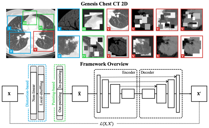
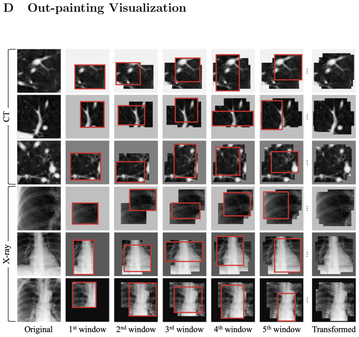
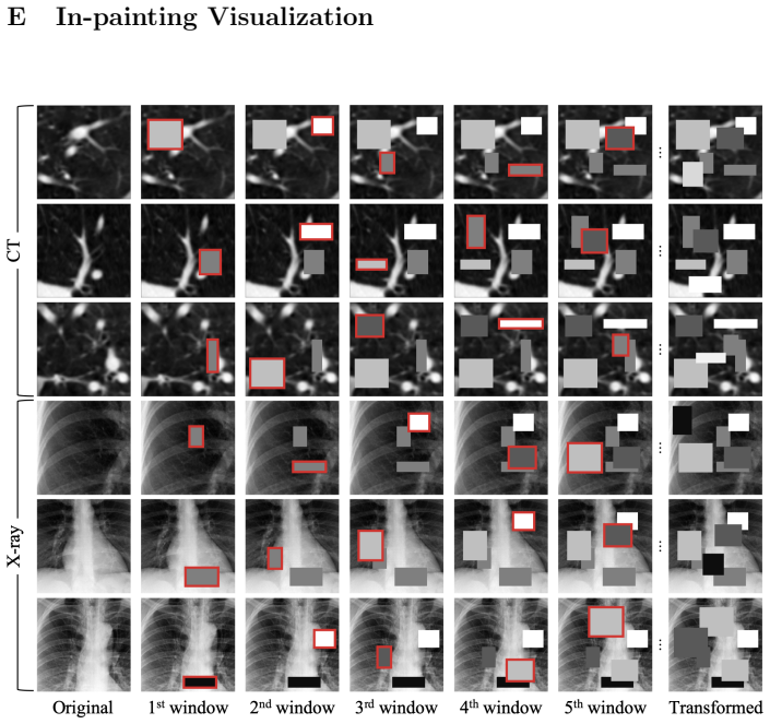
The load-bearing training scheme is image restoration: from an unlabeled image, patches $X$ of arbitrary size are cropped at random locations, transformed into $\tilde{X}$, and an encoder-decoder is trained to recover $X$ with an $L_1$ loss. Four transformations are consolidated under this single objective—a smooth monotonic intensity-remapping curve (appearance), local pixel shuffling inside small windows (texture), masking everything outside a complex window with a random value (global geometry), and filling the window with a constant (local continuity). Because one encoder-decoder handles all transformations, the representation is learned from multiple perspectives at once; the encoder initializes classification targets and the encoder-decoder initializes segmentation targets.
What would settle it
Rerun the five 3D target tasks with trial protocols that resample the training and validation splits each time (for example, repeated k-fold), and check whether Models Genesis still beats both scratch training and 2D ImageNet fine-tuning at p < 0.05; if the advantage disappears, the original significance claim depends on the unresolved protocol.
Extended reading notes
Core claim
The central claim is that a generic source model for 3D medical imaging can be created ex nihilo from unlabeled CT volumes, without any manual annotation, by training an encoder-decoder to restore original image patches from transformed versions. Fine-tuned on five target applications—lung nodule false-positive reduction, lung nodule segmentation, pulmonary embolism false-positive reduction, liver segmentation, and brain tumor segmentation—these models significantly outperform 3D training from scratch in every case, with p-values below 0.05. On the three tasks with a direct 2D comparison, the 3D Models Genesis also outperform fine-tuning from ImageNet and fine-tuning 2D versions of Models Genesis, while 3D scratch models do not consistently do so. The paper attributes the advantage to learning the recurrent anatomy of the chest from CT, arguing that anatomy itself provides supervision and that 3D context is the information 2D transfer paradigms sacrifice.
Load-bearing premise
The central significance claims rest on an unstated experimental detail: the paper says each result comes from 'at least 10 trials' but never says whether a trial resamples the training and validation data or only reinitializes the network on one fixed split; if only the random seed changes, the standard deviations capture seed noise and the reported p-values overstate the evidence.
Editorial extensions
If this is right
- Models Genesis fine-tuning beats 3D training from scratch in every reported 3D task, with nominal p < 0.05 in all five cases.
- Against 2D slice-based baselines, the 3D Models Genesis top both ImageNet fine-tuning and fine-tuning 2D versions of Models Genesis on all tasks compared, a claim that does not hold for 3D scratch models.
- The 2D versions of Models Genesis offer performance equivalent to supervised ImageNet fine-tuning within the same modality, but with zero annotation cost.
- The unified restoration objective is more stable across target tasks than any single transformation, and cross-domain transfer works across diseases, organs, datasets, and modalities, though same-domain transfer is preferred.
Reading between the lines
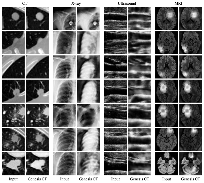
- Inference: the same self-supervised recipe could be applied to MRI or ultrasound archives to build modality-specific source models; the paper lists this as future work, and its cross-modality restoration examples make it a concrete next test.
- Inference: if the advantage is truly anatomical, the margin over 2D transfers should depend on the amount of 3D context available, so ablating slice count or input patch depth on the same tasks would be a direct check of the mechanism.
- Inference: the pretrained encoder should be evaluated as a frozen feature extractor for anomaly detection or scan triage, uses the paper does not explore.
Signed reviews
Editorial analysis
A structured set of objections, weighed in public.
Referee Report
Summary. The paper introduces Models Genesis, a set of self-supervised 3D models pre-trained on unlabeled chest CT images by restoring patches that have been altered with four transformations: non-linear intensity distortion, local pixel shuffling, out-painting, and in-painting. These transformations are unified into a single image-restoration task. The authors fine-tune the pre-trained encoder (and decoder) for downstream tasks and claim that, across five 3D applications, Models Genesis significantly outperform training from scratch, and also outperform 2D approaches including fine-tuning from ImageNet. The paper reports p-values for these gains, compares the unified scheme with individual schemes, provides a 2D version of the method, and releases the pre-trained models publicly.
Significance. If the reported results hold, the paper makes a useful contribution: it shows that self-supervised pre-training directly on 3D medical images can provide a reusable source model that transfers across diseases, organs, datasets, and even modalities, and it is one of the early systematic demonstrations that 3D self-supervised pre-training can beat 2D ImageNet transfer in 3D segmentation and classification tasks. The authors are careful to pre-train on a disjoint subset of LIDC-IDRI so that held-out LIDC-IDRI tests are not contaminated by the proxy-task training data, and they make their pre-trained models publicly available. Reproducibility is also aided by the use of standard architectures (3D U-Net and ResNet-18-based U-Net) and public benchmarks.
major comments (3)
- [Sec. 3, Experiment protocol] The protocol states that performance was measured 'through at least 10 trials' and analyzed with an 'independent two-sample t-test', but it never defines what a trial is. If a trial re-initializes the network on one fixed train/validation/test split, then the trials are not independent draws from the data distribution, the standard deviations in Tables 2 and 4 reflect only seed/optimization noise, and the reported p-values overstate dataset-level significance. This is load-bearing for the smallest and most variable gains in Table 2: BMS (90.16±0.41 vs. 90.60±0.20, p=0.0041) and NCC (94.25±5.07 vs. 98.20±0.51, p=0.0180). The authors must state whether the patient-level split is resampled per trial, report the exact number of trials per condition, and either supply a split-resampling analysis or appropriately qualify the significance claims.
- [Table 3 and Appendix H, Fig. 15] Table 3 reports that the unified framework is not significantly better than the best individual scheme in any of the five tasks (p=0.0848, 0.0520, 0.2102, 0.4249, 0.4276), yet the abstract and Section 4 attribute the performance of Models Genesis to 'our unified self-supervised learning framework' and claim that the unified scheme is more robust. Appendix H, Fig. 15, caption even states 'p<0.05' for this comparison, directly contradicting Table 3. Either the statistical analysis is wrong in one of these places, or the claim that the unified framework is the driver of the gains needs to be substantially softened; the current text overstates what the experiments show.
- [Sec. 4 Conclusion and Table 2 footnotes] The conclusion states that the empirical results are 'surpassing state-of-the-art performances in most of the applications', but the footnotes of Table 2 compare against published results using different metrics and protocols. For LCS, the LiTS winner's 96.60% is a Dice score with postprocessing, while the reported 91.13% is an IoU without postprocessing; for BMS, the BraTS winner's 91.00% uses ensembling while the reported 92.58% does not. Dice and IoU are not interchangeable, so these footnotes do not support a state-of-the-art claim. The authors should either present metric-matched, protocol-matched comparisons or remove the 'surpassing state-of-the-art' wording.
minor comments (5)
- [Sec. 3, Experiment protocol] The exact number of trials is never stated; 'at least 10' is not sufficient to verify the t-test degrees of freedom or to assess whether the standard deviations are stable. Please report the exact n per condition and describe how seeds and data splits are handled.
- [Sec. 1 and Sec. 3] The introduction says Models Genesis 'significantly outperform learning from scratch in all seven target tasks', but Table 2 provides significance tests for only five 3D tasks, and Fig. 2 does not report significance for the 2D tasks. Please align the text with the actual statistical analyses.
- [Table 4] In Table 4 the 2D Genesis columns are numerically below the 2D ImageNet columns for all three tasks (e.g., NCC: 97.45 vs. 97.79), yet the abstract's 'consistently top any 2D approaches' could be read as referring to all Models Genesis variants. The wording should make explicit that the superiority claim is for the 3D Genesis model.
- [Appendix H, Fig. 15 caption] The caption states 'yielding either the best result or comparable performance to the best model (p<0.05)', which is inconsistent with the p-values in Table 3; this needs correction regardless of which analysis is intended.
- [Throughout] There are several typos and formatting issues: 'Genesis Check CT' in the Table 4 caption should be 'Genesis Chest CT', and 'Appendix4' appears without a space. A careful proofread would improve readability.
Circularity Check
No circularity: target performance is measured on held-out external benchmarks after fine-tuning a self-supervised source model; no fitted parameter is renamed as a prediction.
full rationale
The paper's claimed derivation chain is an empirical transfer-learning protocol, not a derivation of a target quantity from an input. Models Genesis are pre-trained on unlabeled CT/X-ray via self-supervised restoration (Sec. 2 and Appendix B–E), and target AUC/IoU is obtained by fine-tuning on held-out public benchmarks (LUNA2016, LIDC-IDRI, LiTS2017, BraTS2013, ChestX-ray8). The protocol explicitly separates pre-training and testing data: 'The reason that we decided not to use all images in LIDC-IDRI and in ChestX-ray8 for training Models Genesis is to avoid test-image leaks between proxy and target tasks, so that we can confidently use the rest images solely for testing Models Genesis as well as the target models.' Thus the headline numbers are not written into the model by construction. The significance statistics rest on an under-specified trial design ('through at least 10 trials' with an 'independent two-sample t-test'), and the caption for Fig. 2 describes Genesis Chest CT 2D as trained on 'LUNA dataset' without stating a disjoint split for the NCC target; these are reproducibility/statistical-reporting gaps, not circularity, because no metric is an algebraic function of a fitted input. Self-citations to Tajbakhsh et al. [9,10] are contextual and not load-bearing, and no uniqueness theorem or ansatz is imported from the authors' prior work. I find no step where a prediction reduces to its input by definition.
Assumptions & free parameters
free parameters (5)
- predefined transformation probabilities =
not reported
- local shuffling window sizes =
2D: 5x5 to 20x20; 3D: unreported
- masked-region cap =
under 1/4 of patch
- pre-training dataset size =
534 CT scans from LIDC-IDRI
- local shuffling window count =
1000 windows per patch
assumptions (4)
- domain assumption Pixel intensities in CT (Hounsfield units) and their relative order in other modalities carry anatomical meaning.
- domain assumption Learning to restore transformed patches produces a representation that transfers to classification and segmentation.
- ad hoc to paper The ten training trials are independent replications.
- domain assumption The held-out test images are disjoint from the 534 pre-training scans.
Cite this review
Pith. "Pith review of Models Genesis: Generic Autodidactic Models for 3D Medical Image Analysis." pith.science (2026). https://pith.science/paper/3MUETIR5
@misc{pith2026190806912,
  author       = {Pith},
  title        = {Pith review of: Models Genesis: Generic Autodidactic Models for 3D Medical Image Analysis},
  year         = {2026},
  howpublished = {\url{https://pith.science/paper/3MUETIR5}},
  note         = {Machine review of arXiv:1908.06912}
}
read the original abstract
Transfer learning from natural image to medical image has established as one of the most practical paradigms in deep learning for medical image analysis. However, to fit this paradigm, 3D imaging tasks in the most prominent imaging modalities (e.g., CT and MRI) have to be reformulated and solved in 2D, losing rich 3D anatomical information and inevitably compromising the performance. To overcome this limitation, we have built a set of models, called Generic Autodidactic Models, nicknamed Models Genesis, because they are created ex nihilo (with no manual labeling), self-taught (learned by self-supervision), and generic (served as source models for generating application-specific target models). Our extensive experiments demonstrate that our Models Genesis significantly outperform learning from scratch in all five target 3D applications covering both segmentation and classification. More importantly, learning a model from scratch simply in 3D may not necessarily yield performance better than transfer learning from ImageNet in 2D, but our Models Genesis consistently top any 2D approaches including fine-tuning the models pre-trained from ImageNet as well as fine-tuning the 2D versions of our Models Genesis, confirming the importance of 3D anatomical information and significance of our Models Genesis for 3D medical imaging. This performance is attributed to our unified self-supervised learning framework, built on a simple yet powerful observation: the sophisticated yet recurrent anatomy in medical images can serve as strong supervision signals for deep models to learn common anatomical representation automatically via self-supervision. As open science, all pre-trained Models Genesis are available at https://github.com/MrGiovanni/ModelsGenesis.
Figures


Figures from the paper (13 more)






Forward citations
Cited by 1 Pith paper
-
Brain Imaging Foundation Models, Are We There Yet? A Systematic Review of Foundation Models for Brain Imaging and Biomedical Research
A systematic review of brain imaging foundation models covering 86 models and 161 datasets, with a performance tournament, dataset atlas, and duplicated-data warnings.
Reference graph
Works this paper leans on
-
[1]
Armato III, S.G., et al. : The lung image database consortium (lidc) and image database resource initiative (idri): a completed reference database of lung nodules on ct scans. Medical physics, 38(2), 915–931 (2011) 19, 21, 24
work page 2011
-
[2]
In: Springer Handbook of Medical Technol- ogy, 311–342 (2011) 13
Buzug, T.M.: Computed tomography. In: Springer Handbook of Medical Technol- ogy, 311–342 (2011) 13
work page 2011
-
[3]
: Self-Supervised GANs via Auxiliary Rotation Loss
Chen, T., et al. : Self-Supervised GANs via Auxiliary Rotation Loss. In: CVPR, 12154-12163 (2019) 12
work page 2019
-
[4]
: ImageNet: A large-scale hierarchical image database
Deng, J., et al. : ImageNet: A large-scale hierarchical image database. In: CVPR, 248–255 (2009) 12
work page 2009
-
[5]
Springer Science & Business Media (2012) 13
Forbes, G.B.: Human body composition: growth, aging, nutrition, and activity. Springer Science & Business Media (2012) 13
work page 2012
-
[6]
TMI, 37(8), 1822–1834 (2018) 26
Gibson, E., et al.: Automatic multi-organ segmentation on abdominal ct with dense v-networks. TMI, 37(8), 1822–1834 (2018) 26
work page 2018
-
[7]
: Niftynet: a deep-learning platform for medical imag- ing
Gibson, E., et al. : Niftynet: a deep-learning platform for medical imag- ing. Computer Methods and Programs in Biomedicine (2018), https://www. sciencedirect.com/science/article/pii/S0169260717311823 26
work page 2018
-
[8]
Hurst, R.T., et al. : Incidence of subclinical atherosclerosis as a marker of car- diovascular risk in retired professional football players.The American journal of cardiology, 105(8), 1107–1111 (2010) 21, 24
work page 2010
Show all 18 references
-
[9]
ACM Transac- tions on Graphics (ToG), 36(4), 107 (2017) 16
Iizuka, S., et al.: Globally and locally consistent image completion. ACM Transac- tions on Graphics (ToG), 36(4), 107 (2017) 16
2017
-
[10]
: Self-supervised visual feature learning with deep neural networks: A survey
Jing, L., et al. : Self-supervised visual feature learning with deep neural networks: A survey. arXiv:1902.06162 (2019) 12
2019 arXiv
-
[11]
: Imagenet classification with deep convolutional neural net- works
Krizhevsky, A., et al. : Imagenet classification with deep convolutional neural net- works. In: NIPS, 1097–1105 (2012) 12
2012
-
[12]
: Revisiting self-supervised visual representation learning
Kolesnikov, A., et al. : Revisiting self-supervised visual representation learning. In: CVPR, 1920–1929 (2019) 12
2019
-
[13]
: The multimodal brain tumor image segmentation benchmark (brats)
Menze, B.H., et al. : The multimodal brain tumor image segmentation benchmark (brats). TMI, 34(10), 1993 (2015) 21, 24
2015
-
[14]
Industrial Press Inc
Mortenson, M.E.: Mathematics for computer graphics applications. Industrial Press Inc. (1999) 13
1999
-
[15]
: Context encoders: Feature learning by inpainting
Pathak, D., et al. : Context encoders: Feature learning by inpainting. In: CVPR, 2536–2544 (2016) 16
2016
-
[16]
Journal of machine learning research 11(Dec), 3371–3408 (2010) 16
Vincent, P., et al.: Stacked denoising autoencoders: Learning useful representations in a deep network with a local denoising criterion. Journal of machine learning research 11(Dec), 3371–3408 (2010) 16
2010
-
[17]
In: CVPR, 2097–2106 (2017) 21, 22, 24
Wang, X., et al.: Chestx-ray8: Hospital-scale chest x-ray database and benchmarks on weakly-supervised classification and localization of common thorax diseases. In: CVPR, 2097–2106 (2017) 21, 22, 24
2017
-
[18]
Journal of digital imaging, 32(2), 290-299 (2019)
Zhou, Z., et al.: Integrating active learning and transfer learning for carotid intima- media thickness video interpretation. Journal of digital imaging, 32(2), 290-299 (2019). 21, 24 27
2019
Reviewed August 14, 2026 · model on record in the stance chip above.
Discussion (0). Continue with ORCID to comment.