REVIEW 5 major objections 5 minor 41 references
Learning Fixed Points in Generative Adversarial Networks: From Image-to-Image Translation to Disease Detection and Localization
T0 review · 5 major / 5 minor · reviewed 2026-08-14 · deepseek-v4-flash
Pith's one-line read Fixed-Point GAN trains a generator to translate any medical image into a healthy one, so subtracting the two reveals diseased regions.
desk verdict A genuine, modestly-scoped extension of StarGAN with an honest but self-admittedly incomplete evaluation; the detection-by-removal idea is clever and worth engaging, but the 'state of the art' claim outruns the evidence. read the letter →
The pith
A machine-rendered reading of the paper's core claim, the machinery that carries it, and where it could break.
The reading
What carries the argument
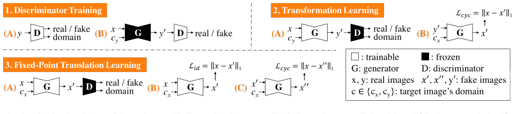
The load-bearing object is fixed-point translation learning. It adds a conditional identity loss $\mathcal{L}_{id} = \mathbb{E}_{x, c_x}[\|G(x, c_x)-x\|_1]$ that penalizes any change when the target domain is the source domain, and it revises the adversarial, domain classification, and cycle consistency losses so that same-domain translations are included; the cycle loss $\mathcal{L}_{cyc} = \mathbb{E}_{x, c_x}[\|G(G(x, c_x), c_x)-x\|_1]$ forces same-domain round trips to return the input. The generator also outputs a residual delta map that is added to the input before a $\tanh$ activation, which makes it structurally easier to leave the image unchanged. Together these components enforce that only disease- or attribute-related pixels are modified.
What would settle it
Inject a synthetic lesion of known location and size into a healthy MRI, translate the image to the healthy domain, and inspect the difference map; if noticeable differences appear outside the injected lesion, or if healthy anatomy is modified, the minimal-transformation assumption is false.
Extended reading notes
Core claim
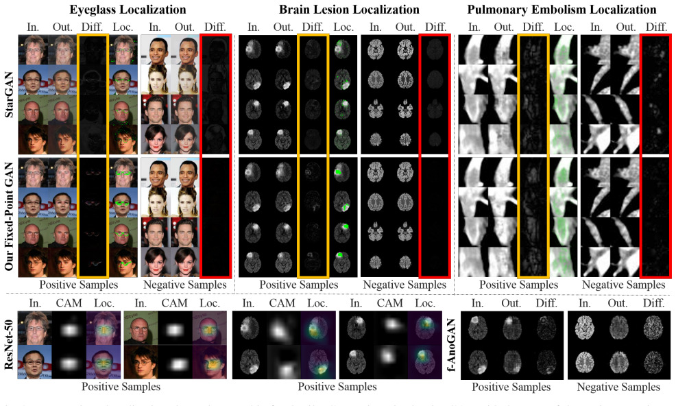
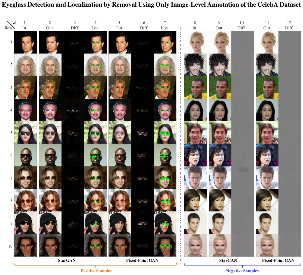
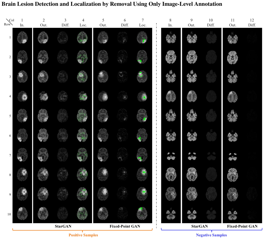
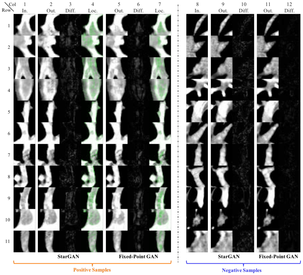
The discovery is a training scheme that endows a GAN with fixed-point translation: when the target domain equals the source domain, the generator is trained to behave as identity, $G(x, c_x) \approx x$, and when the domains differ, it is trained through losses that also cover same-domain pairs, so it learns minimal transformations that avoid touching unrelated content. In the 'virtual healing' application, the generator always translates toward the healthy domain, and the absolute difference between the input and the generated healthy image—the difference map—is used to detect and localize disease. This yields a weakly supervised detection and localization pipeline that needs only image-level healthy/diseased labels.
Load-bearing premise
The pipeline assumes the generator changes only disease-related pixels and preserves all other image content, so the difference between a scan and its healthy translation marks exactly the disease.
Editorial extensions
If this is right
- Healthy images pass through the generator nearly unchanged, so their difference maps are clean and rarely trigger false positives.
- Cross-domain translations, such as changing hair color or removing a lesion, are regularized to modify only task-relevant pixels, which improves translation quality.
- Disease detection and localization can be carried out with only image-level labels by applying color quantization and connected-component analysis to the difference map.
- The same fixed-point training transfers from natural images to brain MRI and CT pulmonary angiography without architectural changes.
- Because the target domain is specified at inference and the source domain is not needed, the method works for images with unknown health status.
Reading between the lines
- The authors note that large objects such as sunglasses and brain lesions are only partially removed, so the difference map is a localization signal rather than a segmentation mask; complete removal would be needed for pixel-level segmentation, which remains open.
- A natural extension is to feed the difference maps back as pseudo-labels to train a supervised segmentation network, potentially improving boundary accuracy.
- Since the conditional identity loss applies only when target equals source, the trained generator is source-domain-independent by construction; this suggests the 'translate to healthy' operation could be applied to any input modality without changing the inference procedure.
- Distribution shift is a testable risk: diseases or artifacts not seen during training could make the generator either fail to remove them or hallucinate changes; a healthy-only calibration set would reveal this failure mode.
Signed reviews
Editorial analysis
A structured set of objections, weighed in public.
Referee Report
Summary. The paper introduces Fixed-Point GAN, an extension of StarGAN that trains a single generator for both cross-domain and same-domain translations. The fixed-point training scheme adds a conditional identity loss for same-domain translation, revises the adversarial and domain-classification losses to cover both same- and cross-domain outputs, and applies cycle consistency to both. The generator is implemented with a residual/delta architecture. The authors evaluate multi-domain translation on CelebA and use the difference between an input image and its translation to the healthy domain for weakly supervised disease detection and localization on BRATS 2013 brain MRI and a pulmonary embolism CTPA dataset, comparing against StarGAN, CAM, ResNet-50-CAM, f-AnoGAN, and Alex et al. The central claim is that the proposed training yields minimal, fixed-point translations, leading to state-of-the-art image translation and superior disease detection/localization with only image-level labels.
Significance. The idea is appealing: a GAN that can 'virtually heal' an image and reveal disease by subtraction would make weakly supervised localization practical, since it requires only image-level labels. The paper includes a useful ablation (Tab. 4) separating the delta-map and fixed-point-training contributions, and the fixed-point component clearly drives most of the gain. The authors also make the implementation publicly available. However, the strongest claims ('outperforms the state of the art', 'surpasses predominant weakly-supervised localization methods') are not fully supported by the evidence as presented, because the minimal-transformation assumption is not quantitatively verified, comparisons are made at non-commensurate operating points, and no uncertainty estimates are given. With revisions that tighten these claims and add targeted analyses, the contribution would be a valuable advance.
major comments (5)
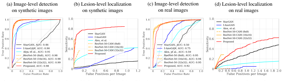
- [Sec. 4.2, Sec. 4.4, Eq. (4), Eq. (5)] The load-bearing minimal-transformation premise is stated but not enforced or quantitatively verified. Eq. (5) penalizes L1 only for same-domain translation, and Eq. (4) cycle consistency penalizes round-trip error; neither prevents the generator from modifying normal structures as long as the reverse map restores them. The paper's own observations—'neither StarGAN nor Fixed-Point GAN can completely remove large objects, like sunglasses or brain lesions' (Sec. 4.4) and 'our method is outperformed at the low false positive range' on real BRATS (Sec. 4.2)—show that the difference map is not guaranteed to be confined to disease. Since image-level detection uses the maximum of the difference map and localization uses connected-component analysis, any residual normal-structure change contributes to false positives. The abstract's unqualified superiority claim should be replaced with a claim conditional on the minimality of the learned transformation, and the paper should report a direct measure of preservation, e.g., the magnitude of the difference map in regions known to be healthy or the per-pixel specificity outside the lesion ground truth.
- [Sec. 4.2, Tab. 4, Fig. 4] The empirical support lacks uncertainty quantification and significance testing. AUC, FROC sensitivity, and IoU are reported as point estimates (e.g., mean IoU 0.2609±0.1283 vs 0.3483±0.2420, AUC 0.9668 vs 0.8832), with no confidence intervals or tests, and the test sets are small (6 real BRATS patients; 127 unique PEs). Many reported gaps, especially those with overlapping standard deviations, could easily arise from noise. Report bootstrap confidence intervals or significance tests, and show error bars on the ROC/FROC curves.
- [Sec. 4.2, f-AnoGAN comparison] The evaluation protocol for f-AnoGAN is selected after inspecting test performance: 'we use the average activation of difference maps as the detection score, because we find it more effective than using the maximum activation ... and also more effective than the anomaly scores proposed in the original work.' Choosing the scoring function based on the test set invalidates the comparison as a fair head-to-head. State the scoring rule before evaluation, or report all considered scores with a multiple-comparison correction, and apply the same selection procedure to all methods.
- [Sec. 4.2, Fig. 4b, Fig. 4d, Fig. 5b] The head-to-head claim against ResNet-50-CAM is made at different operating points: Fixed-Point GAN achieves 84.5% sensitivity at 1 false positive per image, while ResNet-50-CAM achieves 60% at 0.037 false positives per image. These numbers are not comparable. Report sensitivity for all methods at a common false-positive rate, or use a partial-area FROC measure over the same FP range; the same issue appears in the real-image FROC (Fig. 4d) and the PE FROC (Fig. 5b).
- [Sec. 4.1, Eq. (5), Tab. 3] The same-domain L1 comparison is partly by construction. Eq. (5) directly minimizes ||G(x,c_x)-x||_1, and Tab. 3 evaluates exactly this quantity. The fact that Fixed-Point GAN has lower same-domain L1 than StarGAN is therefore a sanity check of the loss rather than independent evidence of translation quality. The paper should present same-domain L1 as a training objective check, and use the independently trained classifier (Tab. 2) and visual inspection as the primary translation-quality evidence; ideally also report a perceptual or human-evaluation metric.
minor comments (5)
- [Abstract, Sec. 4.2, Sec. 4.4] The abstract should be tempered to acknowledge the low-false-positive-range underperformance on real BRATS and the partial-removal failures on large objects, both of which are disclosed in the body of the paper.
- [Sec. 3, Appendix Implementation Details] The main text defines the adversarial loss with the standard log form in Eq. (1), while the appendix states that the actual objective is the Wasserstein loss with gradient penalty in Eq. (8). Clarify which objective is used and keep the notation consistent throughout.
- [Sec. 4.3] For the PE dataset, 'localization' is defined as correct image-level classification of the candidate patch, not as lesion-level localization via connected components; the text should describe this as candidate-level detection rather than localization to avoid overstating the result.
- [Tab. 2] Classification accuracy on generated images is an indirect quality measure and can be biased because the classifier was trained on real images; consider reporting an additional metric, such as a classifier trained on generated images or a perceptual similarity score.
- [Sec. 4.3, Implementation Details] There is a duplicated word in 'sensitivity levels of of 88.9%' in Sec. 4.3, and the code URL is given as https://github.com/jlianglab/Fixed-Point-GAN in the abstract but as http://github.com/jlianglab/Fixed-Point-GAN in the implementation details; these should be unified.
Circularity Check
Same-domain L1 comparison is the trained objective itself; core detection/localization benchmarks remain externally grounded.
-
fitted input called prediction
[Sec. 4.1, Eq. (5), Table 3]
"Lid = { 0, c =cy ; Ex,c[||G(x,c )−x||1], c =cx (5) ... Tab. 3 presents a quantitative comparison between StarGAN and Fixed-Point GAN for the task of same-domain image translation. We use the image-level L1 distance between the input and generated images as the performance metric."
The metric reported in Table 3 is exactly the quantity minimized by the conditional identity loss in Eq. 5: for same-domain translation (c = cx), the loss is E||G(x,c)-x||_1. Fixed-Point GAN is trained to minimize this L1 distance, whereas StarGAN is not. Reporting a lower same-domain L1 distance for Fixed-Point GAN is therefore an optimization outcome, not an independent prediction of superiority. This particular comparison is forced by construction. It does not, however, make the central disease detection/localization claims circular, because those are evaluated with AUC, FROC, and IoU against external baselines such as ResNet-50-CAM and f-AnoGAN, and cross-domain translation quality is judged by an independently trained 40-attribute classifier.
full rationale
The paper's central claims are mostly self-contained and externally benchmarked: cross-domain image-to-image translation is compared against StarGAN using classification accuracy of an independently trained CelebA classifier (Table 2); brain-lesion and pulmonary-embolism detection/localization are evaluated against ResNet-50-CAM, f-AnoGAN, Alex et al., and StarGAN using ROC AUC, FROC, and IoU on BRATS 2013 and a CTPA dataset. These results do not reduce to the training losses. The one clear circular element is the same-domain translation comparison in Table 3: the image-level L1 distance there is the very objective minimized by Eq. 5, so the reported improvement over StarGAN is expected by construction rather than a discovered property. Separately, the paper's load-bearing premise that the generator performs a minimal, disease-only transformation is not guaranteed by the losses; the paper itself concedes in Sec. 4.4 that large objects such as sunglasses and brain lesions are only partially removed, and in Sec. 4.2 that on real BRATS images the method is outperformed at the low false-positive range. These are correctness risks and limitations, not circularity, but they temper the abstract's unqualified 'outperforms' and 'surpasses' wording. Overall, with one-by-construction comparison and otherwise independent benchmarks, a score of 3 is appropriate.
Assumptions & free parameters
free parameters (8)
- lambda_id (identity loss weight) =
10 (CelebA), 0.1 (BRATS 2013), 1 (PE)
- lambda_cyc =
10
- lambda_domain =
1
- lambda_gp =
10
- learning rate =
1e-4
- training iterations =
200K (CelebA, PE), 300K (BRATS 2013)
- connected component area threshold =
10 pixels
- delta map (residual) architecture =
tanh(G(x,c) + x)
assumptions (5)
- domain assumption A diseased image differs from its healthy counterpart by a localized, removable visual pattern such that subtracting the generated healthy image reveals the disease.
- domain assumption The generator can be trained to perform a minimal transformation, changing only disease-related pixels, without altering unrelated structures.
- domain assumption Image-level labels (healthy vs. diseased) are accurate and sufficient for training; the network does not require lesion-level annotations.
- domain assumption Cycle consistency has a feasible inverse mapping for the translation tasks.
- standard math Wasserstein GAN with gradient penalty provides a stable adversarial training objective.
Cite this review
Pith. "Pith review of Learning Fixed Points in Generative Adversarial Networks: From Image-to-Image Translation to Disease Detection and Localization." pith.science (2026). https://pith.science/paper/CO3Q3JCJ
@misc{pith2026190806965,
  author       = {Pith},
  title        = {Pith review of: Learning Fixed Points in Generative Adversarial Networks: From Image-to-Image Translation to Disease Detection and Localization},
  year         = {2026},
  howpublished = {\url{https://pith.science/paper/CO3Q3JCJ}},
  note         = {Machine review of arXiv:1908.06965}
}
read the original abstract
Generative adversarial networks (GANs) have ushered in a revolution in image-to-image translation. The development and proliferation of GANs raises an interesting question: can we train a GAN to remove an object, if present, from an image while otherwise preserving the image? Specifically, can a GAN "virtually heal" anyone by turning his medical image, with an unknown health status (diseased or healthy), into a healthy one, so that diseased regions could be revealed by subtracting those two images? Such a task requires a GAN to identify a minimal subset of target pixels for domain translation, an ability that we call fixed-point translation, which no GAN is equipped with yet. Therefore, we propose a new GAN, called Fixed-Point GAN, trained by (1) supervising same-domain translation through a conditional identity loss, and (2) regularizing cross-domain translation through revised adversarial, domain classification, and cycle consistency loss. Based on fixed-point translation, we further derive a novel framework for disease detection and localization using only image-level annotation. Qualitative and quantitative evaluations demonstrate that the proposed method outperforms the state of the art in multi-domain image-to-image translation and that it surpasses predominant weakly-supervised localization methods in both disease detection and localization. Implementation is available at https://github.com/jlianglab/Fixed-Point-GAN.
Figures

Figures from the paper (12 more)






Reference graph
Works this paper leans on
-
[1]
Generative adver- sarial networks for brain lesion detection
Varghese Alex, Mohammed Safwan KP, Sai Saketh Chen- namsetty, and Ganapathy Krishnamurthi. Generative adver- sarial networks for brain lesion detection. In Medical Imag- ing 2017: Image Processing, volume 10133, page 101330G. International Society for Optics and Photonics, 2017. 4, 7, 8
work page 2017
-
[2]
Wasserstein generative adversarial networks
Martin Arjovsky, Soumith Chintala, and L ´eon Bottou. Wasserstein generative adversarial networks. In Interna- tional Conference on Machine Learning , pages 214–223,
-
[3]
Semi- supervised learning for network-based cardiac mr image seg- mentation
Wenjia Bai, Ozan Oktay, Matthew Sinclair, Hideaki Suzuki, Martin Rajchl, Giacomo Tarroni, Ben Glocker, Andrew King, Paul M Matthews, and Daniel Rueckert. Semi- supervised learning for network-based cardiac mr image seg- mentation. In International Conference on Medical Image Computing and Computer-Assisted Intervention, pages 253–
-
[4]
Visual feature attribu- tion using wasserstein gans
Christian F Baumgartner, Lisa M Koch, Kerem Can Tezcan, Jia Xi Ang, and Ender Konukoglu. Visual feature attribu- tion using wasserstein gans. In Proc IEEE Comput Soc Conf Comput Vis Pattern Recognit, 2017. 4, 19, 21
work page 2017
-
[5]
Jinzheng Cai, Le Lu, Adam P Harrison, Xiaoshuang Shi, Pingjun Chen, and Lin Yang. Iterative attention mining for weakly supervised thoracic disease pattern localization in chest x-rays. In International Conference on Medical Im- age Computing and Computer-Assisted Intervention , pages 589–598. Springer, 2018. 4
work page 2018
-
[6]
Pairedcyclegan: Asymmetric style transfer for apply- ing and removing makeup
Huiwen Chang, Jingwan Lu, Fisher Yu, and Adam Finkel- stein. Pairedcyclegan: Asymmetric style transfer for apply- ing and removing makeup. In 2018 IEEE Conference on Computer Vision and Pattern Recognition (CVPR), 2018. 2
work page 2018
-
[7]
Unsupervised detec- tion of lesions in brain mri using constrained adversarial auto-encoders
Xiaoran Chen and Ender Konukoglu. Unsupervised detec- tion of lesions in brain mri using constrained adversarial auto-encoders. arXiv preprint arXiv:1806.04972, 2018. 4
arXiv 2018
-
[8]
Stargan: Uni- fied generative adversarial networks for multi-domain image-to-image translation
Yunjey Choi, Minje Choi, Munyoung Kim, Jung-Woo Ha, Sunghun Kim, and Jaegul Choo. Stargan: Uni- fied generative adversarial networks for multi-domain image-to-image translation. In The IEEE Conference on Computer Vision and Pattern Recognition (CVPR) , June 2018. The implementation is publicly available at https://github.com/yunjey/StarGAN. 1, 2, 3, 4, 5, 6, 7
work page 2018
Show all 41 references
-
[9]
Generative adversarial nets
Ian Goodfellow, Jean Pouget-Abadie, Mehdi Mirza, Bing Xu, David Warde-Farley, Sherjil Ozair, Aaron Courville, and Yoshua Bengio. Generative adversarial nets. In Advances in neural information processing systems, pages 2672–2680,
-
[10]
Improved training of wasserstein gans
Ishaan Gulrajani, Faruk Ahmed, Martin Arjovsky, Vincent Dumoulin, and Aaron C Courville. Improved training of wasserstein gans. In Advances in Neural Information Pro- cessing Systems, pages 5767–5777, 2017. 21
2017
-
[11]
Deep residual learning for image recognition
Kaiming He, Xiangyu Zhang, Shaoqing Ren, and Jian Sun. Deep residual learning for image recognition. In Proceed- ings of the IEEE conference on computer vision and pattern recognition, pages 770–778, 2016. 7
2016
-
[12]
Self-transfer learn- ing for weakly supervised lesion localization
Sangheum Hwang and Hyo-Eun Kim. Self-transfer learn- ing for weakly supervised lesion localization. In Sebastien Ourselin, Leo Joskowicz, Mert R. Sabuncu, Gozde Unal, and William Wells, editors, Medical Image Computing and Computer-Assisted Intervention – MICCAI 2016, pages 23...
2016
-
[13]
Image-to-image translation with conditional adver- sarial networks
Phillip Isola, Jun-Yan Zhu, Tinghui Zhou, and Alexei A Efros. Image-to-image translation with conditional adver- sarial networks. arXiv preprint arXiv:1611.07004, 2016. 3
2016 arXiv
-
[14]
Learning to discover cross-domain relations with generative adversarial networks
Taeksoo Kim, Moonsu Cha, Hyunsoo Kim, Jung Kwon Lee, and Jiwon Kim. Learning to discover cross-domain relations with generative adversarial networks. In International Con- ference on Machine Learning, pages 1857–1865, 2017. 3
2017
-
[15]
The virtual skeleton database: An open access repository for biomedical research and col- laboration
Michael Kistler, Serena Bonaretti, Marcel Pfahrer, Roman Niklaus, and Philippe B¨uchler. The virtual skeleton database: An open access repository for biomedical research and col- laboration. J Med Internet Res, 15(11):e245, Nov 2013. 7
2013
-
[16]
Photo- realistic single image super-resolution using a generative ad- versarial network
Christian Ledig, Lucas Theis, Ferenc Husz´ar, Jose Caballero, Andrew Cunningham, Alejandro Acosta, Andrew P Aitken, Alykhan Tejani, Johannes Totz, Zehan Wang, et al. Photo- realistic single image super-resolution using a generative ad- versarial network. In CVPR, volume 2, pag...
2017
-
[17]
Thoracic disease identification and lo- calization with limited supervision
Zhe Li, Chong Wang, Mei Han, Yuan Xue, Wei Wei, Li-Jia Li, and Li Fei-Fei. Thoracic disease identification and lo- calization with limited supervision. In Proceedings of the IEEE Conference on Computer Vision and Pattern Recogni- tion, pages 8290–8299, 2018. 4
2018
-
[18]
Computer aided detection of pulmonary embolism with tobogganing and mutiple instance classification in ct pulmonary angiography
Jianming Liang and Jinbo Bi. Computer aided detection of pulmonary embolism with tobogganing and mutiple instance classification in ct pulmonary angiography. InBiennial Inter- national Conference on Information Processing in Medical Imaging, pages 630–641. Springer, 2007. 15, 20
2007
-
[19]
Unsupervised image-to-image translation networks
Ming-Yu Liu, Thomas Breuel, and Jan Kautz. Unsupervised image-to-image translation networks. In Advances in Neural Information Processing Systems, pages 700–708, 2017. 3
2017
-
[20]
Deep learning face attributes in the wild
Ziwei Liu, Ping Luo, Xiaogang Wang, and Xiaoou Tang. Deep learning face attributes in the wild. In Proceedings of the IEEE International Conference on Computer Vision , pages 3730–3738, 2015. 3, 6
2015
-
[21]
The multimodal brain tumor image segmentation benchmark (brats)
Bjoern H Menze, Andras Jakab, Stefan Bauer, Jayashree Kalpathy-Cramer, Keyvan Farahani, Justin Kirby, Yuliya Burren, Nicole Porz, Johannes Slotboom, Roland Wiest, et al. The multimodal brain tumor image segmentation benchmark (brats). IEEE transactions on medical imaging , 34(...
1993
-
[22]
Is object localization for free?-weakly-supervised learning with convolutional neural networks
Maxime Oquab, L ´eon Bottou, Ivan Laptev, and Josef Sivic. Is object localization for free?-weakly-supervised learning with convolutional neural networks. In Proceedings of the IEEE Conference on Computer Vision and Pattern Recogni- tion, pages 685–694, 2015. 4
2015
-
[23]
From image-level to pixel-level labeling with convolutional networks
Pedro O Pinheiro and Ronan Collobert. From image-level to pixel-level labeling with convolutional networks. In Pro- ceedings of the IEEE Conference on Computer Vision and Pattern Recognition, pages 1713–1721, 2015. 4
2015
-
[24]
f-anogan: Fast unsupervised anomaly detection with generative adversarial networks
Thomas Schlegl, Philipp Seeb ¨ock, Sebastian M Waldstein, Georg Langs, and Ursula Schmidt-Erfurth. f-anogan: Fast unsupervised anomaly detection with generative adversarial networks. Medical Image Analysis , 2019. The implemen- tation is publicly available at https://github.co...
2019
-
[25]
Grad-cam: Visual explanations from deep networks via gradient-based localization
Ramprasaath R Selvaraju, Michael Cogswell, Abhishek Das, Ramakrishna Vedantam, Devi Parikh, and Dhruv Batra. Grad-cam: Visual explanations from deep networks via gradient-based localization. In Proceedings of the IEEE In- ternational Conference on Computer Vision, pages 618–626,
-
[26]
Joint weakly and semi-supervised deep learning for localization and classification of masses in breast ultrasound images
Seung Yeon Shin, Soochahn Lee, Il Dong Yun, Sun Mi Kim, and Kyoung Mu Lee. Joint weakly and semi-supervised deep learning for localization and classification of masses in breast ultrasound images. IEEE transactions on medical imaging ,
-
[27]
Deep inside convolutional networks: Visualising image classification models and saliency maps
Karen Simonyan, Andrea Vedaldi, and Andrew Zisserman. Deep inside convolutional networks: Visualising image classification models and saliency maps. arXiv preprint arXiv:1312.6034, 2013. 4
2013 arXiv
-
[28]
Hide-and-seek: Forcing a network to be meticulous for weakly-supervised object and action localization
Krishna Kumar Singh and Yong Jae Lee. Hide-and-seek: Forcing a network to be meticulous for weakly-supervised object and action localization. In 2017 IEEE International Conference on Computer Vision (ICCV), pages 3544–3553. IEEE, 2017. 4
2017
-
[29]
Computer-aided pulmonary embolism detection using a novel vessel-aligned multi-planar image representation and convolutional neural networks
Nima Tajbakhsh, Michael B Gotway, and Jianming Liang. Computer-aided pulmonary embolism detection using a novel vessel-aligned multi-planar image representation and convolutional neural networks. In International Conference on Medical Image Computing and Computer-Assisted Inte...
2015
-
[30]
Computer-aided detection and visualization of pulmonary embolism using a novel, compact, and informa- tive image representation
Nima Tajbakhsh, Jae Y Shin, Michael B Gotway, and Jian- ming Liang. Computer-aided detection and visualization of pulmonary embolism using a novel, compact, and informa- tive image representation. Medical Image Analysis , page 101541, 2019. 8, 20
2019
-
[31]
Convolutional neural networks for medical im- age analysis: Full training or fine tuning? IEEE transactions on medical imaging, 35(5):1299–1312, 2016
Nima Tajbakhsh, Jae Y Shin, Suryakanth R Gurudu, R Todd Hurst, Christopher B Kendall, Michael B Gotway, and Jian- ming Liang. Convolutional neural networks for medical im- age analysis: Full training or fine tuning? IEEE transactions on medical imaging, 35(5):1299–1312, 2016. 8, 20
2016
-
[32]
Attention-guided curriculum learning for weakly supervised classification and localization of thoracic diseases on chest radiographs
Yuxing Tang, Xiaosong Wang, Adam P Harrison, Le Lu, Jing Xiao, and Ronald M Summers. Attention-guided curriculum learning for weakly supervised classification and localization of thoracic diseases on chest radiographs. In International Workshop on Machine Learning in Medical Im...
2018
-
[33]
Chestx- ray8: Hospital-scale chest x-ray database and benchmarks on weakly-supervised classification and localization of com- mon thorax diseases
Xiaosong Wang, Yifan Peng, Le Lu, Zhiyong Lu, Mo- hammadhadi Bagheri, and Ronald M Summers. Chestx- ray8: Hospital-scale chest x-ray database and benchmarks on weakly-supervised classification and localization of com- mon thorax diseases. In Proceedings of the IEEE Conference o...
2017
-
[34]
Deep mr to ct synthesis using unpaired data
Jelmer M Wolterink, Anna M Dinkla, Mark HF Savenije, Pe- ter R Seevinck, Cornelis AT van den Berg, and Ivana Iˇsgum. Deep mr to ct synthesis using unpaired data. InInternational Workshop on Simulation and Synthesis in Medical Imaging , pages 14–23. Springer, 2017. 2
2017
-
[35]
Dualgan: Unsupervised dual learning for image-to-image translation
Zili Yi, Hao (Richard) Zhang, Ping Tan, and Minglun Gong. Dualgan: Unsupervised dual learning for image-to-image translation. In ICCV, pages 2868–2876, 2017. 3
2017
-
[36]
Adversarial complementary learning for weakly supervised object localization
Xiaolin Zhang, Yunchao Wei, Jiashi Feng, Yi Yang, and Thomas S Huang. Adversarial complementary learning for weakly supervised object localization. In Proceedings of the IEEE Conference on Computer Vision and Pattern Recogni- tion, pages 1325–1334, 2018. 4
2018
-
[37]
Learning deep features for discrimi- native localization
Bolei Zhou, Aditya Khosla, Agata Lapedriza, Aude Oliva, and Antonio Torralba. Learning deep features for discrimi- native localization. In Proceedings of the IEEE Conference on Computer Vision and Pattern Recognition , pages 2921– 2929, 2016. 2, 4, 7
2016
-
[38]
Fine-tuning convolutional neural networks for biomedical image analy- sis: Actively and incrementally
Zongwei Zhou, Jae Y Shin, Lei Zhang, Suryakanth R Gu- rudu, Michael B Gotway, and Jianming Liang. Fine-tuning convolutional neural networks for biomedical image analy- sis: Actively and incrementally. InCVPR, pages 4761–4772,
-
[39]
Unpaired image-to-image translation using cycle- consistent adversarial networks
Jun-Yan Zhu, Taesung Park, Phillip Isola, and Alexei A Efros. Unpaired image-to-image translation using cycle- consistent adversarial networks. arXiv preprint, 2017. 3, 4
2017
-
[40]
To- ward multimodal image-to-image translation
Jun-Yan Zhu, Richard Zhang, Deepak Pathak, Trevor Dar- rell, Alexei A Efros, Oliver Wang, and Eli Shechtman. To- ward multimodal image-to-image translation. In Advances in Neural Information Processing Systems , pages 465–476,
-
[2017]
no change
3 Appendix Fig. 6: Now, all humor aside, if a GAN can remove diseases completely from images, it will offer an ideal method for segmenting all diseases, an ambitious goal that has yet to be achieved. Nevertheless, our method is still of great clinical significance in computer-a...
Reviewed August 14, 2026 · model on record in the stance chip above.
Discussion (0). Continue with ORCID to comment.