REVIEW 3 major objections 5 minor 12 references
Endotracheal Tube Detection and Segmentation in Chest Radiographs using Synthetic Data
T0 review · 3 major / 5 minor · reviewed 2026-08-14 · deepseek-v4-flash
Pith's one-line read Synthetic ET tubes composited into real chest X-rays train a combined detector-and-segmenter that reaches 0.99 AUC on a 479-case test set.
desk verdict Solid synthetic-data pipeline for ET tube detection with a strong classification AUC; send to review but demand quantitative segmentation and split transparency. read the letter →
The pith
A machine-rendered reading of the paper's core claim, the machinery that carries it, and where it could break.
The reading
What carries the argument
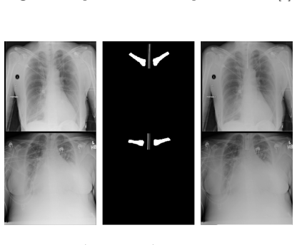
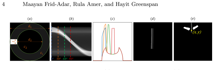
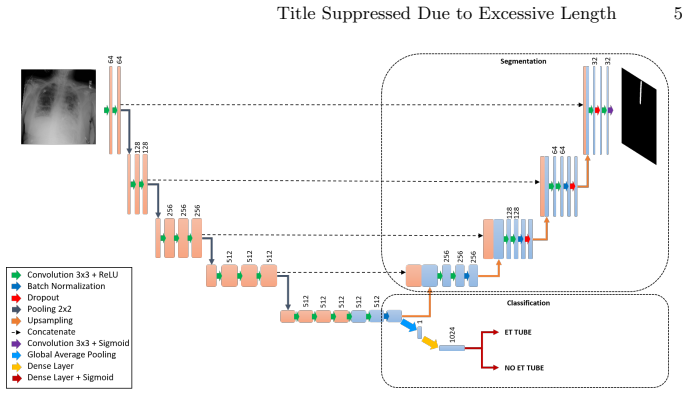
The central mechanism is a parametric synthetic ET tube generator: a hollow tube with a radiopaque marker, parameterized by attenuation coefficients c1=0.1 and c2=1, outer and inner widths d1=160 and d2=100 pixels, and strip thickness t=20, projected with a Radon transform at 0°, 30°, 60°, and 90°, then drawn along a B-spline path placed between the clavicles and blended into real X-ray backgrounds with weights between 0.1 and 0.2. This creates thousands of X-rays with automatic ground-truth masks. The network, ETT-Net, is a two-headed convolutional model with a shared encoder, a U-Net-style decoder (an encoder-decoder segmentation architecture with skip connections) for segmentation, and a global-pooling/dense/sigmoid head for classification; the loss combines binary cross-entropy with a Dice term weighted at 0.1. The two-phase training procedure—synthetic pre-training, then fine-tuning on the model's own high-confidence real cases—is what converts the generator into a clinically sized result.
What would settle it
Have two radiologists draw pixel-level masks of the ET tube on a subset of the 479 test images and measure the model's segmentation with Dice or contour distance; if the masks are not clearly better than a straight tube drawn down the trachea, the high-quality-segmentation claim fails even if classification AUC stays at 0.99.
Extended reading notes
Core claim
The paper's central claim is that the synthetic tube generator is realistic enough to carry the first phase of training: tubes built from physical parameters, projected at a few angles, and blended onto real X-rays yield a network whose synthetic-only phase already reaches 0.96 AUC. Fine-tuning that network on high-confidence real cases selected by the model raises detection to 0.99 AUC with 95.5% sensitivity and 96.5% specificity on a held-out set of 479 real AP chest radiographs (232 with a tube, 247 without). The same network emits a segmentation mask along with the classification, and the authors report the segmentations look accurate on qualitative inspection, though they were not scored against pixel-level manual annotations.
Load-bearing premise
The load-bearing premise is that the hand-designed synthetic tubes, with their chosen attenuation, widths, projection angles, and blending weights, look enough like real ET tubes inside real chest X-rays that a network pretrained on them learns transferable features; if they do not, the first training phase cannot provide a useful starting point for fine-tuning.
Editorial extensions
If this is right
- A tool for tube verification could be built from a public unlabeled chest X-ray collection plus a few dozen hand-set parameters, with no per-image manual annotations.
- Because classification and segmentation come from one network, the same output that says a tube is present also localizes it, which is the information needed to flag a tube sitting too low (bronchial intubation).
- The reported 0.99 AUC is measured against prior systems tested on 60–87 cases, so the result holds on a substantially larger, ten-times-larger evaluation.
- The same synthetic-overlay strategy extends to other tube types and to generating lesions over healthy patients, as the paper states as future work.
Reading between the lines
- The generator's parameters are chosen once by hand; sweeping them (tube width, attenuation contrast, blending weight, projection angles) would reveal how much of the final accuracy is carried by the synthetic phase and could push the synthetic-only model closer to phase-two performance.
- Because phase-two labels are the model's own high-confidence predictions rather than independent expert labels, the fine-tuning set is a self-curated subset; quantifying label noise with a small expert-checked subset would sharpen what the 0.99 AUC means.
- If the same two-phase recipe transfers to other structures, the practical consequence is that annotation-free training could become a default for line and tube detection in medical images, not a special trick for ET tubes.
Signed reviews
Editorial analysis
A structured set of objections, weighed in public.
Referee Report
Summary. The paper proposes ETT-Net, a combined CNN for detecting the presence of endotracheal (ET) tubes and segmenting them in chest radiographs. To avoid manual annotation, the authors synthesize ET tubes over real X-ray images from the public NIH ChestX-ray8 dataset and train the network in two phases: first on synthetic data, then fine-tuned on real images selected automatically by the model's own predictions. The model is evaluated on 479 manually verified AP-view chest radiographs from the NIH dataset, reporting an AUC of 0.987 (rounded to 0.99), sensitivity 95.5%, and specificity 96.5%, with segmentation output shown qualitatively. A DenseNet baseline trained on the same fine-tuning data achieves AUC 0.975.
Significance. If the test-set independence is confirmed, the classification result is a meaningful contribution: the evaluation set of 479 cases is roughly an order of magnitude larger than the 60-87 cases used in previous studies, the method uses public data and avoids manual annotations for training, and the comparison with DenseNet provides a useful ablation. The synthetic ET-tube generation pipeline is also a reusable idea for other medical imaging tasks. The paper does not ship code or split artifacts, and the segmentation claim rests only on qualitative inspection, which tempers the significance. The core value is the classification result and the synthetic-to-real training pipeline, both of which are defensible if the data-leakage concern is resolved.
major comments (3)
- [Section 3.1 and Section 3.2] The manuscript does not demonstrate that the 479-case test set was excluded from the Phase-2 self-training pool. Section 3.1 states that 'we used all NIH dataset AP cases' to select the 7,944 fine-tuning examples, and Section 3.2 states only that the test set 'includes 479 real chest radiographs from the NIH dataset' and asserts independence from 'all training data' without describing any patient-level split, de-duplication, or explicit exclusion mechanism. Because both the Phase-2 training pool and the test set are AP-view images from the same public NIH dataset, unexcluded overlap would directly inflate the reported AUC of 0.987, sensitivity, and specificity. This is load-bearing for the central claim. Please specify the exact split mechanism and provide filename-level or patient-level separation, or re-run Phase 2 after explicitly excluding the test cases; without this, the independence assertion cannot be verified.
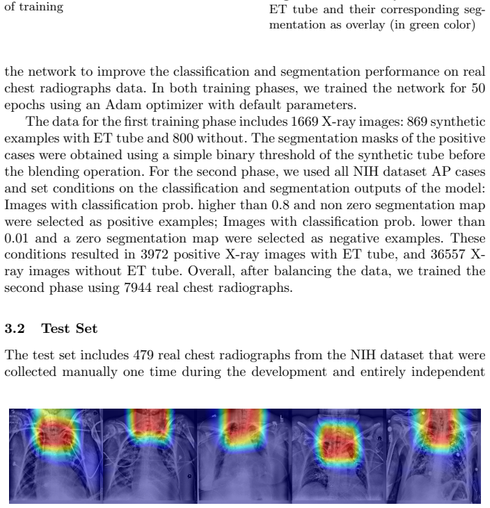
- [Section 3.3 and Figure 6] The segmentation contribution is only qualitatively assessed. The text states that the ground-truth segmentation masks are not pixel-wise accurate and that 'the segmentation output was examined qualitatively,' yet the abstract and conclusion describe the model as outputting 'high quality ET tube segmentation maps.' Since segmentation is half of the paper's claimed contribution, this claim is unsupported. Please add a quantitative evaluation on a subset with expert pixel-level annotations (e.g., Dice or Hausdorff distance), or explicitly delimit the claim to a qualitative demonstration.
- [Section 2.1 and Section 3.1] The synthetic-data parameters and the Phase-2 pseudo-label thresholds are chosen without sensitivity analysis. The final classification result depends on the quality of the automatically selected pseudo-labels in the second phase; a brief ablation varying the selection thresholds (e.g., classification probability 0.8/0.01) and reporting the resulting AUC would strengthen the argument that the fine-tuning procedure is robust to these choices.
minor comments (5)
- [Abstract and Conclusion] The abstract and conclusion state the test set contains 477 real chest radiographs, while Section 3.2 and Table 1 report 479 cases (232 positive and 247 negative). Please correct the mismatch.
- [Table 1] DenseNet (AUC 0.97) and ETT-Net Phase1 (AUC 0.96) are listed with identical sensitivity (89.2%) and specificity (93.0%); please confirm whether this is coincidental or a copy/paste error, and report the operating thresholds clearly.
- [Section 2.2 and Section 3.1] Training details are incomplete: no learning rate, batch size, early stopping criterion, or learning-rate schedule is reported, and it is unclear whether the CLAHE and normalization statistics are computed per image or across the dataset. Please provide these details for reproducibility.
- [Section 2.1] The selection of d1=160, d2=100, and t=20 is justified only as 'based on true physical properties of ET tubes or based on [9],' but the pixel dimensions of a real ET tube depend on the image resolution and source-to-detector distance; please clarify how these values map to physical tube sizes and whether the results are sensitive to them.
- [References] Reference [5] contains a malformed DOI (10.10007/s10278-017-9980-77) and reference [10] has a typo in the arXiv number (1608.069936); please verify and correct both.
Circularity Check
No significant circularity; the central AUC result is measured on an asserted independent test set and does not reduce to the training inputs by construction.
full rationale
The paper's central claim is the 0.99 AUC on 479 real chest radiographs described in Section 3.2 as 'collected manually one time during the development and entirely independent from all training data.' That test evaluation is external to the training procedure, so the metric is not definitionally tied to the synthetic generation parameters or the Phase-2 selected training labels. The synthetic tube parameters {c1=0.1, c2=1, d1=160, d2=100, t=20} in Section 2.1 are stated to be based on physical properties of ET tubes or on Yi et al. [9]; they are not fitted to the test outcomes. The two-phase training uses the model's own confident predictions to select real images for fine-tuning, which is a semi-supervised self-training approach rather than a circular derivation: the final evaluation is against manually verified presence/absence labels, not against the model's own selection scores. The clavicle segmentation method cites the authors' prior U-Net work [7], but the ETT-Net detection and segmentation architecture is trained and evaluated in this paper on the ET tube task, so that self-citation is not load-bearing for the central claim. The one substantive evaluation concern is that the paper does not document a patient-level or image-level exclusion of the 479 test cases from the 7,944 Phase-2 training images drawn from the same NIH AP dataset; that would be a data-leakage or reproducibility issue, but it is not a circularity of the derivation chain, and no equation or fitted parameter reduces to the reported result by construction.
Assumptions & free parameters
free parameters (4)
- ET tube simulation parameters {c1, c2, d1, d2, t} =
{0.1, 1, 160, 100, 20}
- Segmentation loss weight λ =
0.1
- Self-training selection thresholds =
positive > 0.8, negative < 0.01
- Blending weights =
random in [0.1, 0.2]
assumptions (3)
- domain assumption The NIH dataset images without ET tubes can serve as realistic backgrounds, and synthesized tubes on them produce realistic training images.
- domain assumption Clavicle segmentation (Dice 93.1%) is accurate enough to place the synthetic tube in the trachea area.
- domain assumption The model's own predictions at thresholds 0.8/0.01 are reliable enough to create accurate training labels for Phase 2.
Cite this review
Pith. "Pith review of Endotracheal Tube Detection and Segmentation in Chest Radiographs using Synthetic Data." pith.science (2026). https://pith.science/paper/GJSBHKPO
@misc{pith2026190807170,
  author       = {Pith},
  title        = {Pith review of: Endotracheal Tube Detection and Segmentation in Chest Radiographs using Synthetic Data},
  year         = {2026},
  howpublished = {\url{https://pith.science/paper/GJSBHKPO}},
  note         = {Machine review of arXiv:1908.07170}
}
read the original abstract
Chest radiographs are frequently used to verify the correct intubation of patients in the emergency room. Fast and accurate identification and localization of the endotracheal (ET) tube is critical for the patient. In this study we propose a novel automated deep learning scheme for accurate detection and segmentation of the ET tubes. Development of automatic systems using deep learning networks for classification and segmentation require large annotated data which is not always available. Here we present an approach for synthesizing ET tubes in real X-ray images. We suggest a method for training the network, first with synthetic data and then with real X-ray images in a fine-tuning phase, which allows the network to train on thousands of cases without annotating any data. The proposed method was tested on 477 real chest radiographs from a public dataset and reached AUC of 0.99 in classifying the presence vs. absence of the ET tube, along with outputting high quality ET tube segmentation maps.
Figures

Figures from the paper (3 more)

Reference graph
Works this paper leans on
-
[1]
American Journal of Roentgenology 198(3), 563–-571 (2012)
Godoy, M.C., Leitman, B.S., de Groot, P.M., Vlahos, I., Naidich, D.P.: Chest radiography in the icu: part 1, evaluation of airway, enteric, and pleural tubes. American Journal of Roentgenology 198(3), 563–-571 (2012)
work page 2012
-
[2]
Journal of intensive care medicine 18(4), 198–-210 (2003)
Trotman-Dickenson, B.: Radiology in the intensive care unit (part i). Journal of intensive care medicine 18(4), 198–-210 (2003)
work page 2003
-
[3]
In: Medical Imaging 2012: Computer-Aided Diagnosis
Ramakrishna, B., Brown, M., Goldin, J., Cagnon, C., Enzmann, D.: An improved automatic computer aided tube detection and labeling system on chest radiographs. In: Medical Imaging 2012: Computer-Aided Diagnosis. vol. 8315, p. 83150R. Inter-national Society for Optics and Photonics (2012)
work page 2012
-
[4]
International Journal of Computer Assisted Radiology and Surgery11(11), 2049-–2057 (2016)
Chen, S., Zhang, M., Yao, L., Xu, W.: Endotracheal tubes positioning detection in adult portable chest radiography for intensive care unit. International Journal of Computer Assisted Radiology and Surgery11(11), 2049-–2057 (2016)
work page 2016
-
[5]
Journal of Digital Imaging 30(4), 460–-468 (2017)
Lakhani, P.: Deep convolutional neural networks for endotracheal tube position and x-ray image classification: Challenges and opportunities. Journal of Digital Imaging 30(4), 460–-468 (2017). doi:10.10007/s10278-017-9980-77
-
[6]
In: The IEEE Conference on Computer Vision and Pattern Recognition (2017)
Wang, X., Peng, Y., Lu, L., Lu, Z., Bagheri, M., Summers, R.M.: Chestx-ray8: Hospital-scale chest x-ray database and benchmarks on weakly-supervised classification and localization of common thorax diseases. In: The IEEE Conference on Computer Vision and Pattern Recognition (2017)
work page 2017
-
[7]
In: Image Analysis for Moving Organ, Breast, and Thoracic Images, MICCAI
Frid-Adar, M., Ben-Cohen, A., Amer, R., Greenspan, H.: Improving the segmen-tation of anatomical structures in chest radiographs using u-net with an imagenet pre-trained encoder. In: Image Analysis for Moving Organ, Breast, and Thoracic Images, MICCAI. pp. 159–-168. Springer International Publishing, Cham (2018)
work page 2018
-
[8]
Medical Image Analysis 10(1), 19–-40 (2006)
Ginneken, B.V., Stegmann, M.B., Loog, M.: Segmentation of anatomical structures in chest radiographs using supervised methods: a comparative study on a public database. Medical Image Analysis 10(1), 19–-40 (2006)
work page 2006
Show all 12 references
-
[9]
CoRR (2018)
Yi, X., Adams, S., Babyn, P., Elnajmi, A.: Automatic catheter detection in pediatric x-ray images using a scale-recurrent network and synthetic data. CoRR (2018). http://arxiv.org/abs/1806.00921
2018 arXiv
-
[10]
CoRR (2016)
Huang, G., Liu, Z., Weinberger, K.Q.: Densely connected convolutional networks. CoRR (2016). http://arxiv.org/abs/1608.069936
2016 arXiv
-
[11]
, " * write output.state after.block = add.period write
ENTRY address author booktitle chapter doi edition editor eid howpublished institution journal key month note number organization pages publisher school series title type url volume year label INTEGERS output.state before.all mid.sentence after.sentence after.block FUNCTION in...
-
[12]
write newline
" write newline "" before.all 'output.state := FUNCTION n.dashify 't := "" t empty not t #1 #1 substring "-" = t #1 #2 substring "--" = not "--" * t #2 global.max substring 't := t #1 #1 substring "-" = "-" * t #2 global.max substring 't := while if t #1 #1 substring * t #2 gl...
Reviewed August 14, 2026 · model on record in the stance chip above.
Discussion (0). Continue with ORCID to comment.