REVIEW 3 major objections 5 minor 45 references
DirectPET: Full Size Neural Network PET Reconstruction from Sinogram Data
T0 review · 3 major / 5 minor · reviewed 2026-08-14 · deepseek-v4-flash
Pith's one-line read DirectPET reconstructs full-size multi-slice PET volumes directly from sinograms in a single forward pass, producing images quantitatively and qualitatively similar to the clinical OSEM+PSF reference in a fraction of the time.
desk verdict DirectPET is a real architecture contribution that scales direct network PET reconstruction to clinical-size volumes, but its quantitative evaluation is partly circular because the network is trained and scored against the same OSEM+PSF targets using loss metrics. read the letter →
The pith
A machine-rendered reading of the paper's core claim, the machinery that carries it, and where it could break.
The reading
What carries the argument
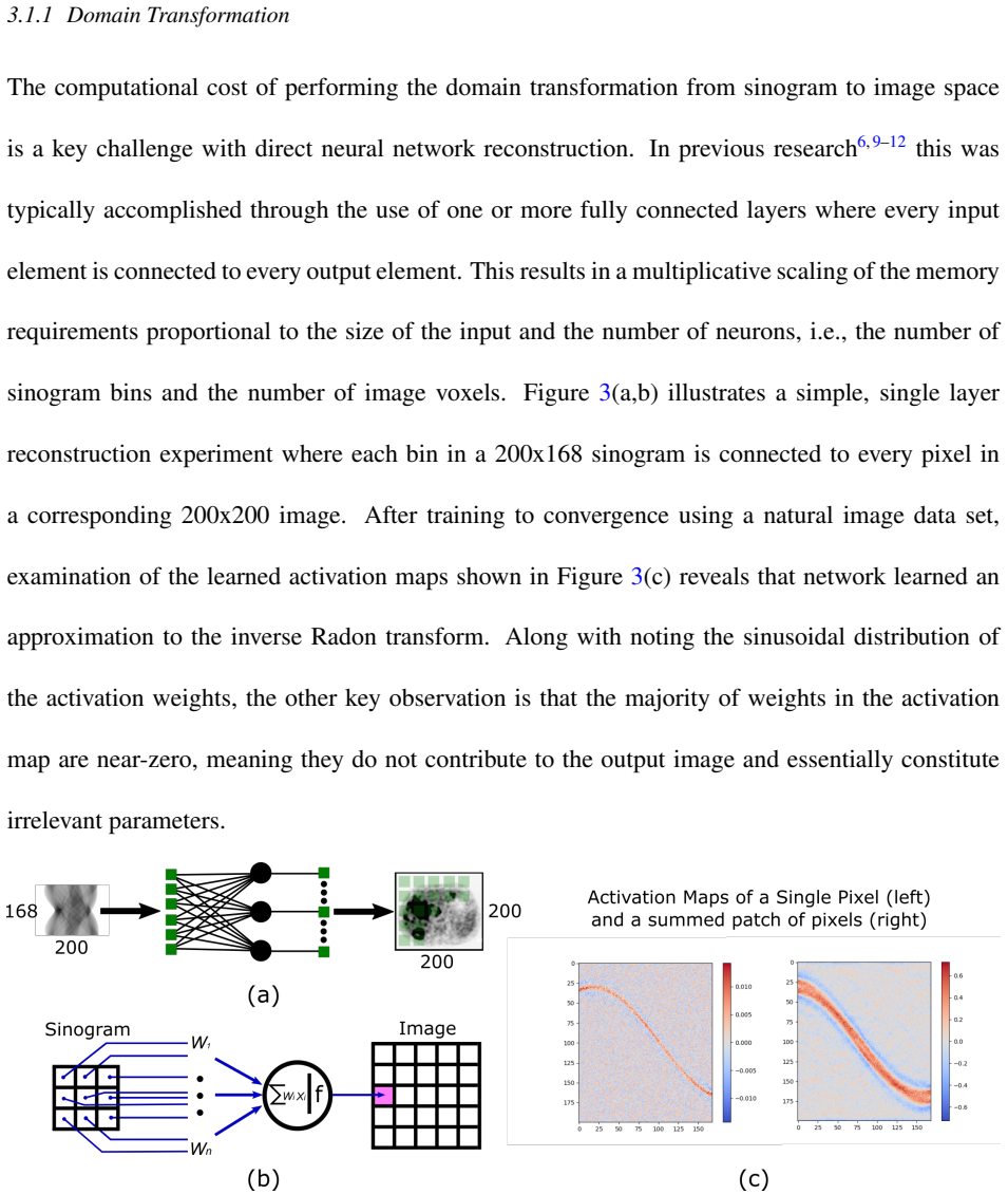
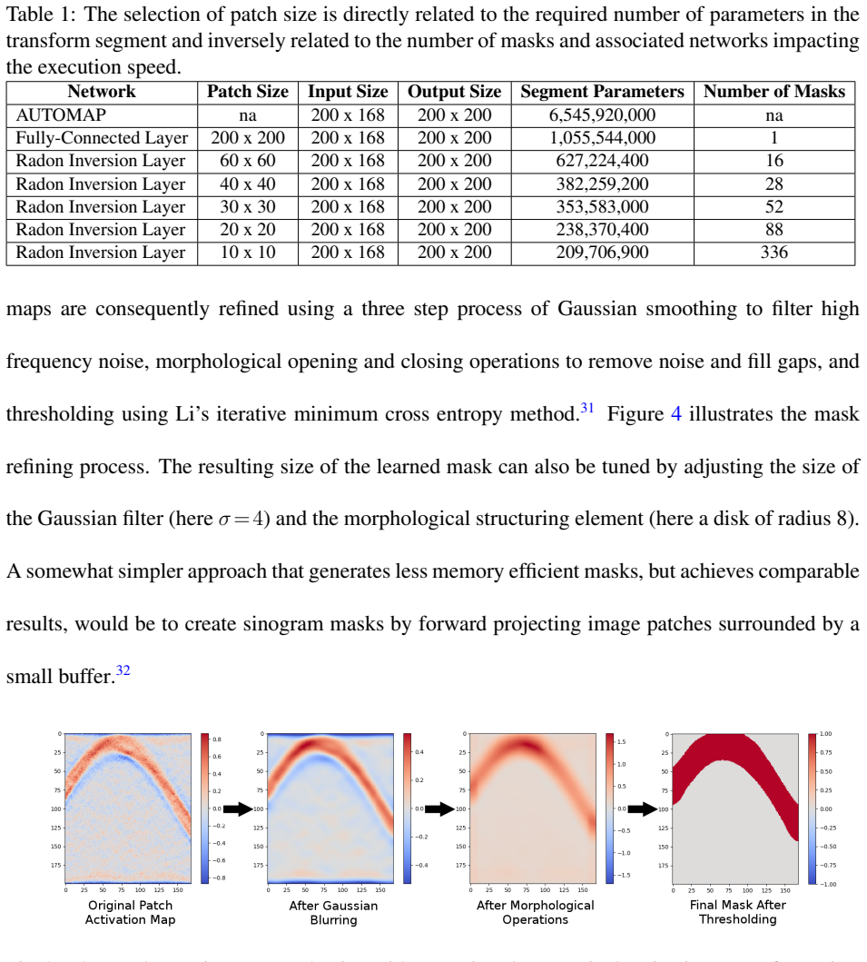
The load-bearing component is the Radon inversion layer, a sparse domain-transformation layer that replaces the dense fully connected mapping from sinogram to image. It partitions the output image into patches, learns a sinogram mask per patch from the activation maps of a small fully connected reconstruction experiment, refines the masks by smoothing, morphological operations, and thresholding, and then connects only the surviving sinogram bins to the neurons of each patch's independent fully connected network. This cuts the transformation parameters from billions to hundreds of millions, making full-size volumes feasible. A convolutional encoder compresses the sinogram before the transform, and a refinement-and-scaling segment with residual blocks and pixel-shuffle upsampling restores full resolution while also ingesting the CT attenuation maps. The training procedure uses a dynamically balanced loss of mean absolute error, multi-scale structural similarity, and a perceptual feature loss computed from a pretrained image-classification network.
What would settle it
Run DirectPET on a NEMA image-quality phantom or on a multi-center patient cohort acquired with a different scanner geometry and radiopharmaceutical, and compare against OSEM+PSF with lesion-level and region-of-interest metrics. If the per-patient MAE, bias, or lesion full-width half-maximum differences exceed the small ranges reported here, or if the network produces artifacts on anatomy outside its training distribution, the claim that DirectPET produces OSEM-equivalent images would be refuted.
Extended reading notes
Core claim
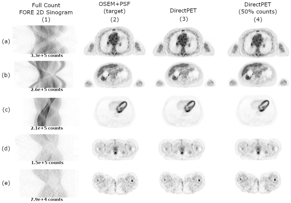
DirectPET is an Encoding–Transformation–Refinement-and-Scaling (ETRS) network that reconstructs full-size multi-slice PET volumes directly from measurement data. Its central claim is that a single trained forward pass can produce 16x400x400 images whose quantitative metrics—mean absolute error, bias, signal-to-noise ratio, and multi-scale structural similarity—match the clinically used OSEM+PSF reconstruction closely enough to be considered equivalent in quality: average MAE 33.07 Bq/ml, average absolute bias 1.82%, MS-SSIM at or above 0.99, and lesion full-width half-maximum within about 1% of the reference. The paper further claims that this equivalence holds when the network is trained on half-count sinograms against full-count targets, producing images nearly identical to the full-count DirectPET and far superior to OSEM+PSF applied to the same half-count data. The authors conclude that image quality in this paradigm depends more on the quality of the training targets than on the raw count level, and that the speed of the method opens the door to clinical use if the remaining distributional and safety questions are resolved.
Load-bearing premise
The argument rests on treating OSEM+PSF reconstructions as both the training targets and the reference standard for image quality; if those images are not faithful to true tracer uptake, or if the ten held-out patients do not represent the wider clinical population, the reported similarities do not establish that DirectPET reconstructs correctly on unseen data.
Editorial extensions
If this is right
- If DirectPET's results hold, an entire 400x400x400 whole-body PET study can be reconstructed in a little more than one second on a GPU, versus tens of seconds for OSEM+PSF; dynamic and gated studies with many frames would shrink from minutes to seconds.
- Because DirectPET-50 matches DirectPET, low-dose acquisitions could be reconstructed at normal-dose quality, potentially reducing patient radiation exposure without changing scanner hardware.
- The dependence of output quality on training targets means improvements in the reference reconstruction, for example maximum a posteriori or non-local-means filtered images, should transfer directly into better network outputs.
- Clinicians could rerun a reconstruction quickly with different settings, provided a small library of networks is trained, replacing the iteration, subset, and filter choices of iterative reconstruction.
- Scatter and attenuation correction are learned rather than explicitly modeled, so the network implicitly encodes scanner physics; this is a direct corollary the paper uses to explain its speed.
Reading between the lines
- The masked-domain-transformation trick should transfer to other tomographic modalities, such as SPECT, CT, or limited-angle imaging, since the masks encode Radon geometry rather than PET-specific physics.
- Because the masks are learned from activation maps, the approach could extend to non-Radon acquisition geometries, such as curved detectors or non-uniform angular sampling, where analytic inversion is hard, potentially by learning masks directly from forward projections.
- A test the paper leaves implicit: train DirectPET on images from a superior reconstruction, such as MAP or a denoised target, and run an observer or lesion-detectability study; the paper's own logic predicts the network will inherit the target's advantages.
- The speed comparison mixes hardware, with OSEM and FBP on a dual-CPU workstation and DirectPET on a GPU workstation, so a controlled same-hardware benchmark would sharpen the factor-of-7 speed claim.
Signed reviews
Editorial analysis
A structured set of objections, weighed in public.
Referee Report
Summary. The paper presents DirectPET, a convolutional neural network that reconstructs 16x400x400 PET image volumes directly from Fourier-rebinned, time-of-flight sinograms together with CT-based attenuation maps. The architecture consists of an encoding segment, a novel patch-wise Radon inversion layer that masks the compressed sinogram and applies small fully connected networks, and a refinement/scaling segment with ResNet blocks. The network is trained on OSEM+PSF reconstructions from 40 whole-body studies and evaluated on 10 held-out patients. The reported results include a reconstruction time of about 4.3 s per field-of-view versus 31 s for OSEM+PSF, a mean absolute error of 33 Bq/ml, MS-SSIM values around 0.99, low bias, and similar lesion line profiles. A half-count variant, DirectPET-50, is trained on thinned raw data with full-count targets and compared against OSEM+PSF-50. The authors acknowledge in Section 4.5 that there is no mathematical or statistical guarantee for unseen data and defer lesion-detectability and observer studies to future work.
Significance. The architectural contribution is significant: direct neural network reconstruction at clinical volume sizes has previously been limited by memory, and the Radon inversion layer with learned masks is a plausible and interesting solution to that bottleneck. The speed advantage is potentially important for dynamic, gated, or interventional PET applications. If the results hold, this would be a substantial step toward practical direct reconstruction. However, the quantitative image-quality evidence is weakened by the fact that the main evaluation metrics, MAE and MS-SSIM, are the same terms that appear in the training loss of Eq. (2), and all image-quality comparisons are made against the training target. The paper therefore demonstrates generalization to held-out patients from the same scanner and reconstruction protocol, but it does not yet establish independent reconstruction accuracy or clinical superiority. The absence of released code or trained models also limits reproducibility. The significance is real but conditional on further validation.
major comments (3)
- [§4.3, Eq. (2) and Fig. 6] The central quantitative evidence for similarity to OSEM+PSF uses MAE and MS-SSIM, both of which are explicit components of the training loss in Eq. (2). The high test-set values (MAE of 33 Bq/ml and MS-SSIM around 0.99) are therefore partly by construction and do not constitute an independent measure of reconstruction accuracy. The authors themselves note in Section 4.3 that the high MAE and MS-SSIM values are driven by these quantities being in the loss function. This is load-bearing for the paper's claim that DirectPET produces images "quantitatively and qualitatively similar" to OSEM+PSF, so the evaluation should be supplemented with at least one independent measure, such as NEMA phantom measurements, lesion contrast or detectability, or image-quality metrics not used in training.
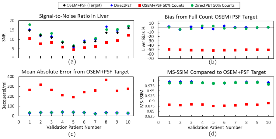
- [§4.3, Fig. 6(b) and §5] The claim that DirectPET-50 provides "superior image quality" in the low-count setting is not supported by the presented comparison. OSEM+PSF-50 is reported to have a negative bias of roughly 50%, which is an unexpectedly large and unexplained systematic effect. It is not shown whether this baseline was tuned for half-count data, for example by adjusting the number of iterations, subsets, regularization, or post-reconstruction filtering. Without a fair and well-optimized low-count baseline, the comparison to OSEM+PSF-50 does not establish that the network maintains image quality under reduced dose. Please explain the observed bias or repeat the comparison with a more appropriate baseline.
- [§4.3, Fig. 7] The spatial-resolution analysis is based on line profiles and full-width half-maximum measurements of only two lesions, which the authors themselves describe as preliminary and somewhat anecdotal. Consequently, the statement that DirectPET preserves spatial resolution is not supported at the level needed for a clinical-viability claim. A quantitative lesion study or a phantom-based resolution measurement would be required to substantiate that claim.
minor comments (5)
- [§4.2] The sentence containing "Is is noteworthy" should read "It is noteworthy."
- [§4.4] The section title "Qualitative Image Image Analysis" contains a duplicated word and should be "Qualitative Image Analysis."
- [§3.1.1] The natural-image data set used to learn the activation maps for mask creation is not identified; please provide a reference or a brief description of that data set.
- [§4.1] The fixed scaling factors for input sinograms and target images (division by 5 and 400, respectively) are stated but not justified; a sentence explaining how these values were chosen would improve reproducibility.
- [§1 and §5] The phrase "path to superior image quality" overstates what is demonstrated; the full-count results show parity with the OSEM+PSF target, and the low-count comparison relies on an apparently unoptimized OSEM+PSF-50 baseline.
Circularity Check
Reported OSEM+PSF similarity and low-dose superiority are partly circular: the network is trained to minimize MAE/MS-SSIM against OSEM+PSF targets and is then scored with those same targets and metrics; the low-dose comparison also uses the full-count training target as the reference.
-
fitted input called prediction
[Section 3.2, Eq. (2); Section 4.3, Fig. 6 (MAE/MS-SSIM evaluation)]
"The loss between reconstructed image ˆx and target image x was thus made to consist of three terms, namely: L(ˆx,x ) =β VGG(ˆx,x ) + (1−α) MAE(ˆx,x ) +α MS-SSIM(ˆx,x ) (2) ... The nearly identical performance of the two neural networks is driven by MAE being a component of their loss function causing them to specifically optimize this measurement in the same way."
The headline test-set numbers (MAE about 33 Bq/ml, MS-SSIM at or above 0.99) are computed against the OSEM+PSF reconstructions that define the network's training targets, using two of the three terms that Eq. (2) explicitly minimizes. The paper itself states that the high MAE and MS-SSIM scores are driven by those terms being in the loss. A held-out split shows the mapping generalizes across patients, but the metric is not independent: it measures how well the trained network reproduces the mean-absolute-error and structural-similarity objective on new inputs from the same scanner and reconstruction protocol, not the accuracy of either DirectPET or OSEM+PSF against true tracer uptake. Consequently the quantitative 'similar to OSEM+PSF' claim is partly forced by construction.
-
fitted input called prediction
[Section 4.1 and Section 4.3, DirectPET-50 low-dose experiment; Section 5 conclusion]
"The second version, which will be referred to as DirectPET-50, was trained with half of the raw counts removed using list-mode thinning while retaining the full count images as training targets. ... This capability was demonstrated by removing half the counts in the raw data through list-mode thinning, training the DirectPET network to reconstruct full count images from half count sinogram data, and comparing the results to OSEM+PSF reconstructions on the decimated data."
DirectPET-50's training targets are the full-count OSEM+PSF images, and the low-dose evaluation compares DirectPET-50 and OSEM+PSF-50 against those same full-count OSEM+PSF images. Since DirectPET-50 is optimized to minimize MAE/MS-SSIM to that reference while OSEM+PSF-50 is an independent reconstruction from decimated data, the reported 'superior' quality of DirectPET-50 (SNR, bias around 2%, MS-SSIM at or above 0.99 versus 0.88) is structurally favored by the choice of reference: the network was trained to land on the reference, and the conventional low-count reconstruction was not. This demonstrates that a fitted mapping can mimic the full-count target, not an independent advantage in clinical accuracy.
full rationale
The central quantitative evidence is not independent of the training objective. The OSEM+PSF reconstructions described in Section 4.1 are both the training targets and the reference standard in Section 4.3, and the loss in Eq. (2) is a weighted sum of VGG, MAE, and MS-SSIM. Therefore test-set MAE around 33 Bq/ml and MS-SSIM at or above 0.99 quantify how well the network generalizes the exact objective it was trained for, not whether either DirectPET or OSEM+PSF matches true tracer uptake. The paper's own text makes this explicit: 'The nearly identical performance of the two neural networks is driven by MAE being a component of their loss function causing them to specifically optimize this measurement in the same way' and 'The fact that MS-SSIM is included in the loss function is what once again leads to similar high performance.' Similarly, the low-dose claim that DirectPET-50 outperforms OSEM+PSF-50 is a consequence of training DirectPET-50 to reproduce full-count OSEM+PSF and then measuring both against that same reference. This is partial circularity, not fabrication: the held-out 10-patient split and the use of an external OSEM+PSF implementation give some independent content, especially for reconstruction speed and cross-patient generalization within the same acquisition protocol. No load-bearing self-citation chain is present; the ETRS and Radon-inversion citations are architectural precedents, not uniqueness theorems. The limitations section further weakens the clinical-reconstruction claim by conceding 'there is no mathematical or statistical guarantee that some unknown new data will be reconstructed with the same image quality' and by deferring lesion-detectability and observer studies to future work. Score 6 reflects that the central similarity claim reduces in part to the training objective while retaining real, but limited, test-set generalization content.
Assumptions & free parameters
free parameters (7)
- Image patch size =
40 x 40 pixels
- Mask Gaussian smoothing sigma =
4
- Mask morphological structuring element radius =
8 (disk)
- Input and target scaling factors =
sinogram scaled by 1/5, image scaled by 1/400
- Learning rate bounds =
eta_min = 0.5e-5, eta_max = 9.0e-5
- Number of input slices =
16
- VGG loss weight beta =
0.5
assumptions (5)
- domain assumption The PET reconstruction mapping from preprocessed sinograms to images is a continuous function with bounded input, so the Universal Approximation Theorem applies.
- domain assumption Sinogram masks learned from a fully-connected layer trained on natural images transfer to PET sinogram geometry.
- domain assumption OSEM+PSF reconstructions are valid as both training targets and reference for image quality.
- domain assumption The held-out test patients are representative of the broader clinical population.
- domain assumption Scatter and attenuation correction can be learned implicitly from training data.
Cite this review
Pith. "Pith review of DirectPET: Full Size Neural Network PET Reconstruction from Sinogram Data." pith.science (2026). https://pith.science/paper/FXDAINDT
@misc{pith2026190807516,
  author       = {Pith},
  title        = {Pith review of: DirectPET: Full Size Neural Network PET Reconstruction from Sinogram Data},
  year         = {2026},
  howpublished = {\url{https://pith.science/paper/FXDAINDT}},
  note         = {Machine review of arXiv:1908.07516}
}
read the original abstract
Purpose: Neural network image reconstruction directly from measurement data is a relatively new field of research, that until now has been limited to producing small single-slice images (e.g., 1x128x128). This paper proposes a novel and more efficient network design for Positron Emission Tomography called DirectPET which is capable of reconstructing multi-slice image volumes (i.e., 16x400x400) from sinograms. Approach: Large-scale direct neural network reconstruction is accomplished by addressing the associated memory space challenge through the introduction of a specially designed Radon inversion layer. Using patient data, we compare the proposed method to the benchmark Ordered Subsets Expectation Maximization (OSEM) algorithm using signal-to-noise ratio, bias, mean absolute error and structural similarity measures. In addition, line profiles and full-width half-maximum measurements are provided for a sample of lesions. Results: DirectPET is shown capable of producing images that are quantitatively and qualitatively similar to the OSEM target images in a fraction of the time. We also report on an experiment where DirectPET is trained to map low count raw data to normal count target images demonstrating the method's ability to maintain image quality under a low dose scenario. Conclusion: The ability of DirectPET to quickly reconstruct high-quality, multi-slice image volumes suggests potential clinical viability of the method. However, design parameters and performance boundaries need to be fully established before adoption can be considered.
Figures


Figures from the paper (9 more)




Reference graph
Works this paper leans on
-
[1]
H. M. Hudson and R. S. Larkin , ``Accelerated image reconstruction using ordered subsets of projection data,'' IEEE Transactions on Medical Imaging 13 , 601--609 (1994)
work page 1994
-
[2]
E. Rapisarda, V. Bettinardi, K. Thielemans, et al. , ``Image-based point spread function implementation in a fully 3d OSEM reconstruction algorithm for PET ,'' Physics in Medicine and Biology 55 , 4131--4151 (2010)
work page 2010
-
[3]
Hornik, ``Approximation capabilities of multilayer feedforward networks,'' Neural Netw
K. Hornik, ``Approximation capabilities of multilayer feedforward networks,'' Neural Netw. 4 , 251--257 (1991)
work page 1991
-
[4]
B. C. Csáji, ``Approximation with artificial neural networks,'' Master's thesis, Faculty Sci., Eötvös Loránd Univ., Budapest, Hungary (2001)
work page 2001
-
[5]
Z. Lu, H. Pu, F. Wang, et al. , ``The expressive power of neural networks: A view from the width,'' in Advances in Neural Information Processing Systems 30 , I. Guyon, U. V. Luxburg, S. Bengio, et al. , Eds., 6231--6239, Curran Associates, Inc. (2017)
work page 2017
-
[6]
B. Zhu, J. Z. Liu, B. R. Rosen, et al. , ``Image reconstruction by domain-transform manifold learning,'' Nature 555 , 487--492 (2018)
work page 2018
-
[7]
I. Häggström, C. R. Schmidtlein, G. Campanella, et al. , `` DeepPET : A deep encoder–decoder network for directly solving the PET image reconstruction inverse problem,'' Medical Image Analysis 54 , 253 -- 262 (2019)
work page 2019
-
[8]
G. Wang, J. C. Ye, K. Mueller, et al. , ``Image reconstruction is a new frontier of machine learning,'' IEEE Transactions on Medical Imaging 37 , 1289--1296 (2018)
work page 2018
Show all 45 references
-
[9]
C. E. Floyd , J. E. Bowsher , M. T. Munley , et al. , ``Artificial neural networks for SPECT image reconstruction with optimized weighted backprojection,'' in Conference Record of the 1991 IEEE Nuclear Science Symposium and Medical Imaging Conference , 2184--2188 vol.3 (1991)
1991
-
[10]
Bevilacqua, D
A. Bevilacqua, D. Bollini, R. Campanini, et al. , ``A new approach to P ositron E mission T omography ( PET ) image reconstruction using A rtificial N eural N etworks ( ANN ),'' International Journal of Modern Physics C 9 (2000)
2000
-
[11]
Paschalis , N
P. Paschalis , N. D. Giokaris , A. Karabarbounis , et al. , `` Tomographic image reconstruction using Artificial Neural Networks ,'' Nuclear Instruments and Methods in Physics Research A 527 , 211--215 (2004)
2004
-
[12]
Argyrou, D
M. Argyrou, D. Maintas, C. Tsoumpas, et al. , ``Tomographic image reconstruction based on artificial neural network (ANN) techniques,'' in 2012 IEEE Nuclear Science Symposium and Medical Imaging Conference Record (NSS/MIC) , 3324--3327 (2012)
2012
-
[13]
Gong , J
K. Gong , J. Guan , C. Liu , et al. , `` PET image denoising using a deep neural network through fine tuning,'' IEEE Transactions on Radiation and Plasma Medical Sciences 3 , 153--161 (2019)
2019
-
[14]
S. J. Kaplan and Y.-M. Zhu, ``Full-dose PET image estimation from low-dose PET image using deep learning: a pilot study,'' Journal of Digital Imaging 32 , 773--778 (2018)
2018
-
[15]
J. Xu , E. Gong , J. Pauly , et al. , `` 200x Low-dose PET Reconstruction using Deep Learning ,'' arXiv e-prints , arXiv:1712.04119 (2017)
2017 arXiv
-
[16]
Ronneberger, P.Fischer, and T
O. Ronneberger, P.Fischer, and T. Brox, ``U-net: Convolutional networks for biomedical image segmentation,'' in Medical Image Computing and Computer-Assisted Intervention (MICCAI) , LNCS 9351 , 234--241, Springer (2015). (available on arXiv:1505.04597 [cs.CV])
2015 arXiv
-
[17]
K. He , X. Zhang , S. Ren , et al. , ``Deep residual learning for image recognition,'' in 2016 IEEE Conference on Computer Vision and Pattern Recognition (CVPR) , 770--778 (2016)
2016
-
[18]
Kang , W
E. Kang , W. Chang , J. Yoo , et al. , ``Deep convolutional framelet denosing for low-dose CT via wavelet residual network,'' IEEE Transactions on Medical Imaging 37 , 1358--1369 (2018)
2018
-
[19]
Han and J
Y. Han and J. C. Ye , ``Framing U-Net via deep convolutional framelets: Application to sparse-view CT ,'' IEEE Transactions on Medical Imaging 37 , 1418--1429 (2018)
2018
-
[20]
Jiao and S
J. Jiao and S. Ourselin , `` Fast PET reconstruction using Multi-scale Fully Convolutional Neural Networks ,'' arXiv e-prints , arXiv:1704.07244 (2017)
2017 arXiv
-
[21]
J. Cui, K. Gong, N. Guo, et al. , `` PET image denoising using unsupervised deep learning,'' European Journal of Nuclear Medicine and Molecular Imaging (2019)
2019
-
[22]
K. Kim , D. Wu , K. Gong , et al. , ``Penalized PET reconstruction using deep learning prior and local linear fitting,'' IEEE Transactions on Medical Imaging 37 , 1478--1487 (2018)
2018
-
[23]
Gong , J
K. Gong , J. Guan , K. Kim , et al. , ``Iterative PET image reconstruction using convolutional neural network representation,'' IEEE Transactions on Medical Imaging 38 , 675--685 (2019)
2019
-
[24]
Chen , K
B. Chen , K. Xiang , Z. Gong , et al. , ``Statistical iterative CBCT reconstruction based on neural network,'' IEEE Transactions on Medical Imaging 37 , 1511--1521 (2018)
2018
-
[25]
Gjesteby, Q
L. Gjesteby, Q. Yang, Y. Xi, et al. , `` Deep learning methods to guide CT image reconstruction and reduce metal artifacts ,'' in Medical Imaging 2017: Physics of Medical Imaging , T. G. Flohr, J. Y. Lo, and T. G. Schmidt, Eds., 10132 , 752 -- 758, International Society for Op...
2017
-
[26]
K. Gong, D. Wu, K. Kim, et al. , `` MAPEM-Net: an unrolled neural network for Fully 3D PET image reconstruction ,'' in 15th International Meeting on Fully Three-Dimensional Image Reconstruction in Radiology and Nuclear Medicine , S. Matej and S. D. Metzler, Eds., 11072 , 109 -...
2019
-
[27]
Adler and O
J. Adler and O. Öktem , ``Learned primal-dual reconstruction,'' IEEE Transactions on Medical Imaging 37 , 1322--1332 (2018)
2018
-
[28]
Defrise, M
M. Defrise, M. E. Casey, C. Michel, et al. , ``Fourier rebinning of time-of-flight PET data,'' Physics in Medicine and Biology 50 , 2749--2763 (2005)
2005
-
[29]
Y. Li, S. Matej, and S. D. Metzler, ``A unified fourier theory for time-of-flight PET data,'' Physics in Medicine and Biology 61 , 601--624 (2015)
2015
-
[30]
Whiteley and J
W. Whiteley and J. Gregor, `` Direct image reconstruction from raw measurement data using an encoding transform refinement-and-scaling neural network ,'' in 15th International Meeting on Fully Three-Dimensional Image Reconstruction in Radiology and Nuclear Medicine , S. Matej ...
2019
-
[31]
Li and C
C. Li and C. Lee, ``Minimum cross entropy thresholding,'' Pattern Recognition 26 (4), 617 -- 625 (1993)
1993
-
[32]
Whiteley and J
W. Whiteley and J. Gregor , ``Efficient neural network image reconstruction from raw data using a radon inversion layer,'' in 2019 IEEE Nuclear Science Symposium and Medical Imaging Conference (NSS/MIC) , 1--2 (2019)
2019
-
[33]
D. E. Rumelhart, G. E. Hinton, and R. J. Williams, ``Parallel distributed processing: Explorations in the microstructure of cognition, vol. 1,'' in Parallel Distributed Processing , D. E. Rumelhart, J. L. McClelland, and C. PDP Research Group, Eds., ch. Learning Internal Repre...
1986
-
[34]
K. He, X. Zhang, S. Ren, et al. , ``Delving deep into rectifiers: Surpassing human-level performance on imagenet classification,'' in Proceedings of the 2015 IEEE International Conference on Computer Vision (ICCV) , ICCV '15 , 1026--1034, IEEE Computer Society, (Washington, DC...
2015
-
[35]
C. Tian, Y. Xu, L. Fei, et al. , ``Deep learning for image denoising: A survey,'' in Genetic and Evolutionary Computing , J.-S. Pan, J. C.-W. Lin, B. Sui, et al. , Eds., 563--572, Springer Singapore, (Singapore) (2019)
2019
-
[36]
Yang , X
W. Yang , X. Zhang , Y. Tian , et al. , ``Deep learning for single image super-resolution: A brief review,'' IEEE Transactions on Multimedia 21 , 1--1 (2019)
2019
-
[37]
W. Shi, J. Caballero, F. Husz \'a r, et al. , ``Real-time single image and video super-resolution using an efficient sub-pixel convolutional neural network,'' 2016 IEEE Conference on Computer Vision and Pattern Recognition (CVPR) , 1874--1883 (2016)
2016
-
[38]
Paszke, S
A. Paszke, S. Gross, S. Chintala, et al. , ``Automatic differentiation in PyTorch ,'' in NIPS-W , (2017)
2017
-
[39]
D. P. Kingma and J. Ba, ``Adam: A method for stochastic optimization,'' International Conference on Learning Representations ICLR('14) (2014)
2014
-
[40]
L. N. Smith, ``Cyclical learning rates for training neural networks,'' 2017 IEEE Winter Conference on Applications of Computer Vision (WACV) , 464--472 (2015)
2015
-
[41]
Zhao , O
H. Zhao , O. Gallo , I. Frosio , et al. , ``Loss functions for image restoration with neural networks,'' IEEE Transactions on Computational Imaging 3 , 47--57 (2017)
2017
-
[42]
Z. Wang, A. C. Bovik, H. R. Sheikh, et al. , ``Image quality assessment: from error visibility to structural similarity,'' IEEE Transactions on Image Processing 13 , 600--612 (2004)
2004
-
[43]
Simonyan and A
K. Simonyan and A. Zisserman , `` Very Deep Convolutional Networks for Large-Scale Image Recognition ,'' arXiv e-prints , arXiv:1409.1556 (2014)
2014 arXiv
-
[44]
Rausch, J
I. Rausch, J. Cal-Gonzalez, D. Dapra, et al. , ``Performance evaluation of the biograph mct flow pet/ct system according to the nema nu2-2012 standard,'' EJNMMI physics 2 , 26 (2015)
2015
-
[45]
write newline
" write newline "" before.all 'output.state := FUNCTION blank.sep after.quote 'output.state := FUNCTION fin.entry output.state after.quoted.block = 'skip 'add.period if write newline FUNCTION new.block output.state before.all = 'skip output.state after.quote = after.quoted.blo...
Reviewed August 14, 2026 · model on record in the stance chip above.
Discussion (0). Continue with ORCID to comment.