REVIEW 2 major objections 6 minor 99 references
The effects of clinically-derived parametric data uncertainty in patient-specific coronary simulations with deformable walls
T0 review · 2 major / 6 minor · reviewed 2026-08-14 · deepseek-v4-flash
Pith's one-line read A 10 percent uncertainty in the heart muscle's squeezing pressure can spread simulated coronary flow and wall shear stress by 27 percent.
desk verdict Solid forward UQ on a deformable coronary model, but the headline 27% flow/TAWSS variability is driven by an assumed 10% intramyocardial pressure input, so the magnitude is conditional even if the ranking is likely right. read the letter →
The pith
A machine-rendered reading of the paper's core claim, the machinery that carries it, and where it could break.
The reading
What carries the argument
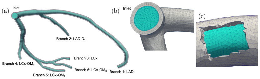
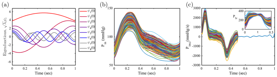
The mechanism is a sub-modeled left coronary artery with Arbitrary-Lagrangian-Eulerian fluid-structure interaction, coupled at six outlets to lumped-parameter coronary boundary conditions that include the intramyocardial pressure and its time derivative. A pulsatile pressure is prescribed at the inlet, so flow is driven by the pressure difference between the inlet and the downstream intramyocardial pressure. Each uncertain input is represented by random variables: inlet pressure and the intramyocardial pressure derivative through Karhunen-Loève expansion of Gaussian processes, Young's modulus through a Gaussian distribution from tensile-test data, and the morphometry exponent through a uniform distribution. Uncertainty is propagated by Monte Carlo, quasi-Monte Carlo, stochastic collocation, and a multi-wavelet stochastic expansion that adaptively refines the stochastic domain. The load-bearing lever is the 10% systolic perturbation of the intramyocardial pressure derivative, because it directly modifies the pressure gradient that drives coronary flow and therefore produces the reported 27% variability in flow and wall shear stress.
What would settle it
An independent clinical estimate of the systolic time-derivative of intramyocardial pressure variability, propagated through the same left coronary model, would confirm or replace the central 27% figure; if the true coefficient of variation is, for example, 5% instead of 10%, the flow and wall shear stress coefficient of variation would be roughly half the reported value.
Extended reading notes
Core claim
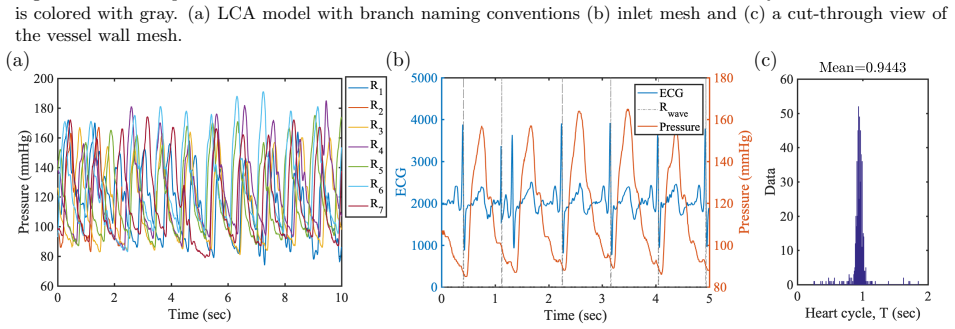
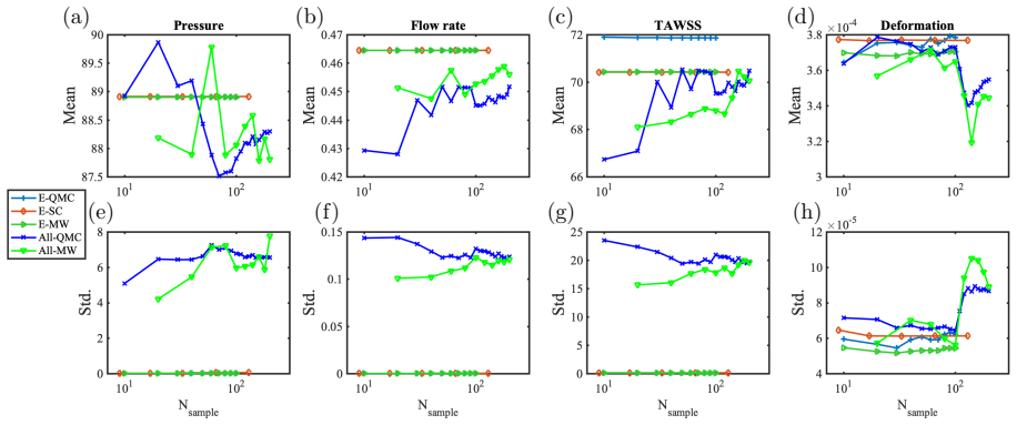
The paper's central quantitative discovery is a separation of uncertainty transmission paths in a left coronary artery sub-model with deformable walls. A 7% coefficient of variation in the inlet pressure waveform, measured from repeated catheterization data in six patients, transmits almost unchanged, about 7%, to outlet pressure and wall deformation, and about 5% to flow rate and time-averaged wall shear stress. A 10% coefficient of variation assumed for the systolic time derivative of intramyocardial pressure produces up to 27% coefficient of variation in flow rate and wall shear stress, while leaving pressure and deformation variability below 3%. Young's modulus uncertainty, modeled as a Gaussian with 17% coefficient of variation from human coronary tensile-test data, affects only wall deformation, also at about 17%, leaving hemodynamics nearly unchanged. Morphometry exponent uncertainty in the range 2.4 to 2.8 has negligible effects. These results indicate which uncertain inputs must be measured more tightly before coronary simulations can report flow and wall shear stress with clinical confidence.
Load-bearing premise
The load-bearing premise is the assumed 10% coefficient of variation for the intramyocardial pressure time derivative during systole; this distribution was assumed rather than inferred from clinical data, and it drives the largest reported output variability.
Editorial extensions
If this is right
- If these results transfer to other coronary anatomies, deterministic coronary simulations should be interpreted with confidence intervals: a 10% uncertainty in the systolic intramyocardial pressure derivative alone can spread time-averaged wall shear stress by up to 27% coefficient of variation.
- Flow and time-averaged wall shear stress variability track each other, while pressure and wall deformation variability track each other, so one member of each pair can serve as a practical proxy for the other in uncertainty reporting.
- Uncertainty in vessel wall stiffness has little bearing on hemodynamic outputs in this small-deformation coronary model, but it must be controlled when wall deformation itself is the quantity of interest.
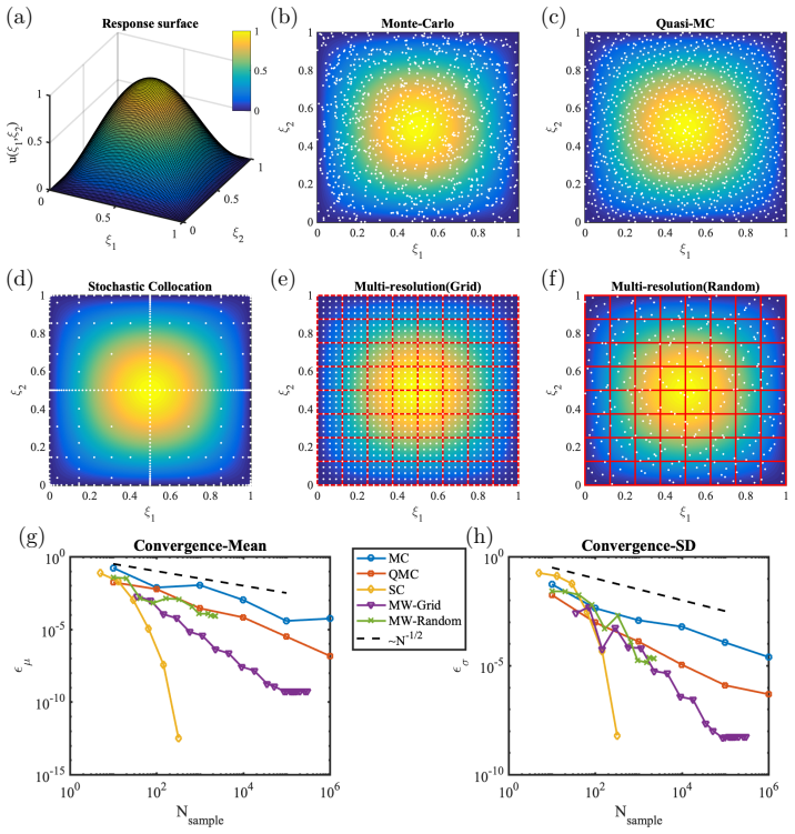
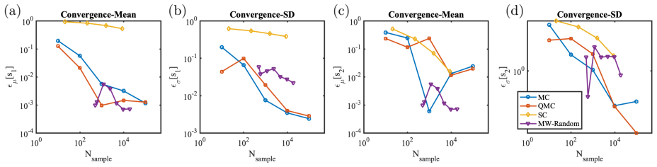
- The multi-wavelet stochastic expansion estimates means and standard deviations accurately with roughly 50 or fewer model evaluations, making uncertainty quantification tractable for deformable coronary sub-models.
- Improving the measurement of intramyocardial pressure, rather than inlet pressure, is the bottleneck for accurate flow and wall shear stress predictions.
Reading between the lines
- If the reported near-linear transmission of inlet pressure variability holds in stenosed vessels, outlet pressure variability could be approximated directly from inlet measurement variability without a full fluid-structure simulation, a shortcut that could be tested on synthetic stenotic geometries.
- Because the intramyocardial pressure derivative was perturbed only during systole, the systolic-window definition is a hidden sensitivity; perturbing the derivative over the full cardiac cycle would test how much of the 27% figure depends on that modeling choice.
- The near-decoupling of hemodynamics from wall mechanics suggests a rigid-wall model with identical pressure boundary conditions may reproduce the flow and wall shear stress variability at lower computational cost, which a direct rigid-wall comparison could verify.
- The 27% spread in time-averaged wall shear stress implies that plaque-progression risk categories based on a single deterministic value may misclassify patients near thresholds, so reporting the full output distribution would be more clinically informative.
Signed reviews
Editorial analysis
A structured set of objections, weighed in public.
Referee Report
Summary. The manuscript performs forward uncertainty quantification for a patient-specific left coronary artery model with deformable walls, using an ALE fluid-structure interaction framework and lumped-parameter network outlet boundary conditions. Stochastic inputs are the inlet coronary pressure waveform (modeled from catheterization data via a Karhunen-Loève expansion), the intramyocardial pressure time derivative (assumed 10% peak-relative standard deviation), the morphometry exponent (uniform on 2.4 to 2.8), and the wall Young's modulus (Gaussian with literature-based moments). Uncertainty propagation is carried out with Monte Carlo, quasi-Monte Carlo, stochastic collocation, and multiwavelet stochastic expansion, first on analytic benchmarks and then on the coronary model using 1203 simulations. The main findings are that 7% input coefficient of variation in inlet pressure propagates to about 7% cv in outlet pressure and wall deformation and about 5% cv in flow and wall shear stress; 10% peak-relative variability in the intramyocardial pressure derivative produces up to 27% cv in flow rate and time-averaged wall shear stress; the morphometry exponent has negligible effect; and Young's modulus uncertainty affects wall deformation only, with about 17% cv. The authors conclude that the multiwavelet method is superior to quasi-Monte Carlo and stochastic collocation for this class of problems.
Significance. If the input distributions are accepted, the paper is a useful demonstration of uncertainty quantification in deformable-wall coronary simulations and provides a fair, sample-counted comparison of propagation methods. The strengths include the use of actual intra-coronary catheterization data for the inlet pressure, a boundary-layer mesh convergence study, explicit reporting of the 1203 simulation count, and systematic benchmarks against analytic and nonlinear test problems. The qualitative ranking, namely that intramyocardial pressure uncertainty dominates flow and wall shear stress variability while wall stiffness uncertainty affects mainly wall mechanics, is clinically plausible and worth reporting. However, the leading quantitative claim of 27% cv in flow and TAWSS is conditional on an assumed 10% peak-relative standard deviation for the intramyocardial pressure derivative, and the paper's own limitation statement concedes that the random input distributions were assumed rather than inferred from clinical data. The title's promise of 'clinically-derived parametric data uncertainty' is therefore not supported for the central quantitative result.
major comments (2)
- [Secs. 3.2, 4.4, and 5] The 10% peak-relative standard deviation assigned to the intramyocardial pressure derivative in Sec. 3.2 is a modeling assumption, not an estimate from clinical data. Section 5 explicitly states that 'the distribution of the random inputs were assumed in this study rather than inferred from available clinical data.' Because dPim/dt appears as a direct source term in the distal-pressure ODE, Eq. (4), the reported 27% cv in flow rate and time-averaged wall shear stress (Sec. 4.4) is approximately proportional to this assumed input amplitude. The authors should either estimate this input from clinical measurements, calibrate it against available data, or explicitly present the 27% as conditional and include a sensitivity sweep over the input amplitude. As written, the paper's leading quantitative result and the title's 'clinically-derived' claim are not supported for this input.
- [Sec. 2] There is an internal inconsistency in the wall thickness specification. The text states a uniform wall thickness h=0.08 mm, then says this is 'consistent with' a coronary wall thickness of 1.0±0.2 mm reported in two echocardiographic studies, and 'larger than' a typical wall thickness equal to 10% of the vessel radius. For a left main diameter of 4 mm, 10% of the radius is 0.2 mm, and the cited 1.0 mm value is 12.5 times larger than 0.08 mm. This inconsistency directly affects the wall deformation quantity of interest and the FSI results. Please correct the value or the citations, and assess how sensitive the wall-mechanics conclusions are to this parameter.
minor comments (6)
- [Sec. 3.2] The phrase '10% cv in the intramyocardial pressure' is misleading because a coefficient of variation is conventionally defined relative to a nonzero mean, whereas the mean of dPim/dt over a cardiac cycle is near zero; the quantity used is a peak-relative standard deviation and should be labeled as such throughout.
- [Secs. 3.4 and 4.6] The Young's modulus input is specified as σ[Es]=0.24 MPa in Sec. 3.4 but as Es∼N(1.48, 0.28^2) in Sec. 4.6; the reported 17% cv in wall deformation is consistent with neither value exactly (16.2% or 18.9%). Please reconcile the two specifications.
- [Sec. 4.1] The functions in Eq. (10) are the Sobol' functions, but the text describes the test as a 'ten dimensional sine response surface'; reword to avoid confusion with the sinusoidal benchmark in Eq. (8).
- [Sec. 4.2] The name 'Kraichnan-Orzag' is misspelled; it should be 'Kraichnan-Orszag'.
- [Sec. 5] The sentence 'The MW showed but showed the best performance on discontinuous response surfaces' contains a typographical repetition, and the abstract's broad claim that multiwavelet expansion is 'superior' to stochastic collocation should be qualified because stochastic collocation outperforms all methods on the smooth sine benchmark (Fig. 7).
- [Sec. 3.1] The selection of the correlation length lc=T/2 is stated after 'examining the covariances for the six patients,' but no quantitative comparison of candidate correlation lengths is shown; please provide the covariance fit or the KL eigenvalue decay to justify this choice.
Circularity Check
No circularity: forward uncertainty propagation with independently benchmarked methods and no output-calibrated inputs; the assumed dPim/dt distribution is a data limitation, not a circular step.
full rationale
This is a forward uncertainty propagation study. Input distributions are constructed from intra-coronary catheterization data (Sec. 3.1: 5-7% cv in coronary pressure), literature values (Sec. 3.3 morphometry exponent, Sec. 3.4 Young's modulus), or an explicitly stated modeling assumption (Sec. 3.2: Pim,t sigma = 10% of its maximum absolute value during the cardiac cycle), and none of these inputs is calibrated against the reported quantities of interest. No equation in the paper defines an input in terms of an output, and the near-linear propagation of variability emphasized in Sec. 4.3 is explained from Poiseuille flow and thick-walled cylinder relations, not from fitting. The 7% inlet-pressure cv propagating to about 7% cv in pressure and deformation, and the 10% dPim/dt input producing up to 27% cv in flow and TAWSS, are sensitivity results rather than predictions obtained by re-inserting the target quantities. The method comparison is anchored to analytic benchmarks with known moments (Secs. 4.1-4.2), so the claim that multiwavelet stochastic expansion performs favorably does not rest solely on self-citation. Self-citations to prior MW work [74, 76, 89] and to the baseline Pim,t waveform [88] provide starting points or methodological settings, but the load-bearing accuracy and ranking claims are independently benchmarked against MC, QMC, SC, and analytic solutions. Section 5 explicitly concedes that 'the distribution of the random inputs were assumed in this study rather than inferred from available clinical data'; this is a limitation on clinical grounding and should be weighed as correctness or validity risk, but it is not circularity because the assumed input amplitude is not derived from the outputs. No step in the derivation chain reduces to its own inputs by construction, so the appropriate finding is no significant circularity.
Assumptions & free parameters
free parameters (4)
- Intramyocardial pressure derivative uncertainty amplitude =
10% of maximum baseline dPim/dt
- KL correlation length for coronary pressure process =
0.5 s (T/2)
- Wall thickness h =
0.08 mm
- Morphometry exponent range =
U(2.4, 2.8)
assumptions (7)
- standard math Incompressible Navier-Stokes equations in ALE form govern blood flow
- domain assumption Blood is a Newtonian fluid
- domain assumption The arterial wall is isotropic, homogeneous, Saint Venant-Kirchhoff hyperelastic with uniform thickness
- domain assumption The diastolic CT configuration is stress-free, with no pre-stress
- domain assumption Coronary pressure variability follows a Gaussian process with exponential covariance, variance 7% of mean, correlation length 0.5 s
- domain assumption Intramyocardial pressure is approximated by left ventricular pressure and its derivative, with 10% variability in systole only
- domain assumption All uncertain inputs are independent
Cite this review
Pith. "Pith review of The effects of clinically-derived parametric data uncertainty in patient-specific coronary simulations with deformable walls." pith.science (2026). https://pith.science/paper/IEKLW34A
@misc{pith2026190807522,
  author       = {Pith},
  title        = {Pith review of: The effects of clinically-derived parametric data uncertainty in patient-specific coronary simulations with deformable walls},
  year         = {2026},
  howpublished = {\url{https://pith.science/paper/IEKLW34A}},
  note         = {Machine review of arXiv:1908.07522}
}
read the original abstract
Cardiovascular simulations are increasingly used for non-invasive diagnosis of cardiovascular disease, to guide treatment decisions, and in the design of medical devices. Quantitative assessment of the variability of simulation outputs due to input uncertainty is a key step toward further integration of cardiovascular simulations in the clinical workflow. In this study, we present uncertainty quantification in computational models of the coronary circulation to investigate the effect of uncertain parameters, including coronary pressure waveform, intramyocardial pressure, morphometry exponent, and the vascular wall Young's modulus. We employ a left coronary artery model with deformable vessel walls, simulated via an ALE framework for FSI, with a prescribed inlet pressure and open-loop lumped parameter network outlet boundary conditions. Stochastic modeling of the uncertain inputs is determined from intra-coronary catheterization data or gathered from the literature. Uncertainty propagation is performed using several approaches including Monte Carlo, Quasi MC, stochastic collocation, and multiwavelet stochastic expansion. Variabilities in QoI, including branch pressure, flow, wall shear stress, and wall deformation are assessed. We find that uncertainty in inlet pressures and intramyocardial pressures significantly affect all resulting QoIs, while uncertainty in elastic modulus only affects the mechanical response of the vascular wall. Variability in the morphometry exponent has little effect on coronary hemodynamics or wall mechanics. Finally, we compare convergence behaviors of statistics of QoIs using several uncertainty propagation methods. From the simulation results, we conclude that the multi-wavelet stochastic expansion shows superior accuracy and performance against Quasi Monte Carlo and stochastic collocation methods.
Figures

Figures from the paper (16 more)





Reference graph
Works this paper leans on
-
[1]
High Performance Computer Applications 6th International Conference , volume 595, 1 2016
2016
-
[2]
Agarwal and N
N. Agarwal and N. Aluru. A domain adaptive stochastic collocation approach for analysis of MEMS under uncertainties. Journal of Computational Physics , 228:7662–7668, 2009
2009
-
[3]
Bangalore Ramachandra, A
A. Bangalore Ramachandra, A. Kahn, and A. Marsden. Patient-specific simulations reveal significant differences in mechanical stimuli in venous and arterial coronary grafts. Journal of cardiovascular translational research, 9(4):279–290, 2016
2016
-
[4]
Baumann, R
S. Baumann, R. Wang, U. J. Schoepf, D. H. Steinberg, J. V. Spearman, R. R. Bayer, C. W. Hamm, and M. Renker. Coronary ct angiography-derived fractional flow reserve correlated with invasive fractional flow reserve measurements –initial experience with a novel physician-driven algorithm. European Radiology, 25(4):1201–1207, 2015
2015
-
[5]
Baumler, V
K. Baumler, V. Vedula, A. Sailer, J. Seo, P. Chiu, G. Mistlbauer, F. Chan, M. Fischbein, A. Marsden, and D. Fleischmann. Fluid–structure interaction simulations of patientspecific aortic dissection.Biomechanics and Modeling in Mechanobiology, pages 1–22, 2020
2020
-
[6]
Bazilevs, V
Y. Bazilevs, V. Calo, J. Cottrell, T. Hughes, A. Reali, and G. Scovazzi. Variational multiscale residual-based turbulence modeling for large eddy simulation of incompressible flows. Computer Methods in Applied Mechanics and Engineering , 197:173–201, 2007
2007
-
[7]
Benjamin, S
E. Benjamin, S. Virani, C. Callaway, A. Chamberlain, A. Chang, S. Cheng, S. Chiuve, M. Cushman, F. Delling, R. Deo, 23 et al. Heart disease and stroke statistics-2018 update: a report from the American Heart Association. Circulation, 137(12):e67, 2018
2018
-
[8]
D. Bergel. The static elastic properties of the arterial wall. Journal of Physiology , 156:445–457, 1961
1961
Show all 99 references
-
[9]
Biehler, M
J. Biehler, M. Gee, and W. Wall. Towards efficient uncertainty quantification in complex and large-scale biomechanical problems based on a Bayesian multi-fidelity scheme. Biomechanics and Modeling in Mechanobiology , 14:485–513, 2015
2015
-
[10]
Biehler and W
J. Biehler and W. Wall. The impact of personalized probabilistic wall thickness models on peak wall stress in abdominal aortic aneurysms. International Journal for Numerical Methods in Biomedical Engineering , 34(e2922), 2017
2017
-
[11]
Blatman and B
G. Blatman and B. Sudret. Adaptive sparse polynomial chaos expansion based on least angle regression. Journal of Computational Physics, 230(6):2345–2367, 2011
2011
-
[12]
Bogren, R
H. Bogren, R. Klipstein, D. Firmin, R. Mohiaddin, S. Underwood, R. Rees, and D. Longmore. Quantitation of ante- grade and retrograde blood flow in the human aorta by magnetic resonance velocity mapping. American Heart Journal, 117(6):1214–1222, 1989
1989
-
[13]
Boileau, S
E. Boileau, S. Pant, C. Roobottom, I. Sazonov, J. Deng, X. Xie, and P. Nithiarasu. Estimating the accuracy of a reduced-order model for the calculation of fractional flow reserve (FFR). International Journal for Numerical Methods in Biomedical Engineering, 34(1):e2908, 2018. e2...
2018
-
[14]
Brault, L
A. Brault, L. Dumas, and D. Lucor. Uncertainty quantification of inflow boundary condition and proximal arterial stiffness–coupled effect on pulse wave propagation in a vascular network. International Journal for Numerical Methods in Biomedical Engineering, 33(e2859), 2017
2017
-
[15]
Changizi and C
M. Changizi and C. Cherniak. Modeling the large-scale geometry of human coronary arteries. Canadian Journal of Physiology and Pharmacology, 78:603–611, 2000
2000
-
[16]
Chen and A
P. Chen and A. Quarteroni. A new algorithm for high-dimensional uncertainty quantification based on dimension-adaptive sparse grid approximation and reduced basis methods. Journal of Computational Physics , 298:176–193, 2015
2015
-
[17]
P. Chen, A. Quarteroni, and G. Rozza. Simulation-based uncertainty quantification of human arterial network hemody- namics. International Journal for Numerical Methods in Biomedical Engineering , 29:698–721, 2013
2013
-
[18]
Coogan, J
J. Coogan, J. Humphrey, and C. Figueroa. Computational simulations of hemodynamic changes within thoracic, coronary, and cerebral arteries following early wall remodeling in response to distal aortic coarctation. Biomech. Model Mechanobi- ology, 12:79–93, 2013
2013
-
[19]
Dodge Jr., B
J. Dodge Jr., B. Brown, E. Bolson, and H. Dodge. Lumen diameter of normal human coronary arteries: influence of age, sex, anatomic variation, and left ventricular hypertrophy or dilation. Circulation, 86(1):232–246, 1992
1992
-
[20]
P. S. Douglas, G. Pontone, M. A. Hlatky, M. R. Patel, B. L. Norgaard, R. A. Byrne, N. Curzen, I. Purcell, M. Gutberlet, G. Rioufol, U. Hink, H. W. Schuchlenz, G. Feuchtner, M. Gilard, D. Andreini, J. M. Jensen, M. Hadamitzky, K. Chiswell, D. Cyr, A. Wilk, F. Wang, C. Rogers, B...
2015
-
[21]
Esmaily-Moghadam, Y
M. Esmaily-Moghadam, Y. Bazilevs, and A. Marsden. A bi-partitioned iterative algorithm for solving linear systems arising from incompressible flow problems. Computer Methods in Applied Mechanics and Engineering , 286:40–62, 2015
2015
-
[22]
Esmaily-Moghadam, T
M. Esmaily-Moghadam, T. Hsia, and A. Marsden. A non-discrete method for computation of residence time in fluid mechanics simulations. Physics of Fluids , 25(11):110802, 2013
2013
-
[23]
Esmaily Moghadam, I
M. Esmaily Moghadam, I. Vignon-Clementel, R. Figliola, and A. Marsden. A modular numerical method for implicit 0D/3D coupling in cardiovascular finite element simulations. Journal of Computational Physics , 244:63–79, 2013
2013
-
[24]
H. Faure. Good permutations for extreme discrepancy. Journal of Number Theory , 42(1):47–56, 1992
1992
-
[25]
Figueroa, I
C. Figueroa, I. Vignon-Clementel, K. Jansen, T. Hughes, and C. Taylor. A coupled momentum method for modeling blood flow in three-dimensional deformable arteries. Computer Methods in Applied Mechanics and Engineering , 195(41- 43):5685–5706, 2006
2006
-
[26]
Fleeter, G
C. Fleeter, G. Geraci, D. Schiavazzi, A. Kahn, M. Eldred, and A. Marsden. Multilevel multifidelity approaches for cardiovascular flow under uncertainty. Technical Report SAND2018-2780O, Center for Computing Research Summer Proceedings, A.D. Baczewski and M.L. Parks, eds., Sandia...
2017
-
[27]
Frauenfelder, M
T. Frauenfelder, M. Lotfey, T. Boehm, and S. Wildermuth. Computational fluid dynamics: Hemodynamic changes in abdominal aortic aneurysm after stent-graft implantation. CardioVascular and Interventional Radiology , 29(4):613–623, Aug 2006
2006
-
[28]
M. H. Friedman, C. B. Bargeron, O. J. Deters, G. M. Hutchins, and F. F. Mark. Correlation between wall shear and intimal thickness at a coronary artery branch. Atherosclerosis, 68(1):27–33, 1987
1987
-
[29]
Geraci, M
G. Geraci, M. Eldred, and G. Iaccarino. A multifidelity control variate approach for the multilevel Monte Carlo technique. Technical report, Stanford University, 2015
2015
-
[30]
Ghanem and P
R. Ghanem and P. Spanos. Stochastic finite elements: a spectral approach . Courier Corporation, 2003
2003
-
[31]
C. M. Gibson, L. Diaz, K. Kandarpa, F. M. Sacks, R. C. Pasternak, T. Sandor, C. Feldman, and P. H. Stone. Relation of vessel wall shear stress to atherosclerosis progression in human coronary arteries. Arteriosclerosis and Thrombosis: A Journal of Vascular Biology , 13(2):310–...
2019
-
[32]
Gijsen, A
F. Gijsen, A. van der Giessen, A. van der Steen, and J. Wentzel. Shear stress and advanced atherosclerosis in human coronary arteries. Journal of Biomechanics , 46(2):240–247, 2013
2013
-
[33]
Gow and C
B. Gow and C. Hadfield. The elasticity of canine and human coronary arteries with reference to postmortem changes. Circulation Research, 45:588–594, 1979
1979
-
[34]
Gradus-Pizlo, B
I. Gradus-Pizlo, B. Bigelow, Y. Mahomed, S. Sawada, K. Rieger, and H. Feigenbaum. Left anterior descending coronary artery wall thickness measured by high-frequency transthoracic and epicardial echocardiography includes adventitia. The 24 American Journal of Cardiology , 9:27–32, 2003
2003
-
[35]
Grande Gutierrez, O
N. Grande Gutierrez, O. Shirinsky, N. Gagarina, G. Lyskina, R. Fukazawa, S. Ogawa, J. Burns, A. Marsden, and A. Kahn. Assessment of coronary artery aneurysms caused by Kawasaki disease using transluminal attenuation gradient analysis of computerized tomography angiograms. The ...
2017
-
[36]
Gundert, A
T. Gundert, A. Marsden, W. Yang, D. Marks, and J. J. LaDisa. Identification of hemodynamically optimal coronary stent designs based on vessel caliber. IEEE Transactions on Biomedical Engineering , 58(7):1992–2002, 2012
1992
-
[37]
Habib, E
S. Habib, E. Parham, M. M. C., S. Jin, D. S. S., M. Charles, T. L. H., Q. A. A., and G. D. P. Coronary artery wall shear stress is associated with progression and transformation of atherosclerotic plaque and arterial remodeling in patients with coronary artery disease. Circula...
2019
-
[38]
J. Halton. On the efficiency of certain quasi-random sequences of points in evaluating multidimensional integrals. Nu- merische Mathematik, 2(1):84–90, 1960
1960
-
[39]
Heroux, R
M. Heroux, R. Bartlett, V. Howle, R. Hoekstra, J. Hu, T. Kolda, R. Lehoucq, K. Long, R. Pawlowski, E. Phipps, A. Salinger, H. Thornquist, R. Tuminaro, J. Willenbring, A. Williams, and K. Stanley. An overview of the Trilinos project. ACM Transactions on Mathematical Software , ...
2005
-
[40]
Hirai, S
T. Hirai, S. Sasayama, T. Kawasaki, and S. Yagi. Stiffness of systemic arteries in patients with myocardial infarction. Circulation, 80:78–86, 1989
1989
-
[41]
Holzapfel, G
G. Holzapfel, G. Sommer, C. Gasser, and P. Regitnig. Determination of layer-specific mechanical properties of human coronary arteries with nonatherosclerotic intimal thickening and related constitutive modeling. American Journal of Physiology-Heart and Circulatory Physiology, 2...
2005
-
[42]
M. C. Hsu and Y. Bazilev. Blood vessel tissue prestress modeling for vascular fluid–structure interaction simulation. Finite Elements in Analysis and Design , 47(6):593–599, 2011
2011
-
[43]
Hutchins, M
G. Hutchins, M. Miner, and J. Boitnott. Vessel caliber and branch-angle of human coronary artery branch-points. Circulation Research, 38:572–576, 1976
1976
-
[44]
Jaquet, L
C. Jaquet, L. Najman, H. Talbot, L. Grady, M. Schaap, B. Spain, H. Kim, I. Vignon-Clementel, and C. Taylor. Generation of patient-specific cardiac vascular networks: a hybrid image-based and synthetic geometric model. IEEE Transactions on Biomedical Engineering, 66:946–955, 2017
2017
-
[45]
Karimi, M
A. Karimi, M. Navidbakhsh, A. Shojaei, and S. Faghihi. Measurement of the uniaxial mechanical properties of healthy and atherosclerotic human coronary arteries. Materials Science and Engineering , C33:2550–2554, 2013
2013
-
[46]
H. Kim, I. Vignon-Clementel, C. Figueroa, J. LaDisa, K. Jansen, J. Feinstein, and C. Taylor. On coupling a lumped param- eter heart model and a three-dimensional finite element aorta model.Annals of Biomedical Engineering, 37(11):2153–2169, 2009
2009
-
[47]
Kissas, Y
G. Kissas, Y. Yang, E. Hwuang, W. Witschey, J. Detre, and P. Perdikaris. Machine learning in cardiovascular flows modeling: Predicting arterial blood pressure from non-invasive 4d flow mri data using physics-informed neural networks. Computer Methods in Applied Mechanics and Eng...
2020
-
[48]
B.-K. Koo, A. Erglis, J.-H. Doh, D. V. Daniels, S. Jegere, H.-S. Kim, A. Dunning, T. DeFrance, A. Lansky, J. Leipsic, and J. K. Min. Diagnosis of Ischemia-Causing Coronary Stenoses by Noninvasive Fractional Flow Reserve Computed From Coronary Computed Tomographic Angiograms: R...
1989
-
[49]
Kraichnan
R. Kraichnan. Direct-interaction approximation for a system of several interacting simple shear waves. Physics of Fluids , 6:1603–1609, 1963
1963
-
[50]
Kural, M
M. Kural, M. Cai, D. Tang, T. Gwyther, J. Zheng, and K. Billiar. Planar biaxial characterization of diseased human coronary and carotid arteries for computational modeling. Journal of Biomechanics , 45:790–798, 2012
2012
-
[51]
Kurata, A
A. Kurata, A. Coenen, M. M. Lubbers, K. Nieman, T. Kido, T. Kido, N. Yamashita, K. Watanabe, G. P. Krestin, and T. Mochizuki. The effect of blood pressure on non-invasive fractional flow reserve derived from coronary computed tomography angiography. European Radiology, 27(4):141...
2017
-
[52]
H. Lan, A. Updegrove, N. Wilson, G. Maher, S. Shadden, and A. Marsden. A re-engineered software interface and workflow for the open-source SimVascular cardiovascular modeling package. Journal of Biomechanical Engineering , 140(024501), 2018
2018
-
[53]
Le Maitre, M
O. Le Maitre, M. Reagan, H. Najm, R. Ghanem, and O. Knio. A atochastic projection method for fluid flow II. random process. Journal of Computational Physics , 181:9–44, 2002
2002
-
[54]
C. Long, A. Marsden, and Y. Bazilevs. Shape optimization of pulsatile ventricular assist devices using FSI to minimize thrombotic risk. Computional Mechanics, 54:921–932, 2014
2014
-
[55]
O. L. Maitre and O. M. Knio. Spectral methods for uncertainty quantification. Springer, 2010
2010
-
[56]
F. Mall. A study of the structural unit of the liver. American Journal of Anatomy , 5:227–308., 1905
1905
-
[57]
A. Marsden. Optimization in cardiovascular modeling. Annual Review of Fluid Mechanics , 46:519–546, 2014
2014
-
[58]
Marsden and M
A. Marsden and M. Esmaily-Moghadam. Multiscale modeling of cardiovascular flows for clinical decision support. Applied Mechanics Reviews, 67:030804, 2015
2015
-
[59]
Mayrovitz and J
H. Mayrovitz and J. Roy. Microvascular blood flow: evidence indicating a cubic dependence on arteriolar diameter. Heart and Circulatory Physiology, 245(6):1031–1038, 1983
1983
-
[60]
Migliavacca, L
F. Migliavacca, L. Petrini, M. Colombo, F. Auricchio, and R. Pietrabissa. Mechanical behavior of coronary stents inves- tigated through the finite element method. Journal of Biomechanics , 35(6):803–811, 2002
2002
-
[61]
J. K. Min, J. Leipsic, M. J. Pencina, D. S. Berman, B.-K. Koo, C. van Mieghem, A. Erglis, F. Y. Lin, A. M. Dunning, P. Apruzzese, M. J. Budoff, J. H. Cole, F. A. Jaffer, M. B. Leon, J. Malpeso, G. B. J. Mancini, S.-J. Park, R. S. Schwartz, L. J. Shaw, and L. Mauri. Diagnostic Ac...
2012
-
[62]
C. Murray. The physiological principle of minimum work. I. the vascular system and the cost of blood volume. Proceedings of the National Acadademy of Science , 12:207–214, 1926
1926
-
[63]
Nichols, M
W. Nichols, M. O’Rourke, and C. Vlachopoulos. McDonald’s Blood Flow in Arteries Theoretical, Experimental, and Clinical Principles. CRC Press, 6th edition, 2011
2011
-
[64]
Orszag and L
S. Orszag and L. Bissonnette. Dynamics properties of truncated Wiener-Hermite expansions. Physics of Fluids , 10(12):2603–2163, 1967
1967
-
[65]
Patel, J
D. Patel, J. Janicki, and T. Carew. Static anisotropic elastic properties of the aorta in living dogs. Circulation Research, XXV:765–779, 1969
1969
-
[66]
Pearson, R
A. Pearson, R. Guo, D. Orsinelli, P. Binkley, and T. Pasierski. Transesophageal echocardiographic assessment of the effects of age, gender, and hypertension on thoracic aortic wall size, thickness, and stiffness. 1994, 128:344–351, American Heart Journal
1994
-
[67]
Perry, M
R. Perry, M. Joseph, D. Chew, P. Aylward, and C. De Pasquale. Coronary artery wall thickness of the left anterior descending artery using high resolution transthoracic echocardiography – normal range of values. Echocardiography, pages 759–764, 2013
2013
-
[68]
Peterson, R
L. Peterson, R. Jensen, and J. Parnell. Mechanical properties of arteries in vivo. Circulation Research, VIII:622–639, 1960
1960
-
[69]
Podessor, F
B. Podessor, F. Neumann, M. Neumann, W. Schreiner, G. Wollnek, and R. Mallinger. Outer radius-wall thickness ratio, a postmortem quantitative histology in human coronary arteries. Acta Anatomica, 163:63–68, 1998
1998
-
[70]
Sankaran, M
S. Sankaran, M. Esmaily-Moghadam, A. Kahn, E. Tseng, J. Guccione, and A. Marsden. Patient-specific multiscale modeling of blood flow for coronary artery bypass graft surgery. Annals of Biomedical Engineering , 40(10):2228–2242, 2012
2012
-
[71]
Sankaran, H
S. Sankaran, H. Kim, G. Choi, and C. Taylor. Uncertainty quantification in coronary blood flow simulations: impact of geometry, boundary conditions and blood viscosity. Journal of biomechanics , 49(12):2540–2547, 2016
2016
-
[72]
Sankaran and A
S. Sankaran and A. Marsden. The impact of uncertainty on shape optimization of idealized bypass graft models in unsteady flow. Physics of Fluids , 22(121902), 2010
2010
-
[73]
Sankaran and A
S. Sankaran and A. Marsden. A stochastic collocation method for uncertainty quantification and propagation in cardio- vascular simulations. Journal of Biomechanical Engineering , 133(3):031001, 2011
2011
-
[74]
Schiavazzi, G
D. Schiavazzi, G. Arbia, C. Baker, A. Hlavacek, T. Hsia, A. Marsden, I. Vignon-Clementel, and the Modeling of Congenital Hearts Alliance (MOCHA) Investigators. Uncertainty quantification in virtual surgery hemodynamics predictions for single ventricle palliation. International ...
2016
-
[75]
Schiavazzi, A
D. Schiavazzi, A. Baretta, G. Pennati, T. Hsia, and A. Marsden. Patient-specific parameter estimation in single-ventricle lumped circulation models under uncertainty. International journal for numerical methods in biomedical engineering , 33(3):e02799, 2017
2017
-
[76]
Schiavazzi, A
D. Schiavazzi, A. Doostan, and G. Iaccarino. Spase multiresolution regression for uncertainty propagation. International Journal for Uncertainty Quantification , 4(4):303–331, 2014
2014
-
[77]
J. Seo, C. Fleeter, A. Kahn, A. Marsden, and D. Schiavazzi. Multi-fidelity Estimators for Coronary Artery Models Under Clinically-informed Data Uncertainty. International Journal for Uncertainty Quantification , In review, 2020
2020
-
[78]
J. Seo, D. Schiavazzi, and A. Marsden. Performance of preconditioned iterative linear solvers for cardiovascular simulations in rigid and deformable vessels. Computational Mechanics, 64:717–739, 2019
2019
-
[79]
T. Sherman. On connecting large vessels to small: the meaning of Murray’s law. Journal of General Physiology, 78(4):431– 453, 1981
1981
-
[80]
S. Smolyak. Quadrature and interpolation formulas for tensor products of certain classes of functions. In Doklady Akademii Nauk, volume 148, pages 1042–1045. Russian Academy of Sciences, 1963
1963
-
[81]
I. Sobol. Uniformly distributed sequences with an additional uniform property. USSR Comput. Math. Math. Phys. , 16:236–242, 1976
1976
-
[82]
Taylor, T
C. Taylor, T. Fonte, and J. Min. Computational fluid dynamics applied to cardiac computed tomography for noninvasive quantification of fractional flow reserve. Journal of the American College of Cardiology , 61:2233–41, 2013
2013
-
[83]
L. H. Timmins, D. S. Molony, P. Eshtehardi, M. C. McDaniel, J. N. Oshinski, D. P. Giddens, and H. Samady. Oscillatory wall shear stress is a dominant flow characteristic affecting lesion progression patterns and plaque vulnerability in patients with coronary artery disease. Jour...
2020
-
[84]
L. H. Timmins, D. S. Molony, P. Eshtehardi, M. C. McDaniel, J. N. Oshinski, H. Samady, and D. P. Giddens. Focal association between wall shear stress and clinical coronary artery disease progression. Annals of Biomedical Engineering, 43(1):94–106, 2015
2015
-
[85]
L. H. Timmins, J. Suo, P. Eshtehardi, D. S. Molony, M. C. McDaniel, J. N. Oshinski, D. P. Giddens, and H. Samady. Com- parison of angiographic and ivus derived coronary geometric reconstructions for evaluation of the association of hemody- namics with coronary artery disease p...
2016
-
[86]
M. Tipping. Sparse Bayesian learning and the relevance vector machine. Journal of Machine Learning Research, 1:211–244, 2001
2001
-
[87]
Towns, T
J. Towns, T. Cockerill, M. Dahan, I. Foster, K. Gaither, A. Grimshaw, V. Hazlewood, S. Lathrop, D. Lifka, G. Peter- son, R. Roskies, J. Scott, and N. Wilkins-Diehr. XSEDE: Accelerating scientific discovery. Computing in Science and Engineering, 16(5):62–74, 2014
2014
-
[88]
J. Tran, D. Schiavazzi, A. Bangalore Ramachandra, A. Kahn, and A. Marsden. Automated tuning for parameter identifi- cation and uncertainty quantification in multi-scale coronary simulations. Computers & Fluids , 142:128–138, 2017. 26
2017
-
[89]
J. Tran, D. Schiavazzi, A. Kahn, and A. Marsden. Uncertainty quantification of simulated biomechanical stimuli in coronary artery bypass grafts. Computer Methods in Applied Mechanics and Engineering , 345:402–428, 2019
2019
-
[90]
Updegrove, N
A. Updegrove, N. Wilson, J. Merkow, H. Lan, A. Marsden, and S. Shadden. SimVascular: an open source pipeline for cardiovascular simulation. Annals of Biomedical Engineering , 45(3):525–541, 2016
2016
-
[91]
A. G. van der Giessen, H. C. Groen, P.-A. Doriot, P. J. de Feyter, A. F. van der Steen, F. N. van de Vosse, J. J. Wentzel, and F. J. Gijsen. The influence of boundary conditions on wall shear stress distribution in patients specific coronary trees. Journal of Biomechanics , 44(6...
2011
-
[92]
Vedula, J
V. Vedula, J. Lee, H. Xu, C. Kuo, T. Hsiai, and A. Marsden. A method to quantify mechanobiologic forces during zebrafish cardiac development using 4-D light sheet imaging and computational modeling. PLoS Computational Biology, 13(10):e1005828, 2017
2017
-
[93]
Wan and G
X. Wan and G. Karniadakis. An adaptive multi-element generalized polynomial chaos method for stochastic differential equations. Journal of Computational Physics , 209:617–642, 2005
2005
-
[94]
Williams and C
C. Williams and C. Rasmussen. Gaussian processes for machine learning , volume 2. MIT press Cambridge, MA, 2006
2006
-
[95]
Xiu and S
D. Xiu and S. Sherwin. Parametric uncertainty analysis of pulse wave propagation in a model of a human arterial network. Journal of Computational Physics , 226:1385–1407, 2007
2007
-
[96]
W. Yang, J. Feinstein, and A. Marsden. Constrained optimization of an idealized Y-shaped baffle for the Fontan surgery at rest and exercise. Computer methods in applied mechanics and engineering , 199(33-36):2135–2149, 2010
2010
-
[97]
Zamir, P
M. Zamir, P. Sinclair, and T. Wonnacott. Relation between diameter and flow in major branches of the arch of the aorta between diameter and flow in major branches of the arch of the aorta. Journal of Biomechanics, 25(11):1303–1310, 1992
1992
-
[98]
Zhou and Y
J. Zhou and Y. Fung. The degree of nonlinearity and anisotropy of blood vessel elasticity. Proceeedings of the National Academy of Science, 94:14255–14260, 1997
1997
-
[99]
Y. Zhou, G. Kassab, and S. Molloi. On the design of the coronary arterial tree: a generalization of Murray’s law. Physics in Medicine & Biology , 44(12):2929, 1999. 27
1999
Reviewed August 14, 2026 · model on record in the stance chip above.
Discussion (0). Continue with ORCID to comment.