REVIEW 3 major objections 7 minor 17 references
Automated Multi-sequence Cardiac MRI Segmentation Using Supervised Domain Adaptation
T0 review · 3 major / 7 minor · reviewed 2026-08-14 · deepseek-v4-flash
Pith's one-line read Fine-tuning on just four late-gadolinium-enhanced cardiac MR scans lets a segmentation network pre-trained on T2 and bSSFP images reach 84.5% average Dice on 40 test subjects.
desk verdict A competent, incremental fine-tuning paper whose 40-subject test-set result is probably real, but whose headline 'significantly outperformed' claim rests on a five-subject validation comparison with no error bars and an inconsistency on training sample count. read the letter →
The pith
A machine-rendered reading of the paper's core claim, the machinery that carries it, and where it could break.
The reading
What carries the argument
The load-bearing mechanism is two-stage transfer learning, a supervised domain adaptation scheme in which a small number of labeled target-domain (LGE) samples guide adaptation of a model trained on source-domain (T2+bSSFP) images. First, an encoder-decoder fully convolutional network with residual connections, skip connections, and a dilated-convolution bottleneck is trained on T2+bSSFP images using a multi-class soft Dice loss. Second, the same architecture is initialized with those learned weights and fine-tuned, with identical hyperparameters, on LGE-MR images. Data augmentation during fine-tuning further lifts validation Dice from 76.6% to 80.7%.
What would settle it
Run the same fine-tuning procedure on an external LGE-MR dataset acquired with a different scanner or field strength, using the same T2+bSSFP pre-trained weights and four annotated subjects for adaptation; if average Dice falls to the source-only level of about 31% rather than remaining near 85%, the adaptation does not generalize to new LGE domains.
Extended reading notes
Core claim
The central claim is that supervised domain adaptation by weight transfer across cardiac MR sequences makes LGE-MR segmentation feasible with very scarce target labels. A fully convolutional encoder-decoder trained on T2+bSSFP images with pixel-wise labels is initialized with those weights and then fine-tuned on just a handful of LGE-MR subjects. On a 40-subject LGE test set, the fine-tuned model obtains an average Dice of 0.844 ± 0.063 (myocardium 0.788, LV 0.912, RV 0.832). In contrast, a model trained from scratch on the same few LGE samples achieves 66.9% average Dice on the validation set, while the un-adapted source model essentially fails on LGE images with 31.3% average Dice. The paper attributes the improvement to better weight initialization and domain-invariant features learned from the source sequences.
Load-bearing premise
The claim rests on the assumption that anatomical features learned from T2 and bSSFP images transfer to LGE images through fine-tuning with only four or five annotated LGE subjects, despite large differences in contrast, resolution, slice thickness, and acquisition protocol.
Editorial extensions
If this is right
- A practical path to LGE-MR segmentation with a handful of labeled subjects is to pre-train on annotated T2/bSSFP images and then fine-tune, reaching 0.844 average Dice on 40 held-out subjects.
- Without fine-tuning, source-sequence knowledge does not transfer by itself: the un-adapted model scores only 0.313 average Dice on LGE images.
- Training from scratch on the same few LGE labels is much weaker, with 0.669 validation Dice, so the method's value lies in weight initialization from multi-sequence data rather than in the architecture alone.
- Data augmentation during fine-tuning adds roughly four Dice points on the validation set, from 0.766 to 0.807, indicating that aggressive augmentation helps in the small-target-label setting.
Reading between the lines
- Beyond the paper, the same fine-tuning recipe could be applied to other scarce cardiac sequences, such as T1 mapping or edema-weighted images, whenever an abundant annotated sequence exists; the paper does not test this.
- A T2-only or bSSFP-only pre-training ablation, which the paper does not report, would reveal whether combining both source sequences is necessary or whether any single annotated sequence suffices.
- Because the network is 2D and slice thickness differs across sequences (5 mm for LGE versus 8–20 mm for source sequences), the transfer may rely on in-plane texture rather than volumetric anatomy; a slice-consistency or 3D evaluation would test this.
- The fine-tuning set contains only five subjects with 5-fold cross-validation, so the paper does not measure how much performance varies with the choice of adaptation subjects; repeated sampling from a larger label pool would quantify that variance.
Signed reviews
Editorial analysis
A structured set of objections, weighed in public.
Referee Report
Summary. The paper proposes a supervised domain adaptation approach for late gadolinium-enhanced (LGE) cardiac MRI segmentation. An encoder-decoder network is first trained on T2-weighted and bSSFP sequences with pixel-level annotations, then fine-tuned on a small number of LGE-MR subjects. The authors evaluate on the MS-CMRSeg 2019 challenge dataset and report validation Dice scores of 80.7% for the adapted network with augmentation versus 66.9% for a from-scratch baseline and 31.3% for a model trained only on T2/bSSFP. On the 40-subject test set they report an average Dice of 84.4%. The central claim is that feature transfer from T2/bSSFP to LGE with only a few target samples significantly outperforms training from scratch on the same LGE data.
Significance. If the reported results are robust, the method is practically relevant because it addresses the high cost of annotating LGE-MR images: the proposed transfer-learning recipe is simple, uses public challenge data, and could reduce the number of required target-domain annotations. The paper also clearly demonstrates the domain-shift problem by showing that a T2/bSSFP-only model performs poorly on LGE images. However, the significance is limited by the lack of statistical validation of the headline comparison, an unresolved inconsistency in the number of LGE training samples, and the absence of any comparison with prior multi-sequence segmentation methods on the same test set.
major comments (3)
- [Abstract and Section 2.4] The abstract states that the domain-adapted network was trained with just four LGE-MR training samples, while Section 2.4 says the model was re-trained with five LGE subjects using 5-fold cross-validation. This is a load-bearing discrepancy because the paper's contribution is specifically that very few target-domain samples are sufficient. The authors must state exactly how many LGE subjects were used, how the folds were constructed, and which fold or aggregation produced the numbers in Table 1.
- [Table 1] The validation comparison in Table 1 reports single average Dice values over five subjects (0.669 for the from-scratch baseline and 0.807 for the proposed method) with no per-subject results, no standard deviation, and no significance test. The abstract's claim that the proposed method 'significantly outperformed' the baseline is therefore not supported by the evidence presented. The authors should report per-fold results, confidence intervals, and a paired statistical test (for example Wilcoxon signed-rank or a bootstrap interval) on the five validation subjects.
- [Section 3, Table 2] The 40-subject test evaluation in Table 2 reports results only for the domain-adapted method. Without a from-scratch baseline or a published multi-sequence method (e.g., MvMM, reference [6]) evaluated on the same test set, the central claim that domain adaptation outperforms no adaptation cannot be assessed on the test data. The 84.4% Dice value is useful, but it does not by itself establish the advantage of the adaptation mechanism. The authors should provide a test-set comparison, or clearly state the challenge ranking and report all compared methods under the same protocol.
minor comments (7)
- [Keywords] The keyword 'Myocardial Infraction' should be 'Myocardial Infarction'.
- [Abstract] The phrase 'the test set comprises of 40 LGE-MR subjects' is grammatically incorrect; 'comprises' should be used without 'of'.
- [Section 2.1] The definition 'domain Dtb = {S, P(X)}' is incomplete: the notation is introduced informally and the label space Y is mentioned before the domain is properly defined. Please tighten the notation for clarity.
- [Section 2.3] The text says 'Since T1 and bSSFP images have very few slices', but the paper only uses T2, bSSFP, and LGE sequences; this should read 'T2 and bSSFP'.
- [Section 3 and Table 1] The text says 'the average Dice improved to 80.9%' while Table 1 reports 0.807 for the same row; use a consistent rounding convention.
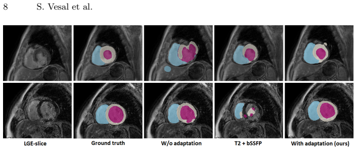
- [Figure 3 caption] The caption contains the typo 'withe color', which should be 'white color'.
- [Equation (1)] The soft Dice loss in Eq. (1) omits the conventional factor of 2 in the numerator, so a perfectly overlapping prediction gives a loss of 0.5 rather than 0. Please clarify whether this is intentional or a typographical error.
Circularity Check
No significant circularity: empirical transfer-learning evaluation against held-out annotations.
full rationale
The central result is an empirical Dice measurement on 40 LGE subjects using challenge ground truth (Table 2), not a derivation from an input. The comparison between fine-tuned and from-scratch networks is an experimental protocol: Section 2.4 describes 5-fold cross-validation on LGE subjects and Section 3 reports the resulting metrics. No equation in the paper defines LGE test performance in terms of the backbone training loss or the T2/bSSFP labels, and no fitted parameter is subsequently renamed as a prediction. The only self-citation is reference [13] for the architectural motif (dilated convolutions), which does not by itself force the reported Dice values. Concerns about validation-set model selection, a per-subject from-scratch baseline without standard deviation, and absent significance testing are statistical-reporting issues, not circularity. The evaluation is self-contained against external annotations, so the paper is not circular.
Assumptions & free parameters
free parameters (4)
- learning_rate =
0.0001
- batch_size =
16 (source), 4 (fine-tuning)
- input_crop_size =
224x224
- dilation_rates =
1 to 8
assumptions (3)
- domain assumption Features learned on T2 and bSSFP images are transferable to LGE images via fine-tuning.
- domain assumption The label space (LV, RV, Myo, background) is consistent across T2, bSSFP, and LGE sequences.
- standard math The multi-class soft Dice loss in Equation (1) is a valid proxy for segmentation quality.
Cite this review
Pith. "Pith review of Automated Multi-sequence Cardiac MRI Segmentation Using Supervised Domain Adaptation." pith.science (2026). https://pith.science/paper/XFSOCFEX
@misc{pith2026190807726,
  author       = {Pith},
  title        = {Pith review of: Automated Multi-sequence Cardiac MRI Segmentation Using Supervised Domain Adaptation},
  year         = {2026},
  howpublished = {\url{https://pith.science/paper/XFSOCFEX}},
  note         = {Machine review of arXiv:1908.07726}
}
abstract
Left ventricle segmentation and morphological assessment are essential for improving diagnosis and our understanding of cardiomyopathy, which in turn is imperative for reducing risk of myocardial infarctions in patients. Convolutional neural network (CNN) based methods for cardiac magnetic resonance (CMR) image segmentation rely on supervision with pixel-level annotations, and may not generalize well to images from a different domain. These methods are typically sensitive to variations in imaging protocols and data acquisition. Since annotating multi-sequence CMR images is tedious and subject to inter- and intra-observer variations, developing methods that can automatically adapt from one domain to the target domain is of great interest. In this paper, we propose an approach for domain adaptation in multi-sequence CMR segmentation task using transfer learning that combines multi-source image information. We first train an encoder-decoder CNN on T2-weighted and balanced-Steady State Free Precession (bSSFP) MR images with pixel-level annotation and fine-tune the same network with a limited number of Late Gadolinium Enhanced-MR (LGE-MR) subjects, to adapt the domain features. The domain-adapted network was trained with just four LGE-MR training samples and obtained an average Dice score of $\sim$85.0\% on the test set comprises of 40 LGE-MR subjects. The proposed method significantly outperformed a network without adaptation trained from scratch on the same set of LGE-MR training data.
Figures


Reference graph
Works this paper leans on
-
[6]
Zhuang, X.: Multivariate mixture model for cardiac segmentation from multi- sequence mri. In: MICCAI. (2016) 581–588
work page 2016
-
[1]
Journal of the American College of Cardiology 55(1) (2009) 1 – 16
Kim, H.W., Farzaneh-Far, A., Kim, R.J.: Cardiovascular magnetic resonance in patients with myocardial infarction: Current and emerging applications. Journal of the American College of Cardiology 55(1) (2009) 1 – 16
work page 2009
-
[2]
Group, E.S.D., Timmis, A., Wilkins, E., Wright, L., Townsend, N., Gale, C., Grobbee, R., Vos, R., Blum, M., Maniadakis, N., Vardas, P., Flather, M., Bax, J., Pinto, F.: European Society of Cardiology: Cardiovascular Disease Statistics
-
[3]
IEEE Transactions on Pattern Analysis and Machine Intelli- gence (2019) 1–1
Zhuang, X.: Multivariate mixture model for myocardial segmentation combining multi-source images. IEEE Transactions on Pattern Analysis and Machine Intelli- gence (2019) 1–1
work page 2019
-
[4]
European Heart Journal - Cardiovascular Imaging 17(1) (05 2015) 76–84
Hammer-Hansen, S., Bandettini, W.P., Hsu, L.Y., Leung, S.W., Shanbhag, S., Mancini, C., Greve, A.M., Kber, L., Thune, J.J., Kellman, P., Arai, A.E.: Mecha- nisms for overestimating acute myocardial infarct size with gadolinium-enhanced cardiovascular magnetic resonance imaging in humans: a quantitative and kinetic study. European Heart Journal - Cardiovas...
work page 2015
-
[5]
Com- puterized Medical Imaging and Graphics (59) (2017) 13–27
Kurzendorfer, T., Forman, C., Schmidt, M., Tillmanns, C., Maier, A., Brost, A.: Fully automatic segmentation of left ventricular anatomy in 3-DLGE-MRI. Com- puterized Medical Imaging and Graphics (59) (2017) 13–27
work page 2017
-
[7]
Computerized Medical Imaging and Graphics 71 (2019) 49 – 57
Liu, J., Xie, H., Zhang, S., Gu, L.: Multi-sequence myocardium segmentation with cross-constrained shape and neural network-based initialization. Computerized Medical Imaging and Graphics 71 (2019) 49 – 57
work page 2019
-
[8]
In: Machine Learning in Medical Imaging
Chen, C., Dou, Q., Chen, H., Heng, P.A.: Semantic-aware generative adversarial nets for unsupervised domain adaptation in chest x-ray segmentation. In: Machine Learning in Medical Imaging. (2018) 143–151
work page 2018
Show all 17 references
-
[9]
In: CVPR
Russo, P., Carlucci, F.M., Tommasi, T., Caputo, B.: From source to target and back: Symmetric bi-directional adaptive gan. In: CVPR. (June 2018)
2018
-
[10]
In: 2017 IEEE International Conference on Computer Vision (ICCV)
Motiian, S., Piccirilli, M., Adjeroh, D.A., Doretto, G.: Unified deep supervised domain adaptation and generalization. In: 2017 IEEE International Conference on Computer Vision (ICCV). (Oct 2017) 5716–5726
2017
-
[11]
IEEE Transactions on Knowl- edge and Data Engineering 22(10) (Oct 2010) 1345–1359
Pan, S.J., Yang, Q.: A survey on transfer learning. IEEE Transactions on Knowl- edge and Data Engineering 22(10) (Oct 2010) 1345–1359
2010
-
[12]
In: MICCAI 2017
Ghafoorian, M., Mehrtash, A., Kapur, T., Karssemeijer, N., Marchiori, E., Pesteie, M., Guttmann, C.R.G., de Leeuw, F.E., Tempany, C.M., van Ginneken, B., Fe- dorov, A., Abolmaesumi, P., Platel, B., Wells, W.M.: Transfer learning for domain adaptation in mri: Application in bra...
2017
-
[13]
In: STACOM
Vesal, S., Ravikumar, N., Maier, A.: Dilated convolutions in neural networks for left atrial segmentation in 3d gadolinium enhanced-mri. In: STACOM. (2018) 319–328
2018
-
[14]
In: CVPR
He, K., Zhang, X., Ren, S., Sun, J.: Deep residual learning for image recognition. In: CVPR. (June 2016) 770–778
2016
-
[15]
In: ICLR
Yu, F., Koltun, V.: Multi-scale context aggregation by dilated convolutions. In: ICLR. (2016)
2016
-
[16]
In: 2016 Fourth International Conference on 3D Vision (3DV)
Milletari, F., Navab, N., Ahmadi, S.: V-net: Fully convolutional neural networks for volumetric medical image segmentation. In: 2016 Fourth International Conference on 3D Vision (3DV). (Oct 2016) 565–571
2016
-
[2017]
European Heart Journal 39(7) (11 2017) 508–579
2017
Reviewed August 14, 2026 · model on record in the stance chip above.
Discussion (0). Continue with ORCID to comment.