REVIEW 2 major objections 5 minor 12 references
Optimal input configuration of dynamic contrast enhanced MRI in convolutional neural networks for liver segmentation
T0 review · 2 major / 5 minor · reviewed 2026-08-14 · deepseek-v4-flash
Pith's one-line read Stacking the six phases of a DCE-MR series as input channels improves CNN liver segmentation over single-phase or separate-phase inputs.
desk verdict Useful, honest feasibility study on input format for DCE-MRI liver segmentation; the channel-stacking result is plausible but the key comparison is confounded by weight sharing and the stats are thinner than the abstract claims. read the letter →
The pith
A machine-rendered reading of the paper's core claim, the machinery that carries it, and where it could break.
The reading
What carries the argument
The load-bearing object is the input tensor: six co-registered DCE-MR phase slices stacked as channels of a single image, so early convolutional kernels compare phase intensities at the same spatial location before any nonlinear feature extraction. Configuration II feeds the same six phases through the network separately and merges feature maps only at the second-to-last layer, with convolutional weights shared across phases. The authors argue that the channel version lets the network learn cross-phase relationships from raw MR intensities, which they identify as the likely reason for its advantage. The two architectures, a dilated fully convolutional network and a compact U-net, serve to show that the effect is not tied to a single network design.
What would settle it
Retrain configuration II on the same data with independently initialized convolutional weights for each of the six phases and compare Dice on the same test set; if per-phase weights erase the gap to configuration III, the central claim about input representation is not supported.
Extended reading notes
Core claim
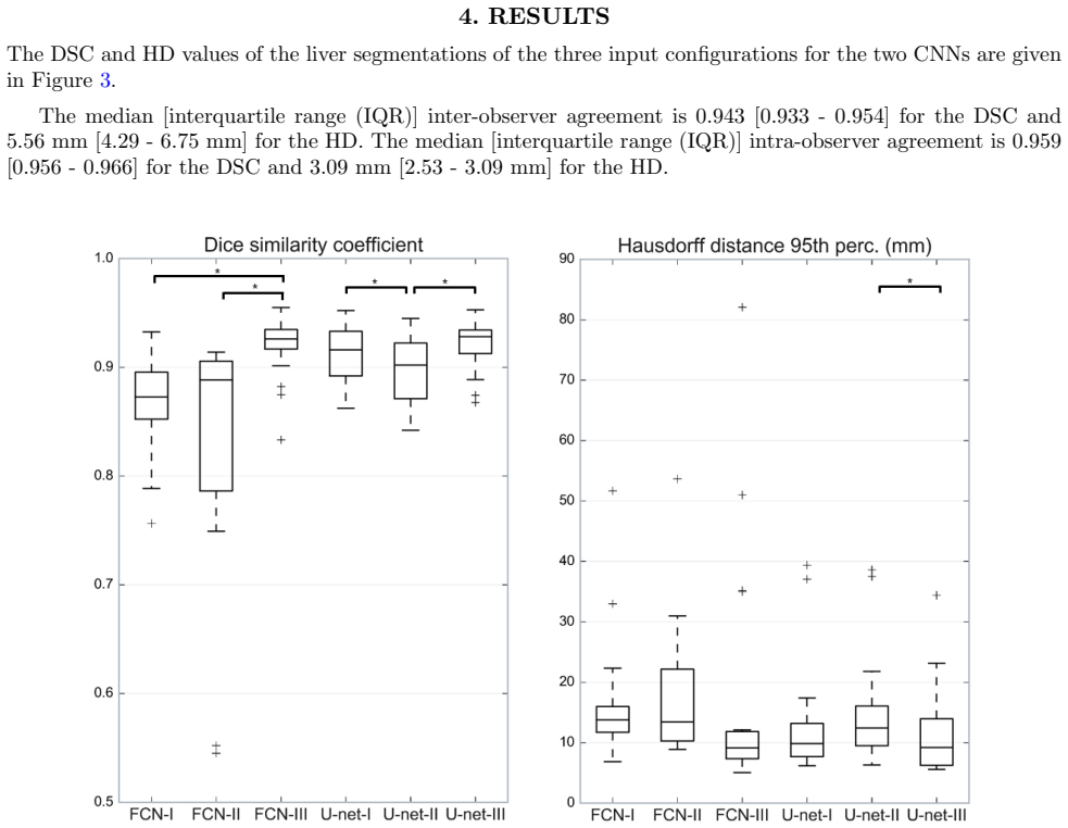
On the paper's own terms, the discovery is that collapsing the six DCE-MR phases into the channel dimension of a single input image is a better way to exploit dynamic contrast information for liver segmentation than either ignoring the dynamics (one phase) or delaying fusion until late feature maps (separate phases). Both the Dice similarity coefficient and the 95th-percentile Hausdorff distance favor configuration III, with significant paired differences over configuration II for both tested networks and over configuration I for the dilated FCN. The two networks did not differ significantly under configuration III, which the authors take as evidence that input configuration matters more than architecture. The authors also report that automatic segmentations remain significantly below inter- and intra-observer agreement, although a radiologist judged the best configuration's outlines satisfactory for further analysis.
Load-bearing premise
The main comparison assumes that running the six phases through shared convolutional weights is a fair way to implement 'separate phase input'; if the six phases instead had their own weights, separate-phase input might perform as well as channel-stacked input.
Editorial extensions
If this is right
- On the tested data, reformatting existing DCE-MR series as multi-channel input improves liver segmentation without any change to network architecture or training procedure.
- Because the two architectures behaved similarly under configuration III, input-format choice should be treated as a first-order design variable in similar segmentation studies.
- The channel-stacked configuration included most liver lesions, showing that the early-fusion input preserves functional contrast information useful beyond the organ boundary.
- Both networks still fall significantly short of human observer agreement, so the input-format gain alone does not close the gap to manual segmentation.
Reading between the lines
- A decisive test of the paper's interpretation would be to retrain configuration II with separate, per-phase convolutional weights; if it then matches configuration III, the reported advantage is a weight-sharing artifact rather than a property of channel input.
- Because the networks were trained on 2D slices and the authors note the concept extends to 3D, a 3D multi-channel version with all phases as channels of a volume is a natural next test that could sharpen or overturn the advantage.
- The same channel-stacking logic plausibly transfers to other multi-phase or multi-sequence imaging tasks, such as DCE-MRI kidney or tumor segmentation or CT perfusion, where early fusion of temporal phases may be broadly beneficial.
Signed reviews
Editorial analysis
A structured set of objections, weighed in public.
Referee Report
Summary. This paper compares three ways of presenting six-phase DCE-MRI data to convolutional neural networks for liver segmentation: (I) a single selected late-arterial-phase image, (II) the six phases processed separately through shared-weight networks and merged late with an extra 1×1 convolution, and (III) the six phases stacked as input channels. Two architectures are considered, a dilated FCN and a small U-net. Using 16 training, 3 validation, and 19 test DCE-MR series, the authors report that configuration III gives significantly higher DSC than configuration II for both networks and than configuration I for the dilated FCN, and that III gives significantly better HD than II in the U-net. They conclude that channel-stacked DCE-MR input is superior to single-phase or separate-phase input regardless of network architecture.
Significance. The question is practically relevant: if the result holds, it provides simple, actionable guidance for designing CNN inputs for DCE-MRI segmentation. The study has genuine strengths: two different architectures are compared, both DSC and a boundary metric are reported, manual annotations are independently generated and checked, and the paper is transparent about the training setup and the acknowledged limitations of configuration II. However, the central comparison is confounded by an asymmetry in the way configuration II and III allocate first-layer weights, and the statistical evidence is weaker than the abstract's categorical conclusion. With an additional ablation or a suitably narrowed claim, this could be a useful feasibility result; in its current form the main conclusion is not fully established.
major comments (2)
- [Section 5] The load-bearing comparison between configuration II (separate phase streams) and configuration III (phase channels) is confounded by weight sharing and first-layer parameter capacity. In configuration II, the same convolutional kernels are applied to every phase and the streams are merged only by a late 1×1 convolution; in configuration III, the first convolutional layer has six input channels and can learn phase-specific kernels at the earliest layer. For the dilated FCN, the first layer in III has 32×6×3×3 weights versus 32×1×3×3 for each stream in II, an excess of 1,440 weights; for the U-net the excess is 5×16×3×3 = 720 weights. The authors themselves state in Section 5 that shared weights in configuration II 'might not be the most optimal setup' and that separately optimizing kernel weights for each phase 'might result in better segmentation results.' The observed advantage of III over II could therefore reflect the addition of early phase-specific capacity or the suboptimal weight-sharing constraint, rather than the channel-input representation itself. I request an additional ablation, for example a separate-weight variant of configuration II or a parameter-matched early-fusion comparison, before the central claim is accepted.
- [Section 4] The statistical support is weaker than the abstract's claim that channel input 'performs better' than the other configurations. The test set has only 19 series; many pairwise comparisons are performed without correction for multiple testing; and several comparisons do not reach significance, including DSC I versus III for the U-net, and most Hausdorff-distance comparisons for the dilated FCN. No p-values or effect sizes are reported, so the reader cannot assess the magnitude or reliability of the differences. The authors should either restrict the conclusion to the comparisons that are significant, pre-specify the primary comparisons, or apply a multiple-comparison correction and report exact p-values.
minor comments (5)
- [Section 2.3] The loss formula is written as (2*X∩Y + s)/(X^2 + Y^2 + s); the denominator appears to be a typo for the sum of the volumes, X+Y, as used in the DSC definition. Please correct.
- [Section 3] The definition of the modified Hausdorff distance is garbled: 'K 95th ranked minimum of the minimum Euclidean distances' should be a directional 95th-percentile Hausdorff distance, and the formula has a misplaced subscript. Please rewrite.
- [Section 5] In the paragraph beginning 'The results did not show a significant difference', configuration III is referred to as configuration II; the text should say 'when using the six phases as channels input image (configuration III)'.
- [Section 2.1] The intensity normalization assumes that the 99.8th percentile corresponds to the contrast-agent peak in the aorta; this is an ad hoc assumption and should be explicitly treated as a free parameter whose influence on the comparison is untested.
- [Section 4] The boxplots in Figure 3 use asterisks without a table of p-values or a key identifying the exact pairs; a supplementary table with all pairwise p-values and effect sizes would improve the reproducibility of the statistical claims.
Circularity Check
No significant circularity: the study is an empirical comparison and its central claim is not derived from its inputs by construction.
full rationale
The paper reports an empirical comparison of three input configurations for CNNs in DCE-MRI liver segmentation. The central claim—that using the six phases as channels outperforms single-phase or separate-phase inputs—is established by training networks and statistically comparing segmentation metrics on a held-out test set. There is no derivation chain in which a predicted quantity is equivalent to a fitted input by definition. The only self-citation is reference [8], a previously published motion-correction method by overlapping authors, used as a preprocessing tool; it does not predefine the outcome of the configuration comparison and is not invoked to justify the main result. The authors also openly acknowledge a potential confound in configuration II, where convolutional kernel weights are shared across phases, noting this 'might not be the most optimal setup.' This is an experimental limitation and a possible alternative explanation for the observed difference, but it is not circularity: the comparison is not forced by the definitions of the configurations. The use of a Dice-like loss and Dice evaluation metric is a standard optimization/evaluation alignment; the paper explicitly notes this and still compares configurations under identical loss, so it does not reduce the conclusion to its inputs. Overall, the derivation is self-contained and the central claim has independent empirical content, so no circularity step is identified.
Assumptions & free parameters
free parameters (6)
- Late arterial phase selected for configuration I =
phase 2 (late arterial phase)
- Intensity normalization cutoff =
99.8th percentile
- Number of phases after averaging per breath hold =
6
- Loss smoothing constant s =
1e-5
- Post-processing threshold =
0.50
- Network hyperparameters =
learning rate 0.001, 500k iterations, batch size 1, kernel counts 32 (FCN) / 16-128 (U-net)
assumptions (5)
- domain assumption Manual liver annotations are accurate enough to serve as ground truth
- domain assumption The 99.8th percentile intensity corresponds to the contrast agent peak in the aorta
- domain assumption PCA-based groupwise registration adequately corrects motion in DCE-MRI
- domain assumption 2D slice-based training on 256x256 slices is representative of 3D liver segmentation
- standard math The paired t-test is valid for DSC values
Cite this review
Pith. "Pith review of Optimal input configuration of dynamic contrast enhanced MRI in convolutional neural networks for liver segmentation." pith.science (2026). https://pith.science/paper/FUKQ6HCS
@misc{pith2026190808251,
  author       = {Pith},
  title        = {Pith review of: Optimal input configuration of dynamic contrast enhanced MRI in convolutional neural networks for liver segmentation},
  year         = {2026},
  howpublished = {\url{https://pith.science/paper/FUKQ6HCS}},
  note         = {Machine review of arXiv:1908.08251}
}
read the original abstract
Most MRI liver segmentation methods use a structural 3D scan as input, such as a T1 or T2 weighted scan. Segmentation performance may be improved by utilizing both structural and functional information, as contained in dynamic contrast enhanced (DCE) MR series. Dynamic information can be incorporated in a segmentation method based on convolutional neural networks in a number of ways. In this study, the optimal input configuration of DCE MR images for convolutional neural networks (CNNs) is studied. The performance of three different input configurations for CNNs is studied for a liver segmentation task. The three configurations are I) one phase image of the DCE-MR series as input image; II) the separate phases of the DCE-MR as input images; and III) the separate phases of the DCE-MR as channels of one input image. The three input configurations are fed into a dilated fully convolutional network and into a small U-net. The CNNs were trained using 19 annotated DCE-MR series and tested on another 19 annotated DCE-MR series. The performance of the three input configurations for both networks is evaluated against manual annotations. The results show that both neural networks perform better when the separate phases of the DCE-MR series are used as channels of an input image in comparison to one phase as input image or the separate phases as input images. No significant difference between the performances of the two network architectures was found for the separate phases as channels of an input image.
Figures


Figures from the paper (2 more)


Reference graph
Works this paper leans on
-
[1]
Lopez-Mir, F., Naranjo, V., Angulo, J., Alca \ n iz, M., and Luna, L., `` Liver segmentation in MRI: A fully automatic method based on stochastic partitions ,'' Computer Methods and Programs in Biomedicine 114 (1), 11--28 (2014)
work page 2014
-
[2]
Huynh, H. T., Le-Trong, N., Bao, P. T., Oto, A., and Suzuki, K., `` Fully automated MR liver volumetry using watershed segmentation coupled with active contouring ,'' International Journal of Computer Assisted Radiology and Surgery 12 (2), 235--243 (2017)
work page 2017
-
[3]
Chartrand, G., Cresson, T., Chav, R., Gotra, A., Tang, A., and De Guise , J. A., `` Liver Segmentation on CT and MR Using Laplacian Mesh Optimization ,'' IEEE Transactions on Biomedical Engineering 64 (9), 2110--2121 (2017)
work page 2017
-
[4]
Masoumi, H., Behrad, A., Pourmina, M. A., and Roosta, A., `` Automatic liver segmentation in MRI images using an iterative watershed algorithm and artificial neural network ,'' Biomedical Signal Processing and Control 7 (5), 429--437 (2012)
work page 2012
-
[5]
(eds) Medical Image Computing and Computer-Assisted Intervention – MICCAI 2015
Ronneberger, O., Fischer, P., and Brox, T., `` U-Net: Convolutional networks for biomedical image segmentation ,'' in [ Navab N., Hornegger J., Wells W., Frangi A. (eds) Medical Image Computing and Computer-Assisted Intervention – MICCAI 2015. MICCAI 2015. Lecture Notes in Computer Science 0.1em ] , 9351 , 234--241 (2015)
work page 2015
-
[6]
Yu, F. and Koltun, V., `` Multi-Scale Context Aggregation by Dilated Convolutions ,'' in [ Fourth International Conference on Learning Representations 0.1em ] , (2016)
work page 2016
-
[7]
Litjens, G., Kooi, T., Bejnordi, B. E., Setio, A. A. A., Ciompi, F., Ghafoorian, M., Laak, J. A. W. M. V. D., Ginneken, B. V., and S \' a nchez, C. I., `` A survey on deep learning in medical image analysis ,'' Medical Image Analysis 42 , 60--88 (2017)
work page 2017
-
[8]
Jansen, M. J. A., Kuijf, H. J., Veldhuis, W. B., Wessels, F. J., van Leeuwen, M., and Pluim, J. P. W., `` Evaluation of motion correction for clinical dynamic contrast enhanced MRI of the liver ,'' Physics in Medicine & Biology 62 (19), 7556--7568 (2017)
work page 2017
Show all 12 references
-
[9]
Milletari, F., Navab, N., and Ahmadi, S.-A., `` V-Net: Fully Convolutional Neural Networks for Volumetric Medical Image Segmentation ,'' in [ 3D Vision (3DV), 2016 Fourth International Conference on 0.1em ] , 1--11 (2016)
2016
-
[10]
Glorot, X. and Bengio, Y., `` Understanding the difficulty of training deep feedforward neural networks ,'' in [ Proceedings of the 13th International Conference on Artificial Intelligence and Statistics (AISTATS) 2010 0.1em ] , 9 , 249--256 (2010)
2010
-
[11]
Jain, A., Duin, R., and Mao, J., `` Statistical pattern recognition: A review ,'' IEEE Transactions on Pattern Analysis and Machine Intelligence 22 (1), 4--37 (2000)
2000
-
[12]
write newline
" write newline "" before.all 'output.state := FUNCTION blank.sep after.quote 'output.state := FUNCTION fin.entry output.state after.quoted.block = 'skip 'add.period if write newline FUNCTION new.block output.state before.all = 'skip output.state after.quote = after.quoted.blo...
Reviewed August 14, 2026 · model on record in the stance chip above.
Discussion (0). Continue with ORCID to comment.