REVIEW 4 major objections 5 minor 18 references
Automatic Rodent Brain MRI Lesion Segmentation with Fully Convolutional Networks
T0 review · 4 major / 5 minor · reviewed 2026-08-14 · deepseek-v4-flash
Pith's one-line read RatLesNet, a 3D fully convolutional network with 0.37M parameters, segments ischemic stroke lesions in rat brain MRI with an average Dice of 0.88 in five-fold cross-validation, outperforming 3D U-Net and VoxResNet, and reaches 0.79 in…
desk verdict A compact 3D FCN for rat lesion segmentation that is genuinely useful; the main superiority claim needs significance testing. read the letter →
The pith
A machine-rendered reading of the paper's core claim, the machinery that carries it, and where it could break.
The reading
What carries the argument
The argument is carried by RatLesNet's dense-block architecture: within each block, every $3\times3\times3$ convolution is concatenated with all preceding feature maps in that block (growth rate 18), $1\times1\times1$ convolutions reset the channel count between blocks, and max-pooling with index-preserving unpooling halves and restores spatial dimensions. This lets the network see the full 3D volume while keeping parameters at 0.37M, which the paper says reduces overfitting relative to 3D U-Net's 19M and VoxResNet's 1.5M parameters. Training uses raw standardized T2 volumes with a mini-batch of 1, cross-entropy loss, and early stopping on validation loss; inference takes about half a second per scan.
What would settle it
Re-run the five-fold cross-validation using a second independent operator's manual segmentations as ground truth; if RatLesNet's average Dice falls to the measured inter-operator level of about 0.73, the reported 0.88 reflects the reference annotator's style rather than true lesion boundaries.
Extended reading notes
Core claim
The central discovery is that a compact dense-block architecture designed for the specific geometry of rodent stroke lesions outperforms larger networks built for anatomical segmentation, despite making no use of preprocessing, skull-stripping, or post-processing. RatLesNet processes each whole $256\times256\times18$ volume with $1\times1\times1$ convolutions for channel adjustment, dense blocks with growth rate 18, and max-pooling/unpooling that reuses pooling indices to restore spatial resolution. In five-fold cross-validation on the 02NOV2016 study, the paper reports an average Dice of 0.88 (0.76 excluding lesion-free sham animals), above the inter-operator Dice of 0.87 (0.73 excluding shams); on generalization to three other studies it averages 0.79, with the best scores on 24-hour post-stroke scans and a lower 0.68 on the 35-day study, where VoxResNet scores 0.71. The paper frames this as the first fully convolutional network specifically designed for rat brain lesion segmentation, with an architecture that accepts any input-channel count and therefore extends to multimodal MRI.
Load-bearing premise
The manual segmentations used for training and evaluation are treated as ground truth, and the four studies are assumed similar enough to transfer; if the reference labels carry one annotator's bias or the studies differ too much, the reported Dice gaps do not show true segmentation accuracy.
Editorial extensions
If this is right
- Preclinical stroke pipelines can replace manual tracing with fully automatic RatLesNet segmentation on raw T2-weighted MRI, eliminating the skull-stripping, bias correction, and hole-filling steps that earlier rodent lesion tools require.
- A whole-volume 3D network with 0.37M parameters trains in about six hours on one GPU and segments a scan in about half a second, making segmentation of hundreds of animals practical.
- Because the input channel count is configurable, the same architecture can be applied to multimodal or multi-sequence MRI without structural changes.
- On 24-hour post-stroke lesions from unseen studies, RatLesNet's average Dice (0.82 and 0.84) exceeds the measured inter-operator agreement of 0.73, indicating automatic segmentations can be at least as reproducible as human ones for that stage.
- The lower Dice of 0.68 on the 35-day study shows the generalization is lesion-stage dependent; adding chronic-stage lesions to training is the direct extension the paper acknowledges.
Reading between the lines
- The paper suggests the parameter gap explains the accuracy gap, but it does not isolate capacity from architecture; a controlled comparison that matches parameter budgets across designs would test that explanation.
- Because the network is trained against a single operator's labels and evaluated on that same reference, the 0.88 Dice may partly reflect learning that operator's delineation style; using an ensemble of expert segmentations as training targets would probe this.
- The dense-block plus $1\times1\times1$ bottleneck pattern that works here for rare lesion voxels is a candidate for other small-lesion or small-animal segmentation tasks where datasets are too small for large 3D networks.
- Adding chronic-stage training examples and a scanner-matched validation set would likely raise the 35-day study's Dice above VoxResNet's 0.71, since the training study contains only 2-hour and 24-hour lesions.
Signed reviews
Editorial analysis
A structured set of objections, weighed in public.
Referee Report
Summary. The paper proposes RatLesNet, a compact 3D fully convolutional network (0.37M parameters) for automatic segmentation of ischemic lesions in T2-weighted rat brain MRI. It is trained and evaluated on 131 scans from four tMCAO studies. In a five-fold cross-validation experiment on one study (02NOV2016), the authors report an average Dice of 0.88, exceeding 3D U-Net (0.64) and VoxResNet (0.85) when all animals are included, and 0.76 vs 0.70 and 0.30 respectively when sham animals are excluded. In a cross-study generalization test, training on 02NOV2016 and testing on the other three studies yields average Dice of 0.79 for RatLesNet, 0.76 for VoxResNet, and 0.62 for 3D U-Net. The authors also claim that RatLesNet exceeds inter-operator variability (average Dice 0.87 including shams). The paper emphasizes that the method requires no skull-stripping, bias-field correction, or post-processing.
Significance. If the reported performance holds, RatLesNet would be a practically valuable tool for preclinical stroke research, where manual segmentation is time-consuming and subjective. The evaluation design is a strength: it includes a multi-study dataset, a realistic cross-study generalization test, and comparisons against two strong 3D FCN baselines. The architecture's small parameter count and ability to process entire volumes without preprocessing are also attractive for reproducibility and clinical translation. However, the central claim of superiority over VoxResNet rests on small average Dice differences with overlapping standard deviations and no statistical testing, and the cross-study result reverses on one of the three test studies. The evidence is promising but not yet conclusive.
major comments (4)
- [Section 3, Table 1 (Cross-validation)] No statistical significance testing or confidence intervals are provided for the comparison between RatLesNet and VoxResNet. The differences are small relative to the reported variability: for example, at 2h without shams, RatLesNet gives 0.67±0.12 vs VoxResNet 0.60±0.16; at 24h without shams, 0.85±0.11 vs 0.79±0.20; and on average without shams, 0.76±0.14 vs 0.70±0.20. Since the same test folds are used for all methods, a paired per-animal test (e.g., Wilcoxon signed-rank test) or bootstrap confidence intervals on the paired differences is needed to support the abstract's claim that RatLesNet is 'quantitatively better' than the compared architectures.
- [Section 2 and Section 3, Table 1 (Inter-operator comparison)] The statement that RatLesNet 'achieves higher Dice coefficients than inter-operator variability' is not supported. The average Dice including sham animals is 0.88 for RatLesNet vs 0.87 for inter-operator agreement, a difference of 0.01; excluding shams it is 0.76 vs 0.73. Moreover, the inter-operator benchmark is based on only one additional manual segmentation of a single study, and RatLesNet is trained on the reference annotator's labels. Exceeding that benchmark may therefore reflect learning the reference annotator's segmentation style rather than improved biological accuracy. The authors should qualify this claim and, if possible, evaluate against multiple independent raters.
- [Section 3, Table 2 (Generalization capability)] The cross-study generalization claim is not robust across studies. RatLesNet outperforms VoxResNet by large margins on 03MAY2016 (0.82 vs 0.77) and 02OCT2017 (0.84 vs 0.78), but on 03AUG2015 VoxResNet achieves the highest Dice (0.71±0.23 vs 0.68±0.26 for RatLesNet). Because no significance testing is provided, the overall claim that RatLesNet 'outperformed the other FCNs at generalizing' rests on averaging over studies and may depend on the particular composition of the test set. The authors should report per-study pairwise statistical comparisons and discuss the time-point difference (35 days for 03AUG2015 vs 2h/24h in the training data) as a likely source of the reversal.
- [Section 3, Cross-validation paragraph] The sentence 'The models that provided the reported results were trained with the best performing learning rate found' is ambiguous and potentially consequential. If the learning rate was selected using the held-out test studies, the generalization results would be invalid. If it was selected using the validation folds inside the cross-validation procedure, this should be stated explicitly. Please clarify the model-selection protocol.
minor comments (5)
- [Abstract] The phrase 'between 3.7% and 38% higher' is ambiguous because Dice improvements can be reported as absolute percentage points or relative percentages. Please specify which convention is used.
- [Section 2 and Table 1] The inter-operator Dice of 0.73 reported in the text is inconsistent with the 'Inter-operator' column in Table 1, which shows an average of 0.87 when shams are included. Please clarify how sham animals (no lesion) enter the inter-operator Dice computation, e.g., whether they are assigned Dice 1 by convention.
- [Section 2, Training] The text states that Adam was used 'with a starting learning rate of 10^-5', but later the models are said to be trained with the 'best performing learning rate found'. Please state the final learning rate(s) used and the search procedure.
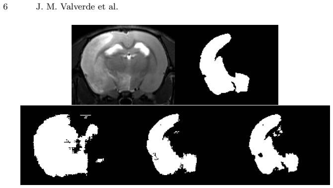
- [Figure 2] The architecture diagram is difficult to interpret: the number of channels at each stage and the positions of the 1x1 convolution bottleneck layers are not annotated. Adding these details would improve reproducibility.
- [Section 3, Figure 3 caption] The phrase 'crispier' in the text describing VoxResNet's predictions is informal; consider using 'sharper' or 'more clearly defined'.
Circularity Check
No circularity: empirical segmentation evaluation against held-out labels and external baselines.
full rationale
RatLesNet is an empirical architecture paper: the network is trained on manual lesion labels and evaluated against held-out labels via 5-fold cross-validation and on three studies not used for training. No network parameter is fitted from the test labels, and the test labels serve only as comparison targets for Dice scoring. The compared methods (3D U-Net and VoxResNet) are external architectures, and the network design is not derived from the target segmentation labels. The reported inter-operator Dice of 0.73 is a descriptive reference for human variability, not an input to the loss function or a fitted target. There are no equations in which a predicted quantity reduces to an input by construction, and no load-bearing self-citation chain is present. The lack of statistical significance testing is a methodological concern about whether the superiority claim is established, not evidence of circularity. The derivation and evaluation chain is therefore self-contained with respect to the paper's core empirical claims.
Assumptions & free parameters
free parameters (4)
- growth rate =
18
- initial learning rate =
1e-5 for RatLesNet; 'best performing' for baselines
- early stopping patience =
5 epochs
- maximum training epochs =
1000
assumptions (3)
- domain assumption The provided manual lesion segmentations are accurate enough to serve as ground truth for training and evaluation.
- domain assumption The four studies are comparable enough for cross-study generalization testing.
- domain assumption Dice coefficient is an appropriate evaluation metric for lesion segmentation with class imbalance.
Cite this review
Pith. "Pith review of Automatic Rodent Brain MRI Lesion Segmentation with Fully Convolutional Networks." pith.science (2026). https://pith.science/paper/VVZAY7Q5
@misc{pith2026190808746,
  author       = {Pith},
  title        = {Pith review of: Automatic Rodent Brain MRI Lesion Segmentation with Fully Convolutional Networks},
  year         = {2026},
  howpublished = {\url{https://pith.science/paper/VVZAY7Q5}},
  note         = {Machine review of arXiv:1908.08746}
}
read the original abstract
Manual segmentation of rodent brain lesions from magnetic resonance images (MRIs) is an arduous, time-consuming and subjective task that is highly important in pre-clinical research. Several automatic methods have been developed for different human brain MRI segmentation, but little research has targeted automatic rodent lesion segmentation. The existing tools for performing automatic lesion segmentation in rodents are constrained by strict assumptions about the data. Deep learning has been successfully used for medical image segmentation. However, there has not been any deep learning approach specifically designed for tackling rodent brain lesion segmentation. In this work, we propose a novel Fully Convolutional Network (FCN), RatLesNet, for the aforementioned task. Our dataset consists of 131 T2-weighted rat brain scans from 4 different studies in which ischemic stroke was induced by transient middle cerebral artery occlusion. We compare our method with two other 3D FCNs originally developed for anatomical segmentation (VoxResNet and 3D-U-Net) with 5-fold cross-validation on a single study and a generalization test, where the training was done on a single study and testing on three remaining studies. The labels generated by our method were quantitatively and qualitatively better than the predictions of the compared methods. The average Dice coefficient achieved in the 5-fold cross-validation experiment with the proposed approach was 0.88, between 3.7% and 38% higher than the compared architectures. The presented architecture also outperformed the other FCNs at generalizing on different studies, achieving the average Dice coefficient of 0.79.
Figures


Reference graph
Works this paper leans on
-
[1]
In: 12th {USENIX} Symposium on Operating Systems Design and Im- plementation ({OSDI} 16)
Abadi, M., Barham, P., Chen, J., Chen, Z., Davis, A., Dean, J., Devin, M., Ghe- mawat, S., Irving, G., Isard, M., et al.: Tensorflow: A system for large-scale machine learning. In: 12th {USENIX} Symposium on Operating Systems Design and Im- plementation ({OSDI} 16). pp. 265–283 (2016)
2016
-
[2]
IEEE transactions on medical imaging 37(7), 1678–1689 (2018)
Arnaud, A., Forbes, F., Coquery, N., Collomb, N., Lemasson, B., Barbier, E.L.: Fully automatic lesion localization and characterization: Application to brain tu- mors using multiparametric quantitative mri data. IEEE transactions on medical imaging 37(7), 1678–1689 (2018)
work page 2018
-
[3]
NeuroImage 170, 446–455 (2018)
Chen, H., Dou, Q., Yu, L., Qin, J., Heng, P.A.: Voxresnet: Deep voxelwise residual networks for brain segmentation from 3d mr images. NeuroImage 170, 446–455 (2018)
work page 2018
-
[4]
Journal of Cerebral Blood Flow & Metabolism 38(8), 1371–1383 (2018)
Choi, C.H., Yi, K.S., Lee, S.R., Lee, Y., Jeon, C.Y., Hwang, J., Lee, C., Choi, S.S., Lee, H.J., Cha, S.H.: A novel voxel-wise lesion segmentation technique on 3.0-t diffusion mri of hyperacute focal cerebral ischemia at 1 h after permanent mcao in rats. Journal of Cerebral Blood Flow & Metabolism 38(8), 1371–1383 (2018)
work page 2018
-
[5]
In: International conference on medical image computing and computer-assisted intervention
C ¸ i¸ cek,¨O., Abdulkadir, A., Lienkamp, S.S., Brox, T., Ronneberger, O.: 3d u-net: learning dense volumetric segmentation from sparse annotation. In: International conference on medical image computing and computer-assisted intervention. pp. 424–432. Springer (2016) 8 J. M. Valverde et al
work page 2016
-
[6]
Journal of neuroscience methods (2019)
De Feo, R., Giove, F.: Towards an efficient segmentation of small rodents brain: a short critical review. Journal of neuroscience methods (2019)
work page 2019
-
[7]
Dice, L.R.: Measures of the amount of ecologic association between species. Ecology 26(3), 297–302 (1945)
work page 1945
-
[8]
Medical image analysis 35, 18–31 (2017)
Havaei, M., Davy, A., Warde-Farley, D., Biard, A., Courville, A., Bengio, Y., Pal, C., Jodoin, P.M., Larochelle, H.: Brain tumor segmentation with deep neural net- works. Medical image analysis 35, 18–31 (2017)
2017
Show all 18 references
-
[9]
In: Proceedings of the IEEE conference on computer vision and pattern recognition
He, K., Zhang, X., Ren, S., Sun, J.: Deep residual learning for image recognition. In: Proceedings of the IEEE conference on computer vision and pattern recognition. pp. 770–778 (2016)
2016
-
[10]
In: Proceedings of the IEEE conference on computer vision and pattern recognition
Huang, G., Liu, Z., Van Der Maaten, L., Weinberger, K.Q.: Densely connected convolutional networks. In: Proceedings of the IEEE conference on computer vision and pattern recognition. pp. 4700–4708 (2017)
2017
-
[11]
arXiv preprint arXiv:1412.6980 (2014)
Kingma, D.P., Ba, J.: Adam: A method for stochastic optimization. arXiv preprint arXiv:1412.6980 (2014)
2014 arXiv
-
[12]
Koizumi, J., Yoshida, Y., Nakazawa, T., Ooneda, G.: Experimental studies of is- chemic brain edema. 1. a new experimental model of cerebral embolism in rats in which recirculation can be introduced in the ischemic area. Jpn J stroke 8, 1–8 (1986)
1986
-
[13]
Frontiers in neuroinformatics 11, 3 (2017)
Mulder, I.A., Khmelinskii, A., Dzyubachyk, O., de Jong, S., Rieff, N., Wermer, M.J., Hoehn, M., Lelieveldt, B.P., van den Maagdenberg, A.M.: Automated is- chemic lesion segmentation in mri mouse brain data after transient middle cerebral artery occlusion. Frontiers in neuroinfo...
2017
-
[14]
In: International MICCAI Brainlesion Workshop
Myronenko, A.: 3d mri brain tumor segmentation using autoencoder regularization. In: International MICCAI Brainlesion Workshop. pp. 311–320. Springer (2018)
2018
-
[15]
In: Proceedings of the IEEE international conference on computer vision
Noh, H., Hong, S., Han, B.: Learning deconvolution network for semantic segmen- tation. In: Proceedings of the IEEE international conference on computer vision. pp. 1520–1528 (2015)
2015
-
[16]
In: International Conference on Medical image computing and computer-assisted intervention
Ronneberger, O., Fischer, P., Brox, T.: U-net: Convolutional networks for biomedi- cal image segmentation. In: International Conference on Medical image computing and computer-assisted intervention. pp. 234–241. Springer (2015)
2015
-
[17]
NeuroImage 186, 713–727 (2019)
Roy, A.G., Conjeti, S., Navab, N., Wachinger, C., Initiative, A.D.N., et al.: Quick- nat: A fully convolutional network for quick and accurate segmentation of neu- roanatomy. NeuroImage 186, 713–727 (2019)
2019
-
[18]
In: 2018 IEEE 15th International Symposium on Biomedical Imaging (ISBI 2018)
Roy, S., Knutsen, A., Korotcov, A., Bosomtwi, A., Dardzinski, B., Butman, J.A., Pham, D.L.: A deep learning framework for brain extraction in humans and an- imals with traumatic brain injury. In: 2018 IEEE 15th International Symposium on Biomedical Imaging (ISBI 2018). pp. 687...
2018
Reviewed August 14, 2026 · model on record in the stance chip above.
Discussion (0). Continue with ORCID to comment.