REVIEW 3 major objections 3 minor 16 references
Accelerated Motion-Aware MR Imaging via Motion Prediction from K-Space Center
T0 review · 3 major / 3 minor · reviewed 2026-08-14 · deepseek-v4-flash
Pith's one-line read A motion-aware 4D MRI method learns breathing motion from a short initial k-space-center training phase and then predicts motion from tiny center patches, cutting acquisition time almost in half and reconstruction from 2 hours to 3…
desk verdict Practical acceleration of motion-aware 4D MRI with real time savings, but the key prediction-generalization and quality claims are not causally validated. read the letter →
The pith
A machine-rendered reading of the paper's core claim, the machinery that carries it, and where it could break.
The reading
What carries the argument
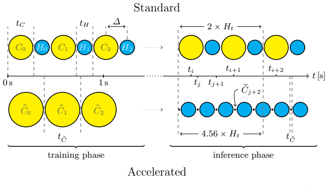
The central mechanism is a learned motion-prediction model built from k-space center patches. In a training phase, full-size center patches are converted to motion fields by non-rigid image registration; these motion fields are then regressed, using a cubic polynomial in the PCA scores of tiny center patches, onto the tiny patches that will be sampled during the accelerated scan. The predicted motion fields are applied to correct peripheral k-space patches before accumulation, and a quadratic-interpolation shift correction with shift $\Delta = 0.5$ compensates for the systematic temporal offset between center and peripheral patch sampling. This model is what lets the sequence replace most full-center measurements with tiny patches, producing the time savings.
What would settle it
Scan a volunteer under free breathing with the accelerated sequence, then have them change breathing depth or cough after the 100-time-point training phase, and compute the motion prediction error against motion fields recovered from full center patches. If the 95th-percentile error consistently exceeds the roughly 4 mm observed in the paper, or if the reconstructed images show visible blurring at the diaphragm, the assumption that trained motion generalizes to later time points is falsified.
Extended reading notes
Core claim
On its own terms, the paper establishes that the motion fields needed for motion-compensated 4D MRI can be inferred from a small subset of k-space center data. During an initial training phase of 100 time points, about 76 seconds, full center patches are acquired and registered to recover motion fields; a cubic regression model is then fit between PCA scores of tiny center patches and these motion fields. In the inference phase, only tiny center patches are sampled and the model predicts the non-rigid motion for the remaining 1400 time points, allowing the peripheral k-space data to be spatially corrected. The paper further introduces a systematic temporal shift correction, shifting motion fields by half a time step using quadratic interpolation to account for the delay between center and peripheral patch acquisition. Experiments on 12 free-breathing volunteers show prediction errors below 2 mm on average and qualitatively equivalent or sharper reconstructions than the standard 11-minute approach.
Load-bearing premise
The load-bearing premise is that the motion observed during the initial training phase is representative of the motion that will occur throughout the rest of the scan; if breathing amplitude, phase, or organ drift changes afterwards, the predicted motion fields will be wrong.
Editorial extensions
If this is right
- Acquisition time for the motion-aware 4D sequence drops from 11.1 to 5.8 minutes, making the protocol more feasible for routine clinical scans of thorax and abdomen.
- Reconstruction time drops from about 2 hours to 3 minutes because full 3D image registration is only needed during the 100-time-point training phase.
- The shift correction increases image sharpness: average total variation rises significantly ($p=0.002$, Cohen's $d=0.86$) across the 12 standard and 6 accelerated acquisitions.
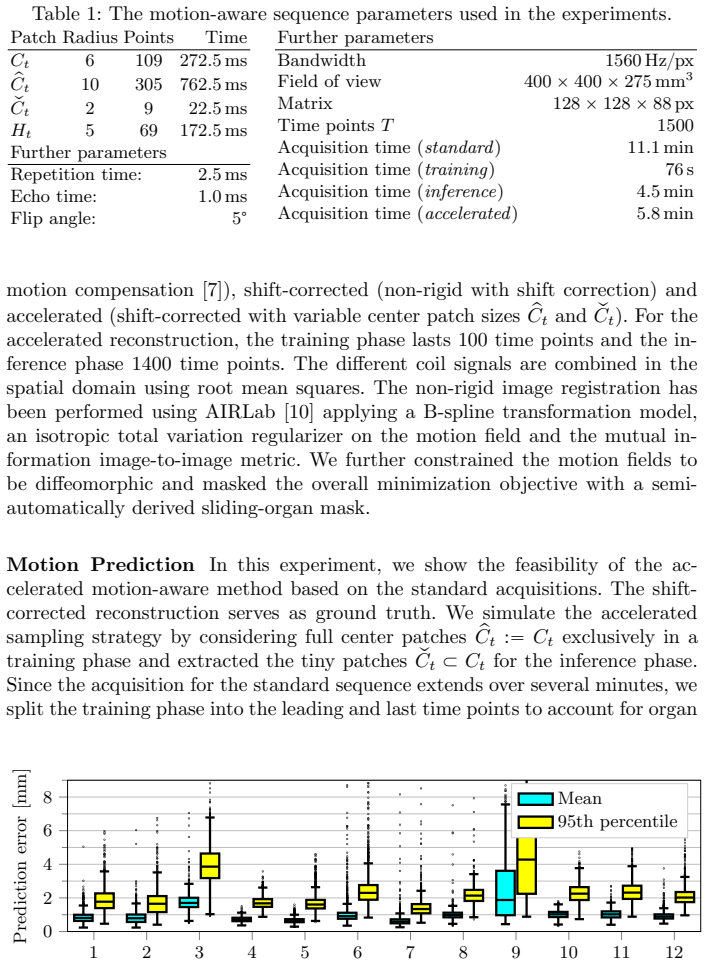
- Motion prediction accuracy stays below 2 mm on average, so the method can track respiratory motion with sub-voxel precision in free-breathing volunteers.
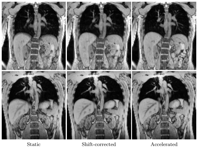
- Because the accelerated method yields equivalent or higher reconstruction quality in less time, motion-aware 4D MRI becomes a plausible option for time-resolved imaging without gating or binning.
Reading between the lines
- The same train-then-predict structure could be extended to cardiac or combined respiratory-cardiac motion, provided a reliable periodic trigger or a longer training phase captures the faster dynamics; the paper only demonstrates respiratory motion.
- The model's reliance on a fixed training phase suggests an online adaptation scheme, periodically re-estimating the regression weights from recently acquired center patches, could make the method robust to drift and amplitude changes without lengthening the scan.
- A direct testable extension would be to train on one volunteer and predict motion for a different scan session or for instructed deep-breathing phases; the paper's training setup uses the same session, so cross-session generalization is not established.
Signed reviews
Editorial analysis
A structured set of objections, weighed in public.
Referee Report
Summary. The manuscript proposes an accelerated version of a motion-aware 4D MR imaging method. In the standard method, the k-space center is sampled repeatedly to estimate non-rigid motion fields that correct peripheral k-space patches, but this requires long acquisition and reconstruction times. The proposed method adds an initial training phase in which larger center patches are acquired, learns a cubic regression model from PCA scores of these patches to motion fields, and during the inference phase predicts motion from much smaller center patches. This reduces the acquisition time from 11.1 min to 5.8 min and the reconstruction time from 2 h to 3 min. The authors also introduce a systematic temporal shift correction based on quadratic interpolation of the motion fields. Experiments on 12 volunteers (6 with the accelerated sequence) report average motion-prediction errors below 2 mm and qualitative reconstruction results that are claimed to be equivalent or better than the standard approach.
Significance. If the claimed gains are validated, the contribution is practically important: a roughly two-fold reduction in acquisition time and two-orders-of-magnitude reduction in reconstruction time would make motion-aware 4D abdominal/thoracic MRI substantially more clinically usable. The acquisition-time and reconstruction-time reductions are precisely stated and consistent with the sequence parameters in Table 1. The shift-correction idea is clearly presented and its quantitative TV-based evaluation is a useful first step. However, the two load-bearing claims, that motion can be predicted forward in time after the training phase and that reconstruction quality is equivalent or higher than the standard method, are not established by the reported experiments. The paper does not provide code or a fully reproducible pipeline, but the experimental setup is described in sufficient detail that the missing validation could in principle be added.
major comments (3)
- [§4, 'Motion Prediction'] The motion-prediction validation does not test the deployed accelerated protocol. The text states that 'we split the training phase into the leading and last time points to account for organ drift,' so the reported sub-2 mm mean error in Figure 3 is an interpolation result: the model has seen data from both ends of the time series. The actual accelerated sequence trains only on the first 100 time points and predicts the remaining 1400 time points (Table 1), which is forward extrapolation. The paper's own conclusion concedes that 'changes in amplitude of the motion after the training phase may compromise the motion prediction though,' and Volunteer 9 is reported to show exactly such a change. As it stands, the motion-prediction claim is therefore not causally validated for the proposed acquisition workflow.
- [§4, 'Accelerated Motion-Aware MR Imaging'] The abstract's claim of 'equivalent to higher reconstruction quality' is not supported by a quantitative or controlled comparison. The text explicitly says 'we cannot make a direct comparison between the shift-corrected and accelerated reconstruction method because they are applied to different acquisitions,' and Figures 4 and 5 are selected qualitative slices. The only quantitative image-quality metric, total variation, is used exclusively in the shift-correction experiment, not in the accelerated-versus-standard comparison. A valid evaluation would need, for example, a forward-split simulation on the standard acquisitions, a quantitative sharpness/artifact metric on the accelerated acquisitions, or a blinded reader study; none is provided.
- [§3.1, Eq. (2)] The regression model in Eq. (2) is trained and evaluated on the same acquisition, so the description as 'motion prediction' overstates what is demonstrated. The training phase provides both the PCA basis and the least-squares weights Ψ for a single subject's scan, and the reported error is computed after training on both temporal ends of that same acquisition. The manuscript should clearly state that the model is a per-acquisition fit and that no cross-subject or cross-session generalization is claimed; the current wording implies a more general predictive model than the experiments establish.
minor comments (3)
- [Table 1] The relation between the training-phase duration (76 s), inference-phase duration (4.5 min), and total accelerated acquisition time (5.8 min) is consistent, but a sentence spelling out that the accelerated time includes the training phase would help readers avoid misreading the 5.8 min as purely inference time.
- [§4, 'Systematic Shift Correction'] The TV-based evaluation is reported as a statistically significant increase with a large effect size, but TV is a proxy for sharpness and can increase with noise or artifacts; a brief discussion of this limitation, or an additional metric, would strengthen the claim that the shift correction improves image quality.
- [§5] There is a typo in the first sentence: 'anaccelerated' should be 'an accelerated'. Also, the discussion of amplitude changes affecting prediction would benefit from a reference to the quantitative error observed for Volunteer 9 in Figure 3.
Circularity Check
The reported sub-2 mm motion-prediction error is measured after training on both leading and last time points, so it is an interpolation fit rather than the forward extrapolation actually used by the accelerated acquisition; the central generalization claim is therefore not independently tested.
-
fitted input called prediction
[Section 4, 'Motion Prediction' experiment; Table 1; Section 5 conclusion]
"For the accelerated reconstruction, the training phase lasts 100 time points and the inference phase 1400 time points. ... Since the acquisition for the standard sequence extends over several minutes, we split the training phase into the leading and last time points to account for organ drift."
The reported prediction error (Figure 3) is obtained with a model trained on both the first and the last time points of the same standard acquisition, so the inference phase lies temporally between two fitted training blocks. In the accelerated sequence actually deployed, the model is trained only on the first 100 time points and must extrapolate 1400 points forward. The validation therefore measures interpolation with both anchors, not the forward extrapolation used by the method; for organ drift the error is partly forced by having seen the endpoint. The paper's own conclusion concedes: 'Changes in amplitude of the motion after the training phase may compromise the motion prediction though.' The central generalization claim is thus not causally tested.
full rationale
The method itself is a supervised regression: PCA-compressed k-space-center patches of the initial phase are used to learn a cubic model for the motion fields, and that regression is subsequently applied to tiny center patches. That is a legitimate fitting procedure, not circular by construction. The 2x acquisition-time reduction is fixed by the sequence design and is independent of prediction accuracy. The circular weakness is in the evaluation of the prediction claim: the motion-prediction experiment in Section 4 departs from the deployed accelerated protocol by including the last time points in the training set 'to account for organ drift.' Consequently, the reported sub-2 mm error is an interpolation result, not the forward-extrapolation result that the accelerated acquisition requires. The paper also states that direct quantitative comparison between the shift-corrected and accelerated reconstructions is impossible because they are applied to different acquisitions, so the equivalent-or-higher reconstruction quality claim rests on qualitative inspection. Those evaluation gaps concern the central accuracy/generalization claims, but they are not definitional circularity of the equations. No load-bearing self-citation or uniqueness-theorem smuggling is present; the prior work [7] serves as a baseline, not as justification of the prediction. Hence a partial-circularity score of 6 is appropriate, reflecting that the headline prediction performance is effectively an in-sample/interpolation fit rather than a validated extrapolation.
Assumptions & free parameters
free parameters (5)
- Regression weight matrix Psi and PCA basis =
least-squares fit on training data
- dpca =
10
- training phase length =
100 time points (76 s)
- tiny center patch radius qCt =
2 (9 k-space points)
- training center patch radius pCt =
10 (305 k-space points)
assumptions (6)
- domain assumption The motion occurring during the acquisition of a single k-space patch is negligible.
- domain assumption Motion estimated from the k-space center patch is representative of motion affecting the peripheral patches.
- domain assumption The breathing motion seen in the initial training phase is representative of the motion during the inference phase.
- domain assumption Total variation is a valid proxy for reconstruction sharpness and quality.
- domain assumption Image registration with B-splines, mutual information, and a sliding-organ mask yields accurate motion fields from the training centers.
- standard math The second-order Taylor expansion with central-difference derivatives approximates the true temporal shift of the motion field.
Cite this review
Pith. "Pith review of Accelerated Motion-Aware MR Imaging via Motion Prediction from K-Space Center." pith.science (2026). https://pith.science/paper/DL67DEAV
@misc{pith2026190809560,
  author       = {Pith},
  title        = {Pith review of: Accelerated Motion-Aware MR Imaging via Motion Prediction from K-Space Center},
  year         = {2026},
  howpublished = {\url{https://pith.science/paper/DL67DEAV}},
  note         = {Machine review of arXiv:1908.09560}
}
read the original abstract
Motion has been a challenge for magnetic resonance (MR) imaging ever since the MR has been invented. Especially in volumetric imaging of thoracic and abdominal organs, motion-awareness is essential for reducing motion artifacts in the final image. A recently proposed MR imaging approach copes with motion by observing the motion patterns during the acquisition. Repetitive scanning of the k-space center region enables the extraction of the patient motion while acquiring the remaining part of the k-space. Due to highly redundant measurements of the center, the required scanning time of over 11 min and the reconstruction time of 2 h exceed clinical applicability though. We propose an accelerated motion-aware MR imaging method where the motion is inferred from small-sized k-space center patches and an initial training phase during which the characteristic movements are modeled. Thereby, acquisition times are reduced by a factor of almost 2 and reconstruction times by two orders of magnitude. Moreover, we improve the existing motion-aware approach with a systematic temporal shift correction to achieve a sharper image reconstruction. We tested our method on 12 volunteers and scanned their lungs and abdomen under free breathing. We achieved equivalent to higher reconstruction quality using the motion-prediction compared to the slower existing approach.
Figures

Figures from the paper (2 more)

Reference graph
Works this paper leans on
-
[1]
Journal of the American College of Radiology 12(7), 689–695 (2015)
Andre, J.B., Bresnahan, B.W., Mossa-Basha, M., Hoff, M.N., Smith, C.P., Anzai, Y., Cohen, W.A.: Toward quantifying the prevalence, severity, and cost associated with patient motion during clinical MR examinations. Journal of the American College of Radiology 12(7), 689–695 (2015)
work page 2015
-
[2]
Magnetic resonance in medicine 54(5), 1273–1280 (2005)
Batchelor, P., Atkinson, D., Irarrazaval, P., Hill, D., Hajnal, J., Larkman, D.: Ma- trix description of general motion correction applied to multishot images. Magnetic resonance in medicine 54(5), 1273–1280 (2005)
work page 2005
-
[3]
Duffy, B.A., Zhang, W., Tang, H., Zhao, L., Law, M., Toga, A.W., Kim, H.: Ret- rospective correction of motion artifact affected structural mri images using deep learning of simulated motion (2018)
work page 2018
-
[4]
Magnetic resonance in medicine 79(2), 826–838 (2018)
Feng, L., Coppo, S., Piccini, D., Yerly, J., Lim, R.P., Masci, P.G., Stuber, M., Sodickson, D.K., Otazo, R.: 5d whole-heart sparse mri. Magnetic resonance in medicine 79(2), 826–838 (2018)
work page 2018
-
[5]
Physics in medicine and biology (2018)
Giger, A.T., Stadelmann, M., Preiswerk, F., Jud, C., De Luca, V., Celicanin, Z., Bieri, O., Salomir, R., Cattin, P.C.: Ultrasound-driven 4D MRI. Physics in medicine and biology (2018)
work page 2018
-
[6]
Magnetic resonance in medicine (2019)
Haskell, M.W., Cauley, S.F., Bilgic, B., Hossbach, J., Splitthoff, D.N., Pfeuffer, J., Setsompop, K., Wald, L.L.: Network accelerated motion estimation and reduc- tion (namer): Convolutional neural network guided retrospective motion correction using a separable motion model. Magnetic resonance in medicine (2019)
work page 2019
-
[7]
In: International Conference on Medical Image Computing and Computer-Assisted Intervention
Jud, C., Nguyen, D., Sandk¨ uhler, R., Giger, A., Bieri, O., Cattin, P.C.: Motion aware MR imaging via spatial core correspondence. In: International Conference on Medical Image Computing and Computer-Assisted Intervention. pp. 198–205. Springer (2018)
work page 2018
-
[8]
Magnetic resonance in medicine 77(5), 1884–1893 (2017)
Luo, J., Addy, N.O., Ingle, R.R., Baron, C.A., Cheng, J.Y., Hu, B.S., Nishimura, D.G.: Nonrigid motion correction with 3D image-based navigators for coronary MR angiography. Magnetic resonance in medicine 77(5), 1884–1893 (2017)
work page 2017
Show all 16 references
-
[9]
Magnetic resonance in medicine 69(3), 621–636 (2013)
Maclaren, J., Herbst, M., Speck, O., Zaitsev, M.: Prospective motion correction in brain imaging: a review. Magnetic resonance in medicine 69(3), 621–636 (2013)
2013
-
[10]
arXiv preprint arXiv:1806.09907 (2018)
Sandk¨ uhler, R., Jud, C., Andermatt, S., Cattin, P.C.: Airlab: Autograd image registration laboratory. arXiv preprint arXiv:1806.09907 (2018)
2018 arXiv
-
[11]
Magnetic resonance in medicine 66(6), 1541–1549 (2011)
Schmidt, J.F., Buehrer, M., Boesiger, P., Kozerke, S.: Nonrigid retrospective res- piratory motion correction in whole-heart coronary MRA. Magnetic resonance in medicine 66(6), 1541–1549 (2011)
2011
-
[12]
Abdominal Radiology 42(1), 306–311 (2017)
Schreiber-Zinaman, J., Rosenkrantz, A.B.: Frequency and reasons for extra se- quences in clinical abdominal MRI examinations. Abdominal Radiology 42(1), 306–311 (2017)
2017
-
[13]
Physics in medicine and biology 52(6), 1547 (2007)
von Siebenthal, M., Szekely, G., Gamper, U., Boesiger, P., Lomax, A., Cattin, P.: 4D MR imaging of respiratory organ motion and its variability. Physics in medicine and biology 52(6), 1547 (2007)
2007
-
[14]
Physics in Medicine & Biology 63(21), 21TR01 (2018)
Stemkens, B., Paulson, E.S., Tijssen, R.H.: Nuts and bolts of 4D-MRI for radio- therapy. Physics in Medicine & Biology 63(21), 21TR01 (2018)
2018
-
[15]
Magnetic Resonance in Medicine 63(1), 91–105 (2010)
White, N., Roddey, C., Shankaranarayanan, A., Han, E., Rettmann, D., Santos, J., Kuperman, J., Dale, A.: Promo: Real-time prospective motion correction in MRI using image-based tracking. Magnetic Resonance in Medicine 63(1), 91–105 (2010)
2010
-
[16]
Journal of Magnetic Resonance Imaging 42(4) (2015)
Zaitsev, M., Maclaren, J., Herbst, M.: Motion artifacts in MRI: a complex problem with many partial solutions. Journal of Magnetic Resonance Imaging 42(4) (2015)
2015
Reviewed August 14, 2026 · model on record in the stance chip above.
Discussion (0). Continue with ORCID to comment.