REVIEW 5 major objections 5 minor 61 references
Anatomy-Guided Radiology Report Generation with Pathology-Aware Regional Prompts
T0 review · 5 major / 5 minor · reviewed 2026-08-12 · deepseek-v4-flash
Pith's one-line read The paper's central claim is that explicitly linking detected lesions to anatomical regions through pathology-aware regional prompts makes chest X-ray report generation more clinically accurate.
desk verdict A competent incremental paper whose clinical-superiority claim outruns its evidence; the method is novel enough and the ablation strong enough to justify review, but the evaluation needs to account for annotation circularity and the expert results don't show clear superiority. read the letter →
The pith
A machine-rendered reading of the paper's core claim, the machinery that carries it, and where it could break.
The reading
What carries the argument
The load-bearing object is the pathology-aware regional prompt: a vector of 29 tokens, one per anatomical region, where each token names the lesion assigned to that region or '[NEG]' if none is found. It is generated by a rule-based mapping from lesion bounding boxes to the 29 anatomical regions via IoU overlap, with only the highest-overlap box kept and with class reduction that removes the redundant 'lung opacity' root whenever a child node is present. A second mechanism is the label-squeeze loss in the multi-label lesion detector, which replaces one-class-per-box labels with a multi-hot class vector per box. Together these provide the decoder with explicit, localized diagnostic guidance on top of the anatomy-level visual features $V_a \in \mathbb{R}^{29 \times 1024}$.
What would settle it
A reader could take one hundred held-out MIMIC-CXR images, have radiologists mark every lesion and its region directly from the images, and then compare both the detector's prompt tokens and the generated report sentences to those marks; if report sentences frequently describe a lesion where the radiologist mark is negative, or miss lesions the radiologist marks positive more often than a baseline does, the claim that regional prompts encode genuine visual diagnosis is refuted.
Extended reading notes
Core claim
The paper's claim is that joint, spatially linked anatomy and pathology information is the missing ingredient in radiology report generation. It replaces fixed patch-level features with anatomy-level features from Faster R-CNN region proposals over 29 chest regions, and adds a YOLOv5-based multi-label lesion detector whose loss permits several diseases in one bounding box. Predicted lesions are assigned to the anatomical region with the highest IoU overlap, and a rule-based label strategy—drop 'lung opacity' as root, keep the more specific second-level node, fall back to '[NEG]'—builds a 29-token prompt that is fed to a BERT decoder along with region features. In Table II the method leads on BLEU-1, ROUGE-L, precision, and F1-score (0.394, 0.302, 0.509, 0.470) and in expert evaluation it scores best on brevity (0.01) and ties for lowest danger (0.03). The paper interprets this as evidence that emulating the radiologist's anatomy-first diagnostic search improves clinical accuracy.
Load-bearing premise
That the automatically constructed Chest ImaGenome annotations—29 region boxes plus lesion labels—are accurate enough to supervise the detectors, to build the prompt tokens, and to support the automated evaluation; if they are noisy or biased, the detectors, prompts, and clinical-efficacy scores all inherit the same bias.
Editorial extensions
If this is right
- The intermediate detections become an audit trail: each generated report sentence can in principle be traced to a detected lesion in a specific region, which would let clinicians spot and correct errors.
- Multi-label association per bounding box models co-occurring findings like 'lung opacity' alongside 'pleural effusion' without forcing a single label choice, a cleaner fit for chest X-rays.
- Adding prompt guidance through a rule-based label strategy improves clinical efficacy without adding trainable parameters to the decoder.
- Seeing all 29 region tokens at once should reduce laterality errors and self-contradictory statements that occur when each region is described by a separate sentence.
- The reported results suggest that structured regional prompts can beat much larger vision-language models on this dataset, at a fraction of the parameter count.
Reading between the lines
- Because the prompt tokens and the CheXbert clinical-efficacy labels both come from the same MIMIC-CXR/Chest ImaGenome annotation stream, the reported F1 gains may partly measure consistency with that dataset's labeling style rather than independent visual truth; an external expert-adjudicated test set would separate the two.
- The rule that always prefers second-level lesion tokens over rarer third-level subtypes keeps prompts reliable but may systematically omit precise diagnoses; a variant that samples or includes third-level tokens when detector confidence is high could recover some of that detail.
- The two-detector-plus-prompt design is not chest-specific: any imaging modality with a region atlas (e.g., CT body-region or mammography reporting) could reuse the same IoU-based prompt construction, though the paper does not claim this.
- The comparison with LLM baselines leaves open whether prompt structure or decoder capacity drives the gap; a controlled experiment varying only the decoder size would answer that.
Editorial analysis
A structured set of objections, weighed in public.
Referee Report
Summary. The paper proposes an anatomy-guided radiology report generation system. A Faster R-CNN anatomical region detector extracts features from 29 chest regions, and a modified multi-label YOLOv5 lesion detector identifies up to 21 pathologies. Detected lesions are assigned to anatomical regions via IoU, generating 29 pathology-aware regional prompt tokens that prefix a BERT-style decoder. The pipeline is trained in two stages on MIMIC-CXR-JPG with Chest ImaGenome scene graphs. The authors report state-of-the-art or competitive results on most NLG and clinical efficacy metrics (Table II), an expert evaluation (Table III), an ablation study (Table IV), and object-detector performance (Tables V-VI).
Significance. If the central claim holds, the combination of anatomy-level visual features with pathology-aware regional prompts is a meaningful step toward explainable and clinically relevant report generation. The architecture is well motivated by radiological workflow, and the internal ablation consistently favors the full prompt system over the no-prompt baseline. The paper also reports detector-level results and qualitative localizations, which are useful for transparency. However, the clinical-efficacy claim is conditional on breaking the circular dependency between the scene-graph annotations, the CheXbert evaluation labels, and the reference reports; the current evidence does not establish that the gains reflect visual diagnosis rather than reproduction of report-derived label statistics.
major comments (5)
- [§IV.A, §III.C.2, §III.D.1, §IV.C.1] The clinical-efficacy claim rests on annotations that are not independent of the evaluation target. Chest ImaGenome scene graphs are automatically constructed from MIMIC-CXR reports (§IV.A), and these scene graphs supply both the lesion-detector training labels (§III.C.2) and the ground-truth prompt labels (§III.D.1). The CheXbert CE labels used as the automated outcome (§IV.C.1) are extracted from the same MIMIC-CXR report stream. The reported Precision/F1 gains could therefore reflect the model learning to reproduce report-derived annotation patterns rather than to diagnose image content. Please provide a concrete test of visual diagnosis: for example, evaluate on a subset with independent radiologist labels, or replace CheXbert with an external labeler on a different corpus, and report whether the PARP advantage persists. A prompt-scrambling control and an oracle-prompt upper bound would also help separate prompt information from decoder memorization.
- [§IV.D, Table II] The headline comparison is not statistically grounded. Most rows in Table II are taken from the original publications rather than re-evaluated under a common protocol; only RGRG*, CheXagent*, and PromptMRG* carry the asterisk denoting re-implementation. Several rows are missing key metrics (e.g., XrayGPT Precision/Recall, MedDr BLEU-2/3 and Precision/Recall/F1), and no confidence intervals or significance tests are reported. The differences that drive the 'outperforms SOTA' claim are sometimes within one or two thousandths (BLEU-2 0.251 vs 0.250; BLEU-4 tie at 0.126). Please re-evaluate all baselines under identical train/test conditions, or at minimum provide paired bootstrap confidence intervals and significance tests for the re-implemented subset, and clearly label missing entries.
- [§IV.C.2, Table III] The formal expert evaluation does not support the claim of clinical superiority. It was performed by two of the paper's authors (M. Komorowski and D. Marshall) on only 100 random samples, with no inter-rater reliability or significance testing reported. In Table III, Ours is not the best on Accuracy (3.51 vs. CheXagent's 3.55) or Rubric (2.26 vs. PromptMRG's 2.32); it only leads on Brevity and ties on Danger. Independent, blinded radiologist assessment on a larger sample, with agreement statistics, is needed before 'formal expert evaluations affirming its potential' can be claimed.
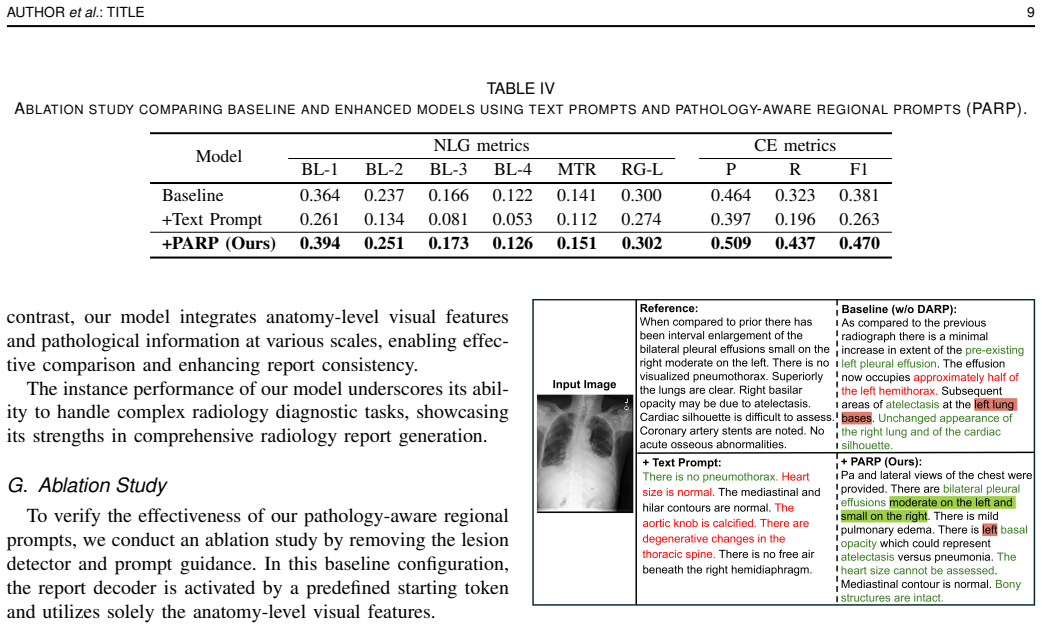
- [§IV.G, Table IV] The ablation is confounded by a training/inference mismatch. The +Text Prompt condition receives free-form textual prompts only at inference, because they have variable length and are not used during training, whereas the +PARP condition uses its prompts in both training and inference. The resulting drop in all metrics (e.g., BLEU-1 from 0.394 to 0.261) may reflect distribution shift rather than the intrinsic superiority of structured token prompts. Please compare against a fixed-length token-prompt baseline that is trained and tested symmetrically, and include a condition with randomly assigned or empty prompts to isolate the contribution of prompt content.
- [§IV.H.2, Table VI] The lesion detector's low recall for the very nodes prioritized in prompt construction undermines the assumed mechanism. Average recall is 0.285 and mAP@0.5 is 0.345; airspace opacity (recall 0.061) and consolidation (recall 0.075) are second-level nodes that, by §III.D.1, would be selected as the prompt token when present. The paper's statement that 'this challenge is unlikely to adversely affect report generation' is not quantified. Please report the fidelity of the constructed inference prompts against ground-truth scene graphs (prompt-level precision/recall per region) and show how prompt errors propagate to CE metrics.
minor comments (5)
- [§III.E, §IV.J] BERT-base has approximately 110 million parameters, not 'around 2B'; please correct this and clarify what 'configured in decoder mode' means for a bidirectional encoder model, including how causal masking is applied.
- [§III.D.2] If [NEG] tokens are used at inference but ground-truth prompts never contain [NEG], the decoder has never learned to handle them; please state whether [NEG] appears in the training prompt labels and, if not, explain how the model is expected to respond to it.
- [§IV.G, Table IV] The phrase 'F1-score increases by 8.9%' should be expressed as 8.9 percentage points (or a relative improvement of about 23%) to avoid ambiguity.
- [Table II] The rows for AdaMatch-Cyclic and MedDr are missing several metrics; please report the missing values or explicitly state why they are unavailable, since the 'most metrics' claim depends on complete comparisons.
- [§IV.A] Please clarify whether 'the official dataset split' refers to the MIMIC-CXR split or the Chest ImaGenome split, as the sample counts in the paper are not immediately matched to a named reference split.
Circularity Check
No significant circularity: the prompt labels are predicted from images at inference and the central comparison is against external baselines; shared report-derived annotation is a validity caveat, not a by-construction reduction.
full rationale
The claimed derivation chain is not circular by construction. Chest ImaGenome supplies bounding-box and lesion labels (Section IV.A), and the multi-label lesion detector is trained on those labels with image input only; at inference the pathology-aware regional prompt is built from detector outputs and anatomical-region overlap (Section III.D.2), not from the reference report. The decoder is trained with ground-truth prompt tokens prefixed to reference reports (Section III.E), which is teacher forcing of a latent summary, but the reported test-time generation uses predicted prompts on held-out images and is evaluated against reference reports and independent baselines. The fact that Chest ImaGenome scene graphs and CheXbert CE labels both derive from MIMIC-CXR reports is a legitimate benchmark-validity concern about shared annotation sources, and the expert evaluation is conducted by two of the authors, but neither makes any model output equal to its input by definition. No fitted parameter is renamed as a prediction, no uniqueness claim is imported from overlapping-author work, and no ansatz is smuggled in via citation. The finding is therefore no significant circularity.
Assumptions & free parameters
free parameters (6)
- lesion class frequency cutoff =
0.5% of training data
- negative image filter fraction =
12%
- lesion-to-region IoU threshold =
0.4
- lesion detector confidence and NMS thresholds =
conf 0.35, NMS IoU 0.45
- lesion detector loss weights =
lambda_cls=0.5, lambda_obj=1.0, lambda_box=0.05
- number of anatomical regions =
29
assumptions (5)
- domain assumption Chest ImaGenome scene graph annotations are accurate and complete for 29 anatomical regions and 21 lesion classes.
- domain assumption IoU overlap between lesion bboxes and anatomical region bboxes is a valid way to localize pathologies to regions.
- domain assumption CheXbert-based CE metrics on 14 observations are valid proxies for clinical accuracy.
- domain assumption The class reduction and rule-based prompt construction retain clinically sufficient information after removing the 'lung opacity' root and tail classes.
- domain assumption The MIMIC-CXR reference reports used as targets are reliable ground truth for report quality.
Cite this review
Pith. "Pith review of Anatomy-Guided Radiology Report Generation with Pathology-Aware Regional Prompts." pith.science (2026). https://pith.science/paper/T7QLQ3IL
@misc{pith2026241110789,
  author       = {Pith},
  title        = {Pith review of: Anatomy-Guided Radiology Report Generation with Pathology-Aware Regional Prompts},
  year         = {2026},
  howpublished = {\url{https://pith.science/paper/T7QLQ3IL}},
  note         = {Machine review of arXiv:2411.10789}
}
read the original abstract
Radiology reporting generative AI holds significant potential to alleviate clinical workloads and streamline medical care. However, achieving high clinical accuracy is challenging, as radiological images often feature subtle lesions and intricate structures. Existing systems often fall short, largely due to their reliance on fixed size, patch-level image features and insufficient incorporation of pathological information. This can result in the neglect of such subtle patterns and inconsistent descriptions of crucial pathologies. To address these challenges, we propose an innovative approach that leverages pathology-aware regional prompts to explicitly integrate anatomical and pathological information of various scales, significantly enhancing the precision and clinical relevance of generated reports. We develop an anatomical region detector that extracts features from distinct anatomical areas, coupled with a novel multi-label lesion detector that identifies global pathologies. Our approach emulates the diagnostic process of radiologists, producing clinically accurate reports with comprehensive diagnostic capabilities. Experimental results show that our model outperforms previous state-of-the-art methods on most natural language generation and clinical efficacy metrics, with formal expert evaluations affirming its potential to enhance radiology practice.
Figures


Figures from the paper (3 more)


Reference graph
Works this paper leans on
-
[1]
Ai-based radiodiagnosis using chest x-rays: A review,
Y . Akhter, R. Singh, and M. Vatsa, “Ai-based radiodiagnosis using chest x-rays: A review,” Front. Big Data, vol. 6, p. 1120989, 2023
work page 2023
-
[2]
Radiologist shortage leaves patient care at risk, warns royal college,
A. Rimmer, “Radiologist shortage leaves patient care at risk, warns royal college,” BMJ-BRIT. MED. J., vol. 359, 2017
2017
-
[3]
Show, attend and tell: Neural image caption generation with visual attention,
K. Xu, J. Ba, R. Kiros, K. Cho, A. Courville, R. Salakhudinov, R. Zemel, and Y . Bengio, “Show, attend and tell: Neural image caption generation with visual attention,” in Proc. Int. Conf. Mach. Learn., 2015, pp. 2048– 2057
work page 2015
-
[4]
Knowing when to look: Adaptive attention via a visual sentinel for image captioning,
J. Lu, C. Xiong, D. Parikh, and R. Socher, “Knowing when to look: Adaptive attention via a visual sentinel for image captioning,” in Proc. IEEE Conf. Comp. Vis. Patt. Recogn. , 2017, pp. 375–383
work page 2017
-
[5]
Imagenet classification with deep convolutional neural networks,
A. Krizhevsky, I. Sutskever, and G. E. Hinton, “Imagenet classification with deep convolutional neural networks,” Advances in Neural Inf. Process. Syst., vol. 25, 2012
work page 2012
-
[6]
A. Vaswani, N. Shazeer, N. Parmar, J. Uszkoreit, L. Jones, A. N. Gomez, Ł. Kaiser, and I. Polosukhin, “Attention is all you need,” Advances in Neural Inf. Process. Syst. , vol. 30, 2017
work page 2017
-
[7]
A survey on deep learning and explainability for automatic report generation from medical images,
P. Messina, P. Pino, D. Parra, A. Soto, C. Besa, S. Uribe, M. And ´ıa, C. Tejos, C. Prieto, and D. Capurro, “A survey on deep learning and explainability for automatic report generation from medical images,” ACM Comput. Surv., vol. 54, no. 10s, 2022
work page 2022
-
[8]
Foundation model for advancing healthcare: Challenges, opportunities, and future directions,
Y . He, F. Huang, X. Jiang, Y . Nie, M. Wang, J. Wang, and H. Chen, “Foundation model for advancing healthcare: Challenges, opportunities, and future directions,” arXiv:2404.03264, 2024
arXiv 2024
Show all 61 references
-
[9]
Promptmrg: Diagnosis-driven prompts for medical report generation,
H. Jin, H. Che, Y . Lin, and H. Chen, “Promptmrg: Diagnosis-driven prompts for medical report generation,” in Proc. AAAI Conf. Artif. Intell., vol. 38, no. 3, 2024, pp. 2607–2615
2024
-
[10]
Camanet: Class activation map guided attention network for radiology report generation,
J. Wang, A. Bhalerao, T. Yin, S. See, and Y . He, “Camanet: Class activation map guided attention network for radiology report generation,” IEEE J. Biomed. Health Inform. , vol. 28, no. 4, pp. 2199–2210, 2024
2024
-
[11]
Generating radiology re- ports via memory-driven transformer,
Z. Chen, Y . Song, T.-H. Chang, and X. Wan, “Generating radiology re- ports via memory-driven transformer,” in Proc.Conf. Empirical Methods Natural Lang. Process., Nov. 2020
2020
-
[12]
Pre- train, prompt, and predict: A systematic survey of prompting methods in natural language processing,
P. Liu, W. Yuan, J. Fu, Z. Jiang, H. Hayashi, and G. Neubig, “Pre- train, prompt, and predict: A systematic survey of prompting methods in natural language processing,” ACM Comput. Surv. , vol. 55, no. 9, 2023
2023
-
[13]
Can prompt learning benefit radiology report generation?
J. Wang, L. Zhu, A. Bhalerao, and Y . He, “Can prompt learning benefit radiology report generation?” arXiv:2308.16269, 2023
2023 arXiv
-
[14]
Advances in deep learning for tuberculosis screening using chest x-rays: the last 5 years review,
K. Santosh, S. Allu, S. Rajaraman, and S. Antani, “Advances in deep learning for tuberculosis screening using chest x-rays: the last 5 years review,” J. Med. Syst. , vol. 46, no. 11, p. 82, 2022
2022
-
[15]
Densely connected convolutional networks,
G. Huang, Z. Liu, L. Van Der Maaten, and K. Q. Weinberger, “Densely connected convolutional networks,” inProc. IEEE Conf. Comp. Vis. Patt. Recogn., 2017, pp. 4700–4708
2017
-
[16]
Deep residual learning for image recognition,
K. He, X. Zhang, S. Ren, and J. Sun, “Deep residual learning for image recognition,” in Proc. IEEE Conf. Comp. Vis. Patt. Recogn. , 2016, pp. 770–778
2016
-
[17]
TSGET: Two-Stage Global Enhanced Transformer for Automatic Radiology Report Generation,
X. Yi, Y . Fu, R. Liu, H. Zhang, and R. Hua, “TSGET: Two-Stage Global Enhanced Transformer for Automatic Radiology Report Generation,” IEEE J. Biomed. Health Inform. , vol. 28, no. 4, pp. 2152–2162, 2024
2024
-
[18]
Memory Guided Transformer With Spatio-Semantic Visual Extractor for Medical Report Generation,
P. Divya, Y . Sravani, C. Vishnu, C. K. Mohan, and Y . W. Chen, “Memory Guided Transformer With Spatio-Semantic Visual Extractor for Medical Report Generation,” IEEE J. Biomed. Health Inform. , vol. 28, no. 5, pp. 3079–3089, 2024
2024
-
[19]
Eye Gaze Guided Cross-Modal Alignment Network for Radiology Report Generation,
P. Peng, W. Fan, Y . Shen, W. Liu, X. Yang, Q. Zhang, X. Wei, and D. Zhou, “Eye Gaze Guided Cross-Modal Alignment Network for Radiology Report Generation,” IEEE J. Biomed. Health Inform. , pp. 1–14, 2024
2024
-
[20]
Swin transformer: Hierarchical vision transformer using shifted windows,
Z. Liu, Y . Lin, Y . Cao, H. Hu, Y . Wei, Z. Zhang, S. Lin, and B. Guo, “Swin transformer: Hierarchical vision transformer using shifted windows,” in Proc. IEEE Int. Conf. Comp. Vis., 2021, pp. 10 012–10 022
2021
-
[21]
Cgftrans: Cross- modal global feature fusion transformer for medical report generation,
L. Xu, Q. Tang, B. Zheng, J. Lv, W. Li, and X. Zeng, “Cgftrans: Cross- modal global feature fusion transformer for medical report generation,” IEEE J. Biomed. Health Inform. , vol. 28, no. 9, pp. 5600–5612, 2024
2024
-
[22]
S3-Net: A Self- Supervised Dual-Stream Network for Radiology Report Generation,
R. Pan, R. Ran, W. Hu, W. Zhang, Q. Qin, and S. Cui, “S3-Net: A Self- Supervised Dual-Stream Network for Radiology Report Generation,” IEEE J. Biomed. Health Inform. , vol. 28, no. 3, pp. 1448–1459, 2024
2024
-
[23]
An image is worth 16x16 words: Transformers for image recognition at scale,
A. Dosovitskiy, L. Beyer, A. Kolesnikov, D. Weissenborn, X. Zhai, T. Unterthiner, M. Dehghani, M. Minderer, G. Heigold, S. Gelly, J. Uszkoreit, and N. Houlsby, “An image is worth 16x16 words: Transformers for image recognition at scale,” axXiv:2010.11929, 2021
2010 arXiv
-
[24]
Xraygpt: Chest radiographs summarization using medical vision-language models,
O. Thawkar, A. Shaker, S. S. Mullappilly, H. Cholakkal, R. M. Anwer, S. Khan, J. Laaksonen, and F. S. Khan, “Xraygpt: Chest radiographs summarization using medical vision-language models,” arXiv:2306.07971, 2023
2023 arXiv
-
[25]
Metransformer: Radiology report generation by transformer with multiple learnable expert tokens,
Z. Wang, L. Liu, L. Wang, and L. Zhou, “Metransformer: Radiology report generation by transformer with multiple learnable expert tokens,” in Proc. IEEE Conf. Comp. Vis. Patt. Recogn. , June 2023, pp. 11 558– 11 567
2023
-
[26]
Long short-term memory,
S. Hochreiter and J. Schmidhuber, “Long short-term memory,” Neural Comput., vol. 9, no. 8, pp. 1735–1780, 1997
1997
-
[27]
Empirical eval- uation of gated recurrent neural networks on sequence modeling,
J. Chung, C. Gulcehre, K. Cho, and Y . Bengio, “Empirical eval- uation of gated recurrent neural networks on sequence modeling,” arXiv:1412.3555, 2014
2014 arXiv
-
[28]
Clinically accurate chest x-ray report generation,
G. Liu, T.-M. H. Hsu, M. McDermott, W. Boag, W.-H. Weng, P. Szolovits, and M. Ghassemi, “Clinically accurate chest x-ray report generation,” in Proc. Mach. Learn. Healthcare Conf., 2019, pp. 249–269. 12 IEEE TRANSACTIONS AND JOURNALS TEMPLATE
2019
-
[29]
When radiology report generation meets knowledge graph,
Y . Zhang, X. Wang, Z. Xu, Q. Yu, A. Yuille, and D. Xu, “When radiology report generation meets knowledge graph,” in Proc. AAAI Conf. Artif. Intell. , vol. 34, no. 07, 2020, pp. 12 910–12 917
2020
-
[30]
BERT: Pre-training of deep bidirectional transformers for language understanding,
J. Devlin, M.-W. Chang, K. Lee, and K. Toutanova, “BERT: Pre-training of deep bidirectional transformers for language understanding,” in Proc. Conf. North Amer. Chapter Assoc. Comput. Linguistics., 2019, pp. 4171– 4186
2019
-
[31]
Language models are unsupervised multitask learners,
A. Radford, J. Wu, R. Child, D. Luan, D. Amodei, I. Sutskever et al., “Language models are unsupervised multitask learners,” OpenAI blog , vol. 1, no. 8, p. 9, 2019
2019
-
[32]
Generating radiology reports via memory-driven transformer,
Z. Chen, Y . Shen, Y . Song, and X. Wan, “Generating radiology reports via memory-driven transformer,” in Proc. 59th Annu. Meeting Assoc. Comput. Linguistics 11th Int. Joint Conf. Natural Lang. Process. , 2021
2021
-
[33]
Clinical-bert: Vision-language pre-training for radiograph diagnosis and reports generation,
B. Yan and M. Pei, “Clinical-bert: Vision-language pre-training for radiograph diagnosis and reports generation,” in Proc. AAAI Conf. Artif. Intell., vol. 36, no. 3, 2022, pp. 2982–2990
2022
-
[34]
On the difficulty of training recurrent neural networks,
R. Pascanu, T. Mikolov, and Y . Bengio, “On the difficulty of training recurrent neural networks,” in Proc. Int. Conf. Mach. Learn. , 2013, pp. 1310–1318
2013
-
[35]
Chexagent: Towards a foundation model for chest x-ray interpretation,
Z. Chen, M. Varma, J.-B. Delbrouck, M. Paschali, L. Blankemeier, D. V . Veen, J. M. J. Valanarasu, A. Youssef, J. P. Cohen, E. P. Reis, E. B. Tsai, A. Johnston, C. Olsen, T. M. Abraham, S. Gatidis, A. S. Chaudhari, and C. Langlotz, “Chexagent: Towards a foundation model for ch...
2024 arXiv
-
[36]
Mistral 7b,
A. Q. Jiang, A. Sablayrolles, A. Mensch, C. Bamford, D. S. Chaplot, D. de las Casas, F. Bressand, G. Lengyel, G. Lample, L. Saulnier, L. R. Lavaud, M.-A. Lachaux, P. Stock, T. L. Scao, T. Lavril, T. Wang, T. Lacroix, and W. E. Sayed, “Mistral 7b,” arXiv:2310.06825, 2023
-
[37]
Eva-clip: Improved training techniques for clip at scale,
Q. Sun, Y . Fang, L. Wu, X. Wang, and Y . Cao, “Eva-clip: Improved training techniques for clip at scale,” arXiv:2303.15389, 2023
2023 arXiv
-
[38]
Meddr: Diagnosis-guided bootstrapping for large-scale medical vision-language learning,
S. He, Y . Nie, Z. Chen, Z. Cai, H. Wang, S. Yang, and H. Chen, “Meddr: Diagnosis-guided bootstrapping for large-scale medical vision-language learning,” arXiv:2404.15127, 2024
2024 arXiv
-
[39]
Bootstrapping large language models for radiology report generation,
C. Liu, Y . Tian, W. Chen, Y . Song, and Y . Zhang, “Bootstrapping large language models for radiology report generation,” in Proc. AAAI Conf. Artif. Intell., vol. 38, no. 17, 2024, pp. 18 635–18 643
2024
-
[40]
Interactive and explainable region-guided radiology report generation,
T. Tanida, P. M ¨uller, G. Kaissis, and D. Rueckert, “Interactive and explainable region-guided radiology report generation,” in Proc. IEEE Conf. Comp. Vis. Patt. Recogn. , 2023, pp. 7433–7442
2023
-
[41]
Finding-aware anatomical tokens for chest x-ray automated reporting,
F. D. Serra, C. Wang, F. Deligianni, J. Dalton, and A. Q. O’Neil, “Finding-aware anatomical tokens for chest x-ray automated reporting,” arXiv:2308.15961, 2023
2023 arXiv
-
[42]
Scene graph aided radiology report generation,
J. Wang, L. Zhu, A. Bhalerao, and Y . He, “Scene graph aided radiology report generation,” arXiv:2403.05687, 2024
2024 arXiv
-
[43]
Faster r-cnn: Towards real-time object detection with region proposal networks,
S. Ren, K. He, R. Girshick, and J. Sun, “Faster r-cnn: Towards real-time object detection with region proposal networks,” Advances in Neural Inf. Process. Syst., vol. 28, 2015
2015
-
[44]
Roberta: A robustly optimized bert pretraining approach,
Y . Liu, M. Ott, N. Goyal, J. Du, M. Joshi, D. Chen, O. Levy, M. Lewis, L. Zettlemoyer, and V . Stoyanov, “Roberta: A robustly optimized bert pretraining approach,” arXiv preprint arXiv:1907.11692 , 2019
1907 arXiv
-
[45]
Medical image understanding with pretrained vision language models: A comprehensive study,
Z. Qin, H. H. Yi, Q. Lao, and K. Li, “Medical image understanding with pretrained vision language models: A comprehensive study,” inProc. Int. Conf. Learn. Representations , 2022
2022
-
[46]
YOLOv5: A state-of-the-art real-time object detection sys- tem,
Ultralytics, “YOLOv5: A state-of-the-art real-time object detection sys- tem,” https://docs.ultralytics.com, 2021
2021
-
[47]
Chest imagenome dataset,
J. Wu, N. Agu, I. Lourentzou, A. Sharma, J. Paguio, J. S. Yao, E. C. Dee, W. Mitchell, S. Kashyap, A. Giovannini et al., “Chest imagenome dataset,” Physio Net, 2021
2021
-
[48]
A survey on long-tailed visual recognition,
L. Yang, H. Jiang, Q. Song, and J. Guo, “A survey on long-tailed visual recognition,” Int. J. Comput. Vision , vol. 130, no. 7, pp. 1837–1872, 2022
2022
-
[49]
Mimic-cxr, a de- identified publicly available database of chest radiographs with free-text reports,
A. E. Johnson, T. J. Pollard, S. J. Berkowitz, N. R. Greenbaum, M. P. Lungren, C.-y. Deng, R. G. Mark, and S. Horng, “Mimic-cxr, a de- identified publicly available database of chest radiographs with free-text reports,” Sci. Data., vol. 6, no. 1, p. 317, 2019
2019
-
[50]
Mimic-cxr-jpg, a large publicly available database of labeled chest radiographs,
A. E. Johnson, T. J. Pollard, N. R. Greenbaum, M. P. Lungren, C.-y. Deng, Y . Peng, Z. Lu, R. G. Mark, S. J. Berkowitz, and S. Horng, “Mimic-cxr-jpg, a large publicly available database of labeled chest radiographs,” arXiv:1901.07042, 2019
1901 arXiv
-
[51]
Decoupled weight decay regularization,
I. Loshchilov and F. Hutter, “Decoupled weight decay regularization,” arXiv:1711.05101, 2017
2017 arXiv
-
[52]
Bleu: a method for automatic evaluation of machine translation,
K. Papineni, S. Roukos, T. Ward, and W.-J. Zhu, “Bleu: a method for automatic evaluation of machine translation,” in Proc. 40th Annu. Meeting Assoc. Comput. Linguistics , 2002, pp. 311–318
2002
-
[53]
Meteor: An automatic metric for mt evaluation with improved correlation with human judgments,
S. Banerjee and A. Lavie, “Meteor: An automatic metric for mt evaluation with improved correlation with human judgments,” in Proc. 6th Workshop Statist. Mach. Transl. , 2005, pp. 65–72
2005
-
[54]
Rouge: A package for automatic evaluation of summaries,
C.-Y . Lin, “Rouge: A package for automatic evaluation of summaries,” in Proc. Text Summarization Branches Out , 2004, pp. 74–81
2004
-
[55]
Combining automatic labelers and expert annotations for accurate radiology report labeling using bert,
A. Smit, S. Jain, P. Rajpurkar, A. Pareek, A. Y . Ng, and M. P. Lungren, “Combining automatic labelers and expert annotations for accurate radiology report labeling using bert,” Proc. Conf. Empirical Methods Natural Lang. Process., 2020
2020
-
[56]
Advancing multimodal medical capabilities of gemini,
L. Yang, S. Xu, A. Sellergren, T. Kohlberger, Y . Zhou, I. Ktena, A. Kiraly, F. Ahmed, F. Hormozdiari, T. Jaroensri et al. , “Advancing multimodal medical capabilities of gemini,” arXiv:2405.03162, 2024
2024 arXiv
-
[57]
Cxr-agent: Vision-language models for chest x-ray interpre- tation with uncertainty aware radiology reporting,
N. Sharma, “Cxr-agent: Vision-language models for chest x-ray interpre- tation with uncertainty aware radiology reporting,” arXiv:2407.08811, 2024
2024 arXiv
-
[58]
Fine-grained image-text alignment in medical imaging enables explainable cyclic image-report generation,
W. Chen, L. Shen, J. Lin, J. Luo, X. Li, and Y . Yuan, “Fine-grained image-text alignment in medical imaging enables explainable cyclic image-report generation,” arXiv:2312.08078, 2024
2024 arXiv
-
[59]
Free dolly: Introducing the world’s first truly open instruction-tuned llm,
M. Conover, M. Hayes, A. Mathur, J. Xie, J. Wan, S. Shah, A. Ghodsi, P. Wendell, M. Zaharia, and R. Xin, “Free dolly: Introducing the world’s first truly open instruction-tuned llm,” Company Blog of Databricks , 2023
2023
-
[60]
Medimageinsight: An open-source embedding model for general domain medical imaging,
N. C. F. Codella, Y . Jin, S. Jain, Y . Gu et al. , “Medimageinsight: An open-source embedding model for general domain medical imaging,” arXiv:2410.06542, 2024
2024 arXiv
-
[61]
Foundation models for generalist medical artificial intelligence,
M. Moor, O. Banerjee, Z. S. H. Abad, H. M. Krumholz, J. Leskovec, E. J. Topol, and P. Rajpurkar, “Foundation models for generalist medical artificial intelligence,” Nature, vol. 616, no. 7956, pp. 259–265, 2023
2023
Reviewed August 12, 2026 · model on record in the stance chip above.
Discussion (0). Continue with ORCID to comment.