REVIEW 3 major objections 5 minor 1 cited by
Active droplets controlled by enzymatic reactions
T0 review · 3 major / 5 minor · reviewed 2026-08-12 · deepseek-v4-flash
Pith's one-line read A two-enzyme system can arrest droplet coarsening, with enzyme concentration and mobility setting the final droplet size.
desk verdict Genuine modeling step with explicit enzyme trajectories; the central trend holds, but the analytic model is a fit and the D-control claim is softened by enzyme self-propulsion. read the letter →
The pith
A machine-rendered reading of the paper's core claim, the machinery that carries it, and where it could break.
The reading
What carries the argument
The load-bearing object is the encounter-time model of droplet destruction. The paper assumes each growing droplet receives a constant protein influx $c$, that droplets are destroyed instantly upon meeting an $E_{B\to A}$ enzyme, and that encounter times are exponentially distributed with mean $t_e$. This reduces the mean droplet size to an exponential-saturation formula, Eq. 4, whose steady-state value is $c t_e/(S \rho_{B\to A})$. The second piece of machinery is the explicit Brownian representation of enzymes, which replaces the artificial density-dependent reaction rates used in previous mean-field droplet models with reaction rates that emerge from enzyme trajectories, and in the hybrid method couples those trajectories to a Cahn-Hilliard-Cook field.
What would settle it
Make an in vitro system with two opposing enzymes acting on a dropletable protein, then measure the steady-state droplet radius as the dispersion-promoting enzyme's diffusion coefficient is varied (for instance by changing solution viscosity). The model predicts that droplet radius decreases monotonically with that diffusion coefficient; a flat or non-monotonic response would contradict the encounter-time mechanism. A second check is to record droplet lifetimes: under the model, they should be exponentially distributed with mean $t_e/(S \rho_{B\to A})$.
Extended reading notes
Core claim
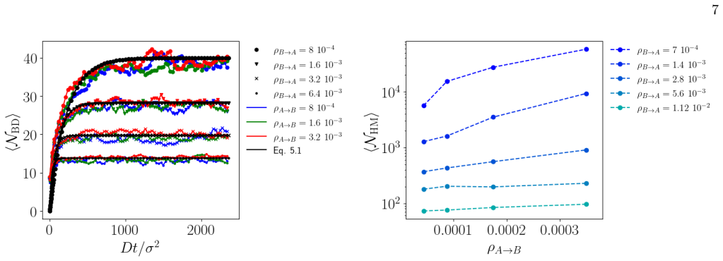
In a model where two enzyme species catalyze opposite conversions between a condensate-prone protein state B and a dispersed state A, the paper's central discovery is that the stochastic, spatially localized action of the enzymes arrests the otherwise uninterrupted growth of droplets. With only the droplet-promoting enzyme present, a single B-rich droplet grows without bound by Ostwald ripening and coalescence. Adding the dispersion-promoting enzyme $E_{B\to A}$ produces a steady state with coexisting droplets; the mean droplet size falls as $\rho_{B\to A}$ increases, and the mean number of droplets per $E_{A\to B}$ enzyme goes through a maximum. Varying the diffusion coefficient of either enzyme changes droplet size the same way: faster enzymes give smaller droplets. The full-Brownian-dynamics data are described by an encounter-destruction picture in which each droplet grows at constant influx and is abruptly emptied when it meets an $E_{B\to A}$ enzyme, yielding the analytic form $\langle N \rangle = (c t_e/S \rho_{B\to A})(1 - \exp(-t S \rho_{B\to A}/t_e))$. The hybrid method, though it shows quantitative differences at large enzyme concentrations, reproduces the same qualitative dependence on enzyme concentration and diffusivity.
Load-bearing premise
The argument assumes an enzyme's bare diffusion coefficient fully controls how often it meets a droplet, yet the simulations show signs that droplet interfaces may actively propel enzymes, which would change the effective encounter rate.
Editorial extensions
If this is right
- In a living cell, raising the expression level of the dispersion-promoting enzyme would shrink condensates continuously, rather than dissolving them abruptly, as long as the droplet-promoting enzyme remains active.
- Tuning enzyme mobility—through viscosity, crowding, or active transport—should provide an independent control over condensate size, separate from changing reaction rates.
- The non-monotonic dependence of droplet number on enzyme ratio means cells can maximize organelle count by balancing the two enzyme activities at an intermediate value.
- The analytic formula predicts that at fixed influx the steady-state droplet size is inversely proportional to the dispersion-enzyme surface concentration, a directly testable scaling law.
Reading between the lines
- If the hint of interface-driven enzyme self-propulsion is confirmed, then an enzyme's effective encounter rate with a condensate is set partly by the condensate itself, so enzyme diffusivity should be treated as a coupled variable rather than a fixed input.
- The encounter-destruction picture implies droplet lifetimes should be exponentially distributed; recording droplet birth-death histories in experiments or simulations would test this renewal mechanism against models where droplets shrink continuously.
- The same machinery could be applied to multiple enzyme species or spatially patterned enzymes, predicting that localizing dispersion enzymes near nucleation sites would create gradients of droplet size across a cell.
- Extending the model to three dimensions with hydrodynamic interactions may change the encounter-time scaling, since the diffusion coefficient of a droplet depends on solvent viscosity and droplet size differently beyond two dimensions.
Signed reviews
Editorial analysis
A structured set of objections, weighed in public.
Referee Report
Summary. Fries et al. propose a two-dimensional model of biocondensate formation in which two enzyme species catalyze opposing interconversions of a two-state protein (A and B), with B proteins attracting each other and forming droplets. Enzymes are explicit Brownian particles, and reactions occur only within a cutoff radius of an enzyme. The authors use two complementary simulation methods: all-particle Brownian dynamics (full BD) and a hybrid method that couples Cahn-Hilliard-Cook dynamics for the protein field with Brownian dynamics for the enzymes (HM). They report that adding EB→A enzymes arrests Ostwald ripening, producing finite steady-state droplet sizes that decrease with increasing EB→A concentration and with increasing enzyme diffusivity, and a non-monotonic number of droplets per EA→B enzyme as a function of EB→A concentration. An analytic model (Eq. 4) with fitted constants reproduces the full BD data for the mean droplet size evolution and steady-state size, and the authors interpret this as evidence that the encounter time between droplets and EB→A enzymes controls droplet size.
Significance. If the conclusions hold, the paper offers a minimal mechanistic picture of how enzyme concentration and transport can select biocondensate size, with explicit enzyme trajectories being a methodological advantage over density-dependent reaction-rate models. The internal agreement between two independent simulation methods for the main qualitative trends (arrested growth, finite steady-state sizes, smaller droplets with faster enzymes, non-monotonic droplet number) is a genuine strength. However, the quantitative status of the central claim is currently limited by the fitted analytic model and by the paper's own observation of interface-induced enzyme self-propulsion, which may contradict the assumption that the imposed diffusion coefficient D is the relevant transport control parameter.
major comments (3)
- [Enzyme diffusivity affects the size of condensates; Eq. 5] The claim that the bare diffusion coefficient D of enzymes controls droplet size is not fully established because the paper reports that enzyme transport near droplets is not ideal Brownian. In the section 'Enzyme diffusivity affects the size of condensates', the authors state that for D = 0.2D0 the fit improves with a slightly higher diffusion coefficient, that MSD 'show a slight increase of the diffusion coefficient' in full BD, and that in the HM 'a first investigation of the mean squared displacements of the enzymes suggests a strong self-propulsion in this range of parameters, which may dominate normal diffusion.' Equations 4-5, however, assume te ∝ 1/(D_EB→A + Ddp), i.e., transport governed by the imposed D. If the effective enzyme mobility is an emergent, interface-driven quantity that is not equal to D, then Fig. 6 does not demonstrate that D alone selects droplet size; the control variable would be an unexplained effective motility. The authors should measure and report effective diffusion coefficients from MSD in both simulation methods and show how they scale with D, or qualify the claim to refer to effective motility rather than the bare diffusion coefficient.
- [Appendix A3, Eqs. 4-7] The analytic model is fitted, not independently predictive. The constants α = 2.7 and β = 900 are fit parameters, and Eq. 7 sets Ddp = D0/⟨N⟩∞, where ⟨N⟩∞ is the measured steady-state droplet size. Because the predicted steady-state size appears in the definition of the droplet diffusivity used in the prediction, the agreement of Eq. 4 with simulation is a consistency check of a two-parameter functional form rather than a validation of the encounter-time mechanism. The paper should state the predictive content explicitly: with α and β fixed from one dataset, does the implicit equation for ⟨N⟩∞ (via Eqs. 4-7) predict the full dependence on ρB→A and D? If so, show that test.
- [Figs. 3, 4, 6; stationary-state definitions] The manuscript does not provide convergence tests for the claimed stationary states or error bars for the plotted means, despite stating that results are averaged over 50 independent realizations. For example, the caption of Fig. 3 says 'the stationary state is assumed to be reached in the interval [1800, 2400] Dt/σ²' without comparing adjacent time windows or showing the time series of the mean and variance. Since the paper makes quantitative claims, including the non-monotonic dependence of droplet number on ρB→A and the dependence on D, the absence of uncertainty quantification makes it difficult to assess the significance of the reported differences and the quality of the fits.
minor comments (5)
- [Models] Typographical errors: 'explicitely' should be 'explicitly', and 'Theses reactions' should be 'These reactions'.
- [Appendix A3, Eq. 24] In Eq. 24, the term c dt²/τ is second order in dt and is neglected without comment; please explicitly state this approximation.
- [Appendix A1e] The text contains 'simularions' for 'simulations'.
- [Figs. 3-6] Full BD and HM are simulated in different parameter regimes (e.g., ρA→B = 1.6×10⁻³ in full BD versus 1.2×10⁻⁴ in HM). The paper should state explicitly that the comparison between the two methods is only qualitative and that quantitative agreement is not expected.
- [Appendix A1a] The droplet detection criterion is described as 'The square root of this threshold, 2σ, is taken as the distance criteria'; this sentence is unclear and should be rewritten.
Circularity Check
Simulation results are self-contained; the analytic model Eq. 4 feeds the measured steady-state droplet size back into the encounter time via Eq. 7, so its fit does not independently confirm the size-selection mechanism.
-
self definitional
[Main text, 'Enzyme diffusivity affects the size of condensates', Eqs. 4-7; Appendix A3, Eqs. 27-28]
"The full BD simulation data shown in Figs. 5-left and 6-left are successfully fitted by Eq. 4, provided that we take c = α sqrt(SρB→A/te), (6) Ddp = D0/⟨N⟩_{t→∞}. (7)"
Eq. 4 is the model for the time-dependent and steady-state droplet size ⟨N⟩, but its parameters include te, and Eq. 5 sets te ∝ 1/(DEB→A + Ddp). Eq. 7 defines Ddp from the measured steady-state size ⟨N⟩_{t→∞}, the very quantity Eq. 4 is supposed to describe and explain. Thus the 'successful fit' of Eq. 4 is not an independent prediction: the target value is built into the rate that controls both the amplitude and the relaxation of the solution. The additional fitted constants α=2.7 and β=900 are calibrated to the same full-BD data, so the agreement cannot independently prove the encounter-time mechanism; it only shows that the functional form can accommodate the data once the outcome is supplied as an input.
full rationale
The core findings of the paper are simulation results: full Brownian dynamics and the hybrid method both show that increasing EB→A enzyme concentration or enzyme diffusivity reduces the steady-state droplet size and changes droplet number (Figs. 3, 4, 6). Those observations are self-contained numerical experiments with a stated model, and no step in the simulation protocol defines the output in terms of itself. The analytic model in Appendix A3 is the only place where a claimed explanation is partly self-referential: Eq. 7 inserts the measured final droplet size into the droplet diffusion coefficient, so Eq. 4 is fitted with the target as an input; the paper itself labels this a fit ('successfully fitted by Eq. 4'), not a first-principles prediction. The same section also acknowledges that at D=0.2D0 the fit works better with a slightly higher enzyme diffusion coefficient and that hybrid-method mean-square displacements 'suggest a strong self-propulsion in this range of parameters, which may dominate normal diffusion'; this is a limitation of the quantitative D-control claim but not a circularity, since the imposed D is varied and the droplet response is measured. The self-citations ([24,25] on interface-driven propulsion, [11] on detailed balance, [27] on enzyme diffusivity) support auxiliary context and are not load-bearing: none of the core simulation results derives from these references. Hence the central claim retains independent content, and the circularity score is moderate rather than severe.
Assumptions & free parameters
free parameters (6)
- alpha =
2.7
- beta =
900
- Ddp (droplet diffusion coefficient) =
D0 / <N>_inf
- Reaction rate k =
10 (in units of D0 / sigma^2)
- Reaction cutoff rcut =
5 sigma
- Hybrid method dimensionless parameters =
See Eqs. 19-22: tau, u, D, c, psi0, Dt, tK, Ra
assumptions (7)
- domain assumption Reverse reactions are negligible for each enzyme: A -> B occurs only near EA->B and B -> A only near EB->A.
- domain assumption Reactions occur only within a fixed cutoff distance rcut of an enzyme, with the random telegraph rate k.
- domain assumption The internal free-energy difference between A and B and the chemical drive Delta-mu are large enough that one reaction direction dominates near each enzyme type.
- ad hoc to paper Droplet-enzyme encounters are memoryless and instantaneously empty the droplet in the analytic model.
- ad hoc to paper The encounter time te depends only on the inverse sum of droplet and enzyme diffusion coefficients.
- domain assumption Cahn-Hilliard-Cook dynamics with a Ginzburg-Landau free energy adequately describes protein phase separation in the hybrid method.
- domain assumption Two-dimensional simulations capture the relevant physics of three-dimensional condensates.
Cite this review
Pith. "Pith review of Active droplets controlled by enzymatic reactions." pith.science (2026). https://pith.science/paper/HTYEV62D
@misc{pith2026241111696,
  author       = {Pith},
  title        = {Pith review of: Active droplets controlled by enzymatic reactions},
  year         = {2026},
  howpublished = {\url{https://pith.science/paper/HTYEV62D}},
  note         = {Machine review of arXiv:2411.11696}
}
read the original abstract
The formation of condensates is now considered as a major organization principle of eukaryotic cells. Several studies have recently shown that the properties of these condensates are affected by enzymatic reactions. We propose here a simple generic model to study the interplay between two enzyme populations and a two-state protein. In one state, the protein forms condensed droplets through attractive interactions, while in the other state, the proteins remain dispersed. Each enzyme catalyzes the production of one of these two protein states only when reactants are in its vicinity. A key feature of our model is the explicit representation of enzyme trajectories, capturing the fluctuations in their local concentrations. The spatially dependent growth rate of droplets naturally arises from the stochastic motion of these explicitly modeled enzymes. Using two complementary numerical methods, (1) Brownian Dynamics simulations, and (2) a hybrid method combining Cahn-Hilliard-Cook diffusion equations with Brownian Dynamics for the enzymes, we investigate how enzyme concentration and dynamics influence the evolution with time, and the steady-state number and size of droplets. Our results show that the concentration and diffusion coefficient of enzymes govern the formation and size-selection of biocondensates.
Figures


Figures from the paper (6 more)



Forward citations
Cited by 1 Pith paper
-
Chemically active droplets in crowded environments
In particle-based and field-based simulations, crowding reduces the size of chemically active droplets while increasing the total volume of the dense phase, driven by depletion, slower diffusion, and faster active reactions.
Reference graph
Works this paper leans on
-
[1]
C. P. Brangwynne, C. R. Eckmann, D. S. Courson, A. Rybarska, C. Hoege, J. Gharakhani, F. J¨ ulicher, and A. A. Hyman, Science 324, 1729 (2009)
2009
-
[2]
S. F. Banani, H. O. Lee, A. A. Hyman, and M. K. Rosen, Nat. Rev. Mol. Cell. Biol. 18, 285 (2017)
work page 2017
- [3]
-
[4]
Zwicker, Curr
D. Zwicker, Curr. Opin. Colloid Interface Sci. 61, 101606 (2022)
2022
-
[5]
F. C. Keber, T. Nguyen, A. Mariossi, C. P. Brangwynne, and M. W¨ uhr, Nat. Cell Biol.26, 346 (2024)
work page 2024
-
[6]
Zwicker, A
D. Zwicker, A. A. Hyman, and F. J¨ ulicher, Phys. Rev. E 92, 012317 (2015)
2015
-
[7]
D. Zwicker, R. Seyboldt, C. A. Weber, A. A. Hyman, and F. J¨ ulicher, Nature Phys.13, 408 (2017)
work page 2017
-
[8]
C. A. Weber, D. Zwicker, F. J¨ ulicher, and C. F. Lee, Rep. Prog. Phys. 82, 064601 (2019)
work page 2019
Show all 39 references
-
[9]
Tjhung, C
E. Tjhung, C. Nardini, and M. E. Cates, Phys. Rev. X 8, 031080 (2018)
2018
-
[10]
Ziethen, J
N. Ziethen, J. Kirschbaum, and D. Zwicker, Phys. Rev. Lett. 130, 248201 (2023)
2023
-
[11]
Berthin, J
R. Berthin, J. Fries, M. Jardat, V. Dahirel, and P. Illien, arXiv:2406.14256 (2024)
2024 arXiv
-
[12]
J. T. Wang, J. Smith, B. C. Chen, H. Schmidt, D. Ra- soloson, A. Paix, B. G. Lambrus, D. Calidas, E. Betzig, and G. Seydoux, eLife 3, 04591 (2014)
2014
-
[13]
Guilhas, J
B. Guilhas, J. C. Walter, J. Rech, G. David, N. O. Wal- liser, J. Palmeri, C. Mathieu-Demaziere, A. Parmeggiani, J. Y. Bouet, A. L. Gall, and M. Nollmann, Molecular Cell 79, 293 (2020)
2020
-
[14]
Linsenmeier, M
M. Linsenmeier, M. Hondele, F. Grigolato, E. Secchi, K. Weis, and P. Arosio, Nat. Commun. 13, 3030 (2022)
2022
-
[15]
H. Wu, X. Chen, Z. Shen, H. Li, S. Liang, Y. Lu, and M. Zhang, Molecular Cell 84, 309 (2024)
2024
-
[16]
W. T. Snead and A. S. Gladfelter, Molecular Cell 76, 295 (2019)
2019
-
[17]
G. A. P´ erez, R. V. Pappu, and D. Milovanovic, Trends in Cell Biology 34, 274 (2024)
2024
-
[18]
I. B. Smokers, B. S. Visser, A. D. Slootbeek, W. T. Huck, and E. Spruijt, Acc. Chem. Res. 57, 1885 (2024)
2024
-
[19]
Alon, An Introduction to Systems Biology: De- sign Principles of Biological Circuits (Chapman and Hall/CRC, 2006)
U. Alon, An Introduction to Systems Biology: De- sign Principles of Biological Circuits (Chapman and Hall/CRC, 2006)
2006
-
[20]
D. L. Ermak, J. Chem. Phys. 62, 4189 (1975)
1975
-
[21]
J. D. Weeks, D. Chandler, and H. C. Andersen, J. Chem. Phys. 54, 5237 (1971)
1971
-
[22]
Smit and D
B. Smit and D. Frenkel, J. Chem. Phys. 94, 5663 (1991)
1991
-
[23]
N. V. Kampen, Physics Reports 24, 171 (1976)
1976
-
[24]
Decayeux, V
J. Decayeux, V. Dahirel, M. Jardat, and P. Illien, Phys. Rev. E 104, 034602 (2021)
2021
-
[25]
Decayeux, J
J. Decayeux, J. Fries, V. Dahirel, M. Jardat, and P. Il- lien, Soft Matter 19, 8997 (2023)
2023
-
[26]
J. W. Cahn, J. Chem. Phys. 30, 1121 (1959)
1959
-
[27]
Agudo-Canalejo, T
J. Agudo-Canalejo, T. Adeleke-Larodo, P. Illien, and R. Golestanian, Acc. Chem. Res. 51, 2365 (2018)
2018
-
[28]
Feng and M
M. Feng and M. K. Gilson, Annu. Rev. Biophys. 49, 87 (2020)
2020
-
[29]
Ghosh, A
S. Ghosh, A. Somasundar, and A. Sen, Annu. Rev. Con- 14 dens. Matter Phys. 12, 177 (2021)
2021
-
[30]
Szent-Gy¨ orgyi, Science124, 873 (1956)
A. Szent-Gy¨ orgyi, Science124, 873 (1956)
1956
-
[31]
Utilization of binding energy and coupling rules for active transport and other coupled vectorial pro- cesses,
W. P. Jencks, “Utilization of binding energy and coupling rules for active transport and other coupled vectorial pro- cesses,” in Methods in Enzymology , Vol. 171 (Academic Press, 1989) pp. 145–164
1989
-
[32]
Mitchell, FEBS Letters 43, 189 (1974)
P. Mitchell, FEBS Letters 43, 189 (1974)
1974
-
[33]
Shacter, P
E. Shacter, P. B. Chock, and E. R. Stadtman, J. Biol. Chem. 259, 12260 (1984)
1984
-
[34]
R. A. Alberty, Biochemical Education 28, 12 (2000)
2000
-
[35]
Tanaka and T
H. Tanaka and T. Araki, Phys. Rev. Lett. 85, 1338 (2000)
2000
-
[36]
J. W. Cahn and J. E. Hilliard, J. Chem. Phys. 31, 688 (1959)
1959
-
[37]
Cook, Acta Metallurgica 18, 297 (1970)
H. Cook, Acta Metallurgica 18, 297 (1970)
1970
-
[38]
P. L. Barclay and J. R. Lukes, Phys. Fluids 31, 092107 (2019)
2019
-
[39]
R. C. Ball and R. L. H. Essery, J. Phys.: Condens. Matter 2, 10303 (1990)
1990
Reviewed August 12, 2026 · model on record in the stance chip above.
Discussion (0). Continue with ORCID to comment.