REVIEW 5 major objections 5 minor 21 references
Multimodal 3D Brain Tumor Segmentation with Adversarial Training and Conditional Random Field
T0 review · 5 major / 5 minor · reviewed 2026-08-12 · deepseek-v4-flash
Pith's one-line read A 3D generative adversarial network with a conditional random field post-processing layer outperforms classical segmentation networks on BraTS-2018 brain tumor MRI.
desk verdict Incremental architecture combo with a promising idea, but Table 3 is internally unreliable and the claimed outperformance is unestablished. read the letter →
The pith
A machine-rendered reading of the paper's core claim, the machinery that carries it, and where it could break.
The reading
What carries the argument
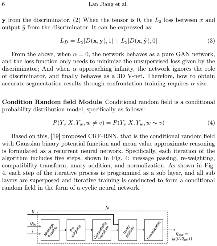
Three components carry the argument. First, the V-Net generator is built with residual blocks that use Pseudo-3D convolutions, decomposing a 3D convolution into a 2D spatial filter followed by a 1D depth filter as in $(I+DS)x_t = x_{t+1}$, which lowers parameter count. Second, a CRF-RNN layer is inserted as post-processing to enforce label smoothness through iterative message passing, re-weighting, compatibility transform, unary addition, and normalization. Third, the discriminator receives the original image $x$ alongside either the ground truth $y$ or the prediction $\hat{y}$, and the losses are $L_G = L_2[D(x,\hat{y}),1] + \alpha\,\mathrm{GDL}(y,\hat{y})$ and $L_D = L_2[D(x,y),1] + L_2[D(x,\hat{y}),0]$, with α controlling the balance between adversarial and dice-based learning.
What would settle it
Retrain U-Net, GAN, FCN, and 3D V-Net on the same BraTS-2018 10-fold splits with the identical preprocessing, patch extraction, augmentation, and hyperparameter budget used for 3D-vGAN, reporting per-fold metrics. If any baseline reaches or exceeds DSC 82.13% or specificity 99.97%, or if the reported baseline numbers change materially, the claimed outperformance is not supported.
Extended reading notes
Core claim
The paper claims that adding a CRF-RNN layer inside the generator, using Pseudo-3D convolutions in the residual blocks, and feeding the original MRI image as extra guidance to the discriminator yields better tumor segmentation than conventional networks. The loss function balances an adversarial L2 term with a generalized dice loss via a scalar α; at α=5 the model reaches its reported optimum, with specificity above 99.8%. The authors present this as evidence that the proposed architecture, not just adversarial training or V-Net alone, drives the accuracy gain.
Load-bearing premise
The central claim rests on Table 3 being a fair comparison: the four baselines are assumed to have been trained and evaluated with the same preprocessing, 10-fold splits, and tuning effort as 3D-vGAN, but the paper provides no baseline implementation details and two baseline rows share identical specificity and Hausdorff values, so the performance gap could reflect evaluation or tuning differences rather than the architecture.
Editorial extensions
If this is right
- If the reported performance holds under a fair comparison, combining CRF-RNN with adversarial training is a straightforward way to sharpen tumor boundaries in 3D medical volumes without switching to a fundamentally different architecture.
- The Pseudo-3D design reduces memory and computation relative to full 3D convolutions, which could make 3D segmentation feasible on smaller GPUs.
- The very high specificity suggests the model is conservative about false positives, which matters for clinical screening tools, though the Dice score indicates volume overlap still has room to improve.
- The α parameter gives practitioners a single dial to shift between adversarial sharpening and dice-based volume accuracy, and the paper shows performance degrades when α is too small or too large.
Reading between the lines
- Ablating the CRF-RNN layer while keeping the adversarial components would show how much of the boundary improvement comes from the CRF alone; the paper does not report this.
- The baseline comparison in Table 3 lacks implementation details, and the identical specificity and Hausdorff values for U-Net and 3D V-Net suggest those rows may share a common coarse evaluation step, so the margins could shrink under equal tuning.
- On the BraTS-2018 benchmark, state-of-the-art models typically report DSC above 85%, so the practical takeaway is not that 3D-vGAN sets a new standard, but that old-fashioned CNN architectures can still be pushed with CRF and adversarial refinement.
Editorial analysis
A structured set of objections, weighed in public.
Referee Report
Summary. The paper proposes 3D-vGAN, a multimodal 3D generative adversarial network for brain tumor segmentation. The generator is a V-Net with Pseudo-3D residual blocks, a CRF-RNN post-processing layer, and the discriminator receives the original image as additional guidance. The training loss combines an L2 adversarial term with a weighted generalized dice loss (Eq. 2). Using BraTS-2018, the authors report DSC 82.13%, sensitivity 84.42%, specificity 99.97%, and Hausdorff distance 11.89 mm, and claim that 3D-vGAN outperforms U-net, GAN, FCN, and 3D V-net. The manuscript also includes a parameter sweep over the weighting coefficient alpha.
Significance. The general idea of combining adversarial training, CRF-based post-processing, and 3D volumetric segmentation is relevant to medical imaging, and the paper gives a clear statement of the loss functions and the CRF-RNN algorithm. If the reported results were properly supported, the method would be of interest to the brain-tumor segmentation community. However, the paper ships no code or trained models, and the central outperformance claim rests entirely on Table 3, which is internally inconsistent and is not backed by error bars, significance tests, or ablations. The three claimed contributions (Pseudo-3D V-Net, CRF post-processing, and original-image guidance to the discriminator) are never isolated, so the significance of the method is currently unsubstantiated.
major comments (5)
- [Table 3] The U-net and 3D V-net rows report identical specificity (95.79%) and identical Hausdorff distance (26.31 mm). Since these are two different architectures trained and evaluated independently, this duplication is implausible and suggests a transcription or copying error. Because this table is the sole evidence for the headline claim of outperformance, the table must be corrected and the baseline evaluation must be described with per-fold results and implementation details.
- [Tables 3 and 4] The 3D-vGAN row of Table 3 (DSC 82.13, sensitivity 84.42, specificity 99.97, Hausdorff 11.89) is not reconciled with the alpha=5 row of Table 4, which reports per-subregion ET/WT/TC values of DSC 82.67/92.15/90.97, sensitivity 82.11/92.16/91.03, and Hausdorff 28.99/3.42/3.19. No averaging convention is stated, and the whole-tumor Hausdorff value of 28.99 is much larger than the 11.89 in Table 3. The reader cannot verify which quantity is being reported or how the subregion metrics were combined.
- [Section 3.1] The text states that the BraTS-2018 dataset 'contains 100 three-dimensional images', whereas the official BraTS-2018 training set contains 285 cases. If a 100-case subset was used, the selection procedure and the cross-validation splits must be described; otherwise the results are not externally comparable with the BraTS-2018 benchmark and the claim of using the BraTS-2018 dataset is unverifiable.
- [Section 2 / Table 2] Table 2 lists three possible backbones (ResNet-50, ResNet-101, ResNet-152) for the V-Net generator, but the paper never states which backbone was used in the experiments. The method is therefore underspecified. Additionally, Eq. (1) defines the Pseudo-3D unit but refers to a nonlinear residual function F that does not appear in the equation, and the mapping from Eq. (1) to the residual blocks in Fig. 2 is not explained.
- [Section 3.3 / Table 4] The parameter alpha is selected as the value that gives the best performance on the same BraTS-2018 data that is later used to report the final metrics in Table 3. No separate validation set, nested cross-validation, or statistical significance testing is reported, so part of the reported superiority may be attributable to selection on the test data. Furthermore, no ablation experiments isolate the contributions of the CRF-RNN module, the original-image guidance to the discriminator, or the Pseudo-3D blocks, so the claimed contribution of each component is not supported.
minor comments (5)
- [Section 2, Eq. (1)] The notation in Eq. (1) should be defined carefully: the residual function is described as F, but the displayed equation uses only I, D, and S, and the meaning of the operation DS is not specified.
- [Section 2, Algorithm 2] Algorithm 2 says 'Update the discriminator by descending the loss' followed by LG, but LG is the generator loss; this should read 'Update the generator'. Also, the formula for LD in Algorithm 2 is written as L2[D(x, y), 1] + L2[D(x, ˆy, 0)], which is missing a parenthesis compared with Eq. (3).
- [Section 3.1] The z-score preprocessing description states that 'x represents the value of a single sample, v represents the average of all samples', but the formula uses mu and sigma; the notation should be consistent and the garbled character '120590' should be replaced with sigma.
- [Table 1] The performance entries in Table 1 mix inconsistent formats, for example 'DICE: 0.9401%, 0.9463%' should be 0.9401 and 0.9463 rather than percentages, and 'Dice: 0.830 ± 0.154' uses a different convention; the table should be harmonized.
- [Section 1.1 / References] Reference [12] cites the original BraTS benchmark paper by Menze et al., but the experiments use BraTS-2018; the authors should cite the specific BraTS-2018 challenge dataset release.
Circularity Check
No derivational circularity; the only minor self-referential element is that alpha=5 is selected by a sweep on the same BraTS-2018 benchmark and then presented as the method's headline result.
-
fitted input called prediction
[Section 3.3 (parameter experiments), Table 4 vs. Table 3]
"We perform the segmentation on the BraTS2018 dataset for different α. The differences are reported in Table 4; it can be clearly seen when α = 5 the performance of the model reached the optimal values."
The claim that 3D-vGAN outperforms the baselines rests on the Table 3 row for 3D-vGAN, which uses the configuration whose alpha was selected as the best of the sweep in Section 3.3 on the same BraTS-2018 data. The reported superiority is therefore the tuned optimum of a hyperparameter rather than an independent, out-of-sample prediction of the method. The effect is mild rather than definitional: Table 3 reports aggregate metrics while Table 4 reports per-subregion metrics, so the result is not algebraically forced, but the evaluation is not protected from selection bias.
full rationale
This paper is an empirical architecture study rather than a derivation: the losses in Eqs. (2)-(3) are standard adversarial and generalized-dice losses, and no reported metric is defined in terms of the target claim. The cited components, Pseudo-3D [15] and CRF-RNN [19], are external prior works, not self-citations, and no uniqueness theorem is used to force the architecture. The alpha=5 sweep is a genuine selection-on-the-evaluation-benchmark concern, but it does not make the comparison true by construction because the Table 3 aggregate metrics are not identical to the Table 4 per-region optimum and the baseline comparison could in principle go the other way. The internal inconsistencies in Table 3 and the dataset-size mismatch are reproducibility and correctness problems outside the circularity dimension. Hence the only circular-adjacent element is the tuned hyperparameter presented as headline performance, giving a low score.
Assumptions & free parameters
free parameters (2)
- alpha (α) in LG = L2[D(x, yhat), 1] + α * GDL(y, yhat) =
5 (selected from sweep over 0, 1, 5, 10, 25 in Table 4)
- Training hyperparameters =
lr=1e-4, batch size=4, beta1=0.5, beta2=0.999, dropout=0.2, epochs=200
assumptions (6)
- domain assumption Pseudo-3D factorization (I + DS)x approximates full 3D convolution well enough to preserve segmentation accuracy.
- domain assumption CRF-RNN post-processing improves boundary detail and spatial smoothness of the segmentation output.
- domain assumption Adversarial training with the original image as auxiliary discriminator input improves segmentation over the dice loss alone.
- domain assumption The four baseline networks were implemented and evaluated equivalently to 3D-vGAN.
- domain assumption The BraTS-2018 subset used contains 100 images selected without bias and split by 10-fold cross-validation.
- standard math Mean-field CRF inference as implemented in Algorithm 1 converges to the conditional distribution defined by Eq. 4.
Cite this review
Pith. "Pith review of Multimodal 3D Brain Tumor Segmentation with Adversarial Training and Conditional Random Field." pith.science (2026). https://pith.science/paper/KARJFGF5
@misc{pith2026241114418,
  author       = {Pith},
  title        = {Pith review of: Multimodal 3D Brain Tumor Segmentation with Adversarial Training and Conditional Random Field},
  year         = {2026},
  howpublished = {\url{https://pith.science/paper/KARJFGF5}},
  note         = {Machine review of arXiv:2411.14418}
}
read the original abstract
Accurate brain tumor segmentation remains a challenging task due to structural complexity and great individual differences of gliomas. Leveraging the pre-eminent detail resilience of CRF and spatial feature extraction capacity of V-net, we propose a multimodal 3D Volume Generative Adversarial Network (3D-vGAN) for precise segmentation. The model utilizes Pseudo-3D for V-net improvement, adds conditional random field after generator and use original image as supplemental guidance. Results, using the BraTS-2018 dataset, show that 3D-vGAN outperforms classical segmentation models, including U-net, Gan, FCN and 3D V-net, reaching specificity over 99.8%.
Figures


Figures from the paper (4 more)


Reference graph
Works this paper leans on
-
[1]
Ieee Access8, 153589–153598 (2020)
Ali, M., Gilani, S.O., Waris, A., Zafar, K., Jamil, M.: Brain tumour image segmen- tation using deep networks. Ieee Access8, 153589–153598 (2020)
work page 2020
-
[2]
Alrashedy, H.H.N., Almansour, A.F., Ibrahim, D.M., Hammoudeh, M.A.A.: Brain- GAN: brain mri image generation and classification framework using GAN archi- tectures and cnn models. Sensors22(11), 4297 (2022)
work page 2022
-
[3]
In: Medical Image ComputingandComputer-AssistedIntervention–MICCAI2016:19thInternational Conference, Athens, Greece, October 17-21, 2016, Proceedings, Part II 19
Çiçek, Ö., Abdulkadir, A., Lienkamp, S.S., Brox, T., Ronneberger, O.: 3d u-net: learning dense volumetric segmentation from sparse annotation. In: Medical Image ComputingandComputer-AssistedIntervention–MICCAI2016:19thInternational Conference, Athens, Greece, October 17-21, 2016, Proceedings, Part II 19. pp. 424–
2016
-
[4]
Cirillo, M.D., Abramian, D., Eklund, A.: Vox2vox: 3D-GAN for brain tumour segmentation. In: Brainlesion: Glioma, Multiple Sclerosis, Stroke and Traumatic Brain Injuries: 6th International Workshop, BrainLes 2020, Held in Conjunction with MICCAI 2020, Lima, Peru, October 4, 2020, Revised Selected Papers, Part I
work page 2020
-
[5]
Pattern Recognition124, 108434 (2022)
Fang, L., Wang, X.: Brain tumor segmentation based on the dual-path network of multi-modal mri images. Pattern Recognition124, 108434 (2022)
work page 2022
-
[6]
pp. 274–284. Springer (2021)
work page 2021
-
[7]
Communications of the ACM 63(11), 139–144 (2020)
Goodfellow, I., Pouget-Abadie, J., Mirza, M., Xu, B., Warde-Farley, D., Ozair, S., Courville, A., Bengio, Y.: Generative adversarial networks. Communications of the ACM 63(11), 139–144 (2020)
2020
-
[8]
BMC medical imaging 22, 1–18 (2022)
Guan, X., Yang, G., Ye, J., Yang, W., Xu, X., Jiang, W., Lai, X.: 3d agse-vnet: an automatic brain tumor mri data segmentation framework. BMC medical imaging 22, 1–18 (2022)
work page 2022
Show all 21 references
-
[9]
Biomedical Signal Processing and Control80, 104246 (2023)
Güven, S.A., Talu, M.F.: Brain mri high resolution image creation and segmen- tation with the new GAN method. Biomedical Signal Processing and Control80, 104246 (2023)
2023
-
[10]
Medical image analysis35, 18–31 (2017)
Havaei, M., Davy, A., Warde-Farley, D., Biard, A., Courville, A., Bengio, Y., Pal, C., Jodoin, P.M., Larochelle, H.: Brain tumor segmentation with deep neural net- works. Medical image analysis35, 18–31 (2017)
2017
-
[11]
He,K.,Zhang,X.,Ren,S.,Sun,J.:Deepresiduallearningforimagerecognition.In: Proceedings of the IEEE conference on computer vision and pattern recognition. pp. 770–778 (2016) Multimodal 3D Brain Tumor Segmentation 13
2016
-
[12]
In: Proc
Maas, A.L., Hannun, A.Y., Ng, A.Y., et al.: Rectifier nonlinearities improve neural network acoustic models. In: Proc. icml. vol. 30, p. 3. Atlanta, GA (2013)
2013
-
[13]
IEEE transactions on medical imaging 34(10), 1993–2024 (2014)
Menze, B.H., Jakab, A., Bauer, S., Kalpathy-Cramer, J., Farahani, K., Kirby, J., Burren, Y., Porz, N., Slotboom, J., Wiest, R., et al.: The multimodal brain tumor image segmentation benchmark (brats). IEEE transactions on medical imaging 34(10), 1993–2024 (2014)
2014
-
[14]
Biomedical Signal Processing and Control74, 103537 (2022)
Neelima, G., Chigurukota, D.R., Maram, B., Girirajan, B.: Optimal deepMRSeg based tumor segmentation with GAN for brain tumor classification. Biomedical Signal Processing and Control74, 103537 (2022)
2022
-
[15]
Biomedical Signal Processing and Control55, 101641 (2020)
Nema, S., Dudhane, A., Murala, S., Naidu, S.: Rescuenet: An unpaired GAN for brain tumor segmentation. Biomedical Signal Processing and Control55, 101641 (2020)
2020
-
[16]
In: proceedings of the IEEE International Conference on Computer Vision
Qiu, Z., Yao, T., Mei, T.: Learning spatio-temporal representation with pseudo- 3d residual networks. In: proceedings of the IEEE International Conference on Computer Vision. pp. 5533–5541 (2017)
2017
-
[17]
arXiv preprint arXiv:1511.06434 (2015)
Radford,A.,Metz,L.,Chintala,S.:Unsupervisedrepresentationlearningwithdeep convolutional generative adversarial networks. arXiv preprint arXiv:1511.06434 (2015)
2015 arXiv
-
[18]
In: Medical image computing and computer-assisted intervention–MICCAI 2015: 18th international conference, Munich, Germany, Oc- tober 5-9, 2015, proceedings, part III 18
Ronneberger, O., Fischer, P., Brox, T.: U-net: Convolutional networks for biomed- ical image segmentation. In: Medical image computing and computer-assisted intervention–MICCAI 2015: 18th international conference, Munich, Germany, Oc- tober 5-9, 2015, proceedings, part III 18....
2015
-
[19]
Expert Systems with Applications38(8), 10049–10053 (2011)
Zhang, Y., Dong, Z., Wu, L., Wang, S.: A hybrid method for mri brain image classification. Expert Systems with Applications38(8), 10049–10053 (2011)
2011
-
[20]
In: Pro- ceedings of the IEEE international conference on computer vision
Zheng, S., Jayasumana, S., Romera-Paredes, B., Vineet, V., Su, Z., Du, D., Huang, C., Torr, P.H.: Conditional random fields as recurrent neural networks. In: Pro- ceedings of the IEEE international conference on computer vision. pp. 1529–1537 (2015)
2015
-
[21]
Computers in Biology and Medicine144, 105387 (2022)
Zhu, L., He, Q., Huang, Y., Zhang, Z., Zeng, J., Lu, L., Kong, W., Zhou, F.: DualMMP-GAN: Dual-scale multi-modality perceptual generative adversarial net- work for medical image segmentation. Computers in Biology and Medicine144, 105387 (2022)
2022
Reviewed August 12, 2026 · model on record in the stance chip above.
Discussion (0). Continue with ORCID to comment.