REVIEW 3 major objections 4 minor 67 references
Learning Volumetric Neural Deformable Models to Recover 3D Regional Heart Wall Motion from Multi-Planar Tagged MRI
T0 review · 3 major / 4 minor · reviewed 2026-08-12 · deepseek-v4-flash
Pith's one-line read A volumetric neural deformable model with a hybrid point transformer maps sparse 2D apparent-motion cues from multi-planar tagged MRI into dense 3D true heart-wall motion, reporting a mean error of 0.954 mm on a synthetic cardiac test set.
desk verdict A genuinely new learning-based method for 3D heart wall motion recovery, but the evaluation is entirely synthetic, so the title's promise about real tagged MRI remains unproven. read the letter →
The pith
A machine-rendered reading of the paper's core claim, the machinery that carries it, and where it could break.
The reading
What carries the argument
The central object is the volumetric neural deformable model (υNDM), an extension of a two-surface neural deformable model to a full wall with multiple layers, combining global deformation parameter functions $q_g = (a_1, a_2, a_3, \tau)^\top$ with a diffeomorphic local point flow $q_d$. The carrying mechanism is the hybrid point transformer: a point cross-attention layer treats each material point as a query and nearby SPAMM datapoints as keys and values, so sparse apparent-motion cues from short-axis and long-axis planes are upsampled and fused into dense true-motion hints; stacked point self-attention encoder-decoder layers then refine the hints into a latent motion code, from which MLPs predict the global parameters and a conditional neural ODE predicts the local diffeomorphic flow. Marginal-space learning gradually increases deformation freedom during training, and a two-stage scheme with a longer sequential rollout reduces accumulated drift during inference. The full mapping is written as $\Psi(M(t_q), A(t_q, t_{q+1})) \mapsto (\hat{M}(t_{q+1}), q^\upsilon_N(t_q, t_{q+1}))$.
What would settle it
Take a real multi-planar tagged MRI sequence of a heart or a mechanical phantom with known 3D motion, run the network trained on the synthetic dataset, and measure the distance between predicted and known material-point trajectories; if the error is much larger than the 0.954 mm reported here, or the deformed mesh shows frequent self-intersections, the synthetic distribution does not represent real apparent motion.
Extended reading notes
Core claim
The paper's central claim is that dense 3D true heart-wall motion can be recovered from sparse multi-planar 2D apparent motion cues by learning a volumetric neural deformable model. The model represents the left-ventricular wall as a layered ellipsoidal mesh whose geometry is governed by global deformation parameter functions (aspect ratios and twisting) and whose local deformation is a diffeomorphic point flow integrated by a neural ODE. A hybrid point transformer maps the observed SPAMM datapoint pairs $\{S(t_q), S(t_{q+1})\}$ into per-material-point motion: cross-attention with material points as queries and SPAMM datapoints as keys and values upsamples and fuses the sparse cues from SAX and LAX views into dense true-motion hints, and hierarchical self-attention refines these hints into a latent motion code that the network decodes into global and local deformation parameters. The authors report that this learned mapping recovers 3D material-point trajectories with a mean absolute error of 0.954 mm over a cardiac cycle, compared with 1.180 mm for the iterative meshless deformable model and 1.532 mm for the geometry-only neural deformable model, while keeping the self-intersection face ratio at 0.096 on the synthetic test set.
Load-bearing premise
The whole evaluation rests on the synthetic data being realistic: ground-truth motion is produced by linearly interpolating between end-diastolic and end-systolic wall geometries with manually chosen time-varying scalars, and the apparent-motion cues are computed exactly by clipping the mesh with imaging planes, with only a radiologist's visual review rather than quantitative comparison to real tagged MRI.
Editorial extensions
If this is right
- A fixed set of material points is deformed sequentially through a cardiac cycle, so material correspondence across all 20 phases is established automatically without a separate tracking or registration step.
- The learned fusion of SAX and LAX apparent-motion cues with mixed coordinates outperforms the classical practice of feeding only in-plane components from SAX and through-plane components from LAX, as shown in ablation A4.
- Global parameters capture twisting and regional shape change while the diffeomorphic local field keeps the deformed mesh nearly fold-free, with a self-intersection ratio of 0.096 on the synthetic test set.
- Two-stage training with a rollout length of five reduces accumulation drift, making sequential inference over a full cardiac cycle stable.
- The simulation framework generates full-cardiac-cycle 3D wall motion with known ground truth, which the authors use both to train the network and to quantify recovery accuracy, and the same tool could be reused to evaluate future methods.
Reading between the lines
- Editorial inference: the cross-attention fusion design is not specific to cardiac tagging; the same query-key-value pattern could be applied to any 2D-to-3D motion task where two orthogonal image-plane families provide complementary displacement components, such as tissue phase mapping or DENSE imaging, provided synthetic training data with ground truth can be generated.
- Editorial inference: the paper's simulation uses hand-set temporal scalars and linear interpolation between two cardiac phases; a natural stress test would be to generate training data with a learned cardiac motion model or with noise consistent with real SPAMM tag fading, then check whether the reported accuracy survives.
- Editorial inference: because SPAMM datapoints do not coincide with material points and the method fuses all coordinates from both plane families, the paper implicitly claims that learned attention can replace the hand-coded separation of in-plane and through-plane cues used by classical deformable models; a direct test would be to compare on real data where through-plane motion is large.
Editorial analysis
A structured set of objections, weighed in public.
Referee Report
Summary. The paper proposes a learning-based framework, volumetric neural deformable models (υNDMs), to recover dense 3D heart wall motion from sparse 2D apparent motion cues in multi-planar tagged MRI. The heart wall is represented as a volumetric deformable model with global deformation parameters (ellipsoid aspect ratios and twisting angle) and a local diffeomorphic point flow. A hybrid point transformer with cross-attention between material points and SPAMM datapoints, followed by self-attention encoder–decoder layers, is trained to map apparent motion cues to true 3D material point trajectories. Since no real tagged-MRI ground truth is available, the authors construct a synthetic dataset by fitting two-layer υNDMs to ED and ES segmentations from a public CMR dataset, interpolating intermediate frames with hand-set temporal scalars, and computing SPAMM datapoints by mesh-plane clipping. On a synthetic test set of 200 subjects the method reports MAE 0.954 mm (SI 0.096), outperforming a reimplemented meshless deformable model (MDM) and NDM. Ablations examine the contributions of global/local deformations, k-NN number, training stages, and separated vs. mixed cue fusion.
Significance. If the synthetic results transfer to real tagged MRI, the approach would be a valuable contribution: it is fast (0.147 s per time point), provides dense correspondence-preserving motion fields, and introduces a new architecture for fusing multi-planar apparent motion cues. The simulation framework itself could be useful for benchmarking if its realism is established. However, the current evidence is only a simulation-to-simulation proof of concept; the central claim that the method recovers 3D motion from multi-planar tagged MRI is not yet supported by quantitative results on real data. The paper is honest about the synthetic nature of its evaluation, but the strength of the claim in the title and abstract exceeds the evidence.
major comments (3)
- [§3.2 Step 3, Eq. (6) and Supplementary §7, §8; Table 1] The evaluation is confined to a synthetic dataset generated by the authors' own simulation, and the ground-truth motion lies within the same parametric family the network is designed to invert. In Step 3, intermediate frames are produced by linearly interpolating ED and ES υNDMs with hand-set temporal scalars s_x(t_q), s_y(t_q), s_z(t_q) (Supplementary Eqs. 13–15). The network outputs global parameters (a1, a2, a3, τ) plus a local diffeomorphic flow, which can represent exactly this type of interpolation. The test set is drawn from the same 500-subject simulation pipeline used for training. Consequently, the reported MAE of 0.954 mm (Table 1) is an in-distribution fitting result and does not establish that the network recovers true physiological heart wall motion. Supplementary Section 8 explicitly concedes that missing intermediate cardiac phases in the CMR dataset can make the synthetic data deviate from the real distribution. The paper provides only radiological QC of the simulated shapes, no quantitative comparison to any real tagged-MRI motion measurements. The title and abstract claim recovery from multi-planar tagged MRI; that claim is not supported by the current experiments.
- [§3.2 Step 4 and §3.3] The apparent motion cues fed to the network are exact SPAMM datapoints computed by mesh-plane clipping, with one-to-one correspondences and no noise, tag fading, tracking errors, or through-plane artifacts. In real tagged MRI, SPAMM datapoint extraction is noisy and correspondences between time frames are not perfect; in practice these signals are obtained by tracking algorithms such as those cited in the introduction (e.g., DeepTag). The paper does not evaluate robustness to these realistic degradations. Since the stated task is to recover motion from multi-planar tagged MRI, the evaluation should include either real tagged-MRI data or a realistic noise/correspondence-error model applied to the synthetic inputs. Without such evidence, the method's performance on clean, exactly-corresponding synthetic datapoints does not predict its clinical utility.
- [§4.3, Table 1] The comparison to MDM may be confounded by the fact that MDM is a reimplementation whose hyperparameters (e.g., phyxel kernel radius) are acknowledged to be tuned on the training dataset, with no sensitivity analysis or cross-validation reported. The paper states that tuning on training data cannot guarantee optimal test performance, but it does not quantify how sensitive the MDM results are to its parameters. This is a secondary issue, but it weakens the claim that the proposed method outperforms conventional deformable-model approaches; the heading 'state-of-the-art' for the reimplemented MDM is not substantiated.
minor comments (4)
- [§4.2, Eq. (12)] The MAE metric excludes the starting time point, which is reasonable, but the caption of Table 1 says 'averaged over a whole cardiac cycle' without mentioning this exclusion; please clarify the caption or the metric definition.
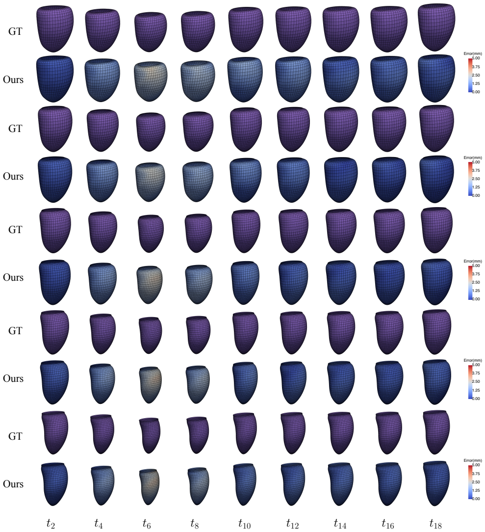
- [§4.5.1, Fig. 8] The color scale in Fig. 8 for error maps is not defined in the caption (the same color bar appears to cover 0–5 mm); a clear color bar and a statement of the displayed cardiac phases would improve readability.
- [§3.3, Eq. (8)] The notation 'we take M as e in Eq. (2)(3)' is confusing; the distinction between the undeformed material points M and the ellipsoid primitive e should be spelled out when defining the global deformation step M'(t_q) = M(t_q) ∘ q_g.
- [Abstract and §1] The claim that this is the first learning-based approach for 3D true heart wall motion recovery from 2D apparent motion cues is a novelty assertion that is difficult to verify and not essential; consider softening it to 'a learning-based approach' and citing related learned motion-recovery works where applicable.
Circularity Check
No significant circularity: the synthetic benchmark is self-consistent but not circular; the absence of real-tagged-MRI validation is an external-validity limitation, not a derivation-level circularity.
full rationale
The paper's derivation chain is not circular. Ground-truth material points M(t_q) are generated by interpolating ED/ES υNDM fits (Eq. 6, Supp. Eqs. 13–15), and SPAMM datapoints S(t_q) are computed from the same meshes by mesh-plane clipping (Step 4). The network Ψ is then trained with an ℓ₂ position loss (Eq. 11) against these independently generated labels, on a disjoint subject split (200/100/200), and it must solve a genuine inverse problem from sparse exact cues: the apparent cues are a function of the ground truth, but the ground truth is not a function of the network output or of the network's fitted parameters. The shared υNDM representation between simulator and network means the target motion lies in the model class, but this does not make the prediction equivalent to its inputs by construction, and the ablation results (Tables 2–3) show non-trivial dependence on architecture choices. The cited prior work [39] for the ES twisting value and [55] for NDM is published, externally reviewable support rather than an unverified self-citation chain. Supplementary Section 8 explicitly concedes that missing intermediate cardiac phases can make synthetic data deviate from real data distributions, and validation is radiologist QC rather than quantitative real-MRI evaluation; this is an external-validity caveat about the simulation-to-simulation nature of the demonstration, not evidence that any equation reduces to its own input.
Assumptions & free parameters
free parameters (5)
- Temporal interpolation scalars s_x(tq), s_y(tq), s_z(tq) =
hand-set arrays in Supplementary Eqs. 13-15
- ES twisting parameter function tau_es(u,w) =
not specified numerically; set to normal subject value from [39]
- Loss weights lambda_d, lambda_s =
0.1, 0.05
- k-NN neighbor count k in cross-attention =
64
- Shape parameters a0, a1, a2, a3 fitted to ED/ES point clouds =
not reported
assumptions (4)
- domain assumption Interpolation between ED and ES υNDMs with the given scalars produces physiologically realistic heart wall motion.
- domain assumption SPAMM datapoints computed by mesh-plane clipping with linear interpolation are a sufficient representation of tagged MRI apparent motion cues.
- standard math Diffeomorphic point flow preserves the topology of the heart wall under deformation.
- domain assumption The hybrid point transformer can learn the mapping from apparent motion cues to true motion with sufficient accuracy given the training data.
Cite this review
Pith. "Pith review of Learning Volumetric Neural Deformable Models to Recover 3D Regional Heart Wall Motion from Multi-Planar Tagged MRI." pith.science (2026). https://pith.science/paper/C74KOY7D
@misc{pith2026241115233,
  author       = {Pith},
  title        = {Pith review of: Learning Volumetric Neural Deformable Models to Recover 3D Regional Heart Wall Motion from Multi-Planar Tagged MRI},
  year         = {2026},
  howpublished = {\url{https://pith.science/paper/C74KOY7D}},
  note         = {Machine review of arXiv:2411.15233}
}
abstract
Multi-planar tagged MRI is the gold standard for regional heart wall motion evaluation. However, accurate recovery of the 3D true heart wall motion from a set of 2D apparent motion cues is challenging, due to incomplete sampling of the true motion and difficulty in information fusion from apparent motion cues observed on multiple imaging planes. To solve these challenges, we introduce a novel class of volumetric neural deformable models ($\upsilon$NDMs). Our $\upsilon$NDMs represent heart wall geometry and motion through a set of low-dimensional global deformation parameter functions and a diffeomorphic point flow regularized local deformation field. To learn such global and local deformation for 2D apparent motion mapping to 3D true motion, we design a hybrid point transformer, which incorporates both point cross-attention and self-attention mechanisms. While use of point cross-attention can learn to fuse 2D apparent motion cues into material point true motion hints, point self-attention hierarchically organised as an encoder-decoder structure can further learn to refine these hints and map them into 3D true motion. We have performed experiments on a large cohort of synthetic 3D regional heart wall motion dataset. The results demonstrated the high accuracy of our method for the recovery of dense 3D true motion from sparse 2D apparent motion cues. Project page is at https://github.com/DeepTag/VolumetricNeuralDeformableModels.
Figures


Figures from the paper (8 more)


Reference graph
Works this paper leans on
-
[1]
Dense: displacement encoding with stimulated echoes in cardiac functional mri
Anthony H Aletras, Shujun Ding, Robert S Balaban, and Han Wen. Dense: displacement encoding with stimulated echoes in cardiac functional mri. Journal of magnetic resonance (San Diego, Calif.: 1997), 137(1):247, 1999. 2
work page 1997
-
[2]
Myocardial strain imaging: review of general principles, validation, and sources of discrepancies
Mihaela Silvia Amzulescu, Mathieu De Craene, Hel ´en`e Langet, Agnes Pasquet, David Vancraeynest, Anne- Catherine Pouleur, Jean-Louis Vanoverschelde, and BL Ger- ber. Myocardial strain imaging: review of general principles, validation, and sources of discrepancies. European Heart Journal-Cardiovascular Imaging, 20(6):605–619, 2019. 1
work page 2019
-
[3]
Mr imaging of motion with spatial modulation of magnetization
Leon Axel and Lawrence Dougherty. Mr imaging of motion with spatial modulation of magnetization. Radiology, 171 (3):841–845, 1989. 1
work page 1989
-
[4]
Tagged mag- netic resonance imaging of the heart: a survey
Leon Axel, Albert Montillo, and Daniel Kim. Tagged mag- netic resonance imaging of the heart: a survey. Medical im- age analysis, 9(4):376–393, 2005. 3
work page 2005
-
[5]
Wenjia Bai, Wenzhe Shi, Antonio de Marvao, Timothy JW Dawes, Declan P O’Regan, Stuart A Cook, and Daniel Rueckert. A bi-ventricular cardiac atlas built from 1000+ high resolution mr images of healthy subjects and an anal- ysis of shape and motion. Medical image analysis , 26(1): 133–145, 2015. 4, 6, 7, 1
work page 2015
-
[6]
Functional analysis, Sobolev spaces and partial differential equations
Haim Brezis and Haim Br ´ezis. Functional analysis, Sobolev spaces and partial differential equations. Springer, 2011. 4
work page 2011
-
[7]
Analysis of 3-d myocardial motion in tagged mr images using nonrigid image registration
Raghavendra Chandrashekara, Raad H Mohiaddin, and Daniel Rueckert. Analysis of 3-d myocardial motion in tagged mr images using nonrigid image registration. IEEE Transactions on Medical Imaging, 23(10):1245–1250, 2004. 3
work page 2004
-
[8]
Qi Chang, Zhennan Yan, Meng Ye, Kanski Mikael, Subhi Al’Aref, Leon Axel, and Dimitris N Metaxas. An unsuper- vised 3d recurrent neural network for slice misalignment cor- rection in cardiac mr imaging. In International Workshop on Statistical Atlases and Computational Models of the Heart , pages 141–150. Springer, 2021. 1
work page 2021
Show all 67 references
-
[9]
Neural ordinary differential equa- tions
Ricky TQ Chen, Yulia Rubanova, Jesse Bettencourt, and David K Duvenaud. Neural ordinary differential equa- tions. Advances in neural information processing systems , 31, 2018. 4
2018
-
[10]
Automated 3d motion tracking using gabor filter bank, robust point matching, and deformable models
Ting Chen, Xiaoxu Wang, Sohae Chung, Dimitris Metaxas, and Leon Axel. Automated 3d motion tracking using gabor filter bank, robust point matching, and deformable models. IEEE Transactions on Medical Imaging , 29(1):1–11, 2009. 3
2009
-
[11]
Population-based studies of myocardial hypertrophy: high resolution cardiovascular magnetic reso- nance atlases improve statistical power
Antonio de Marvao, Timothy JW Dawes, Wenzhe Shi, Christopher Minas, Niall G Keenan, Tamara Diamond, Giu- liana Durighel, Giovanni Montana, Daniel Rueckert, Stu- art A Cook, et al. Population-based studies of myocardial hypertrophy: high resolution cardiovascular magnetic reso-...
2014
-
[12]
Three-dimensional my- ocardial strain reconstruction from tagged mri using a cylin- drical b-spline model
Xiang Deng and Thomas S Denney. Three-dimensional my- ocardial strain reconstruction from tagged mri using a cylin- drical b-spline model. IEEE Transactions on Medical Imag- ing, 23(7):861–867, 2004. 3
2004
-
[13]
Reconstruction of 3-d left ventricular motion from planar tagged cardiac mr im- ages: An estimation theoretic approach
Thomas S Denney and Jerry L Prince. Reconstruction of 3-d left ventricular motion from planar tagged cardiac mr im- ages: An estimation theoretic approach. IEEE transactions on medical imaging, 14(4):625–635, 1995. 3
1995
-
[14]
Model-free reconstruction of three-dimensional myocardial strain from planar tagged mr images
Thomas S Denney Jr and Elliot R McVeigh. Model-free reconstruction of three-dimensional myocardial strain from planar tagged mr images. Journal of Magnetic Resonance Imaging, 7(5):799–810, 1997. 3
1997
-
[15]
Vari- ational problems on flows of diffeomorphisms for image matching
Paul Dupuis, Ulf Grenander, and Michael I Miller. Vari- ational problems on flows of diffeomorphisms for image matching. Quarterly of applied mathematics , pages 587– 600, 1998. 4
1998
-
[16]
Computer Graphics: Principles and Prac- tice
James D Foley. Computer Graphics: Principles and Prac- tice. Addison-Wesley, 1996. 4
1996
-
[17]
Visual- ization of multidirectional regional left ventricular dynamics by high-temporal-resolution tissue phase mapping
Daniela F ¨oll, Bernd Jung, Felix Staehle, Elfriede Schilli, Christoph Bode, J¨urgen Hennig, and Michael Markl. Visual- ization of multidirectional regional left ventricular dynamics by high-temporal-resolution tissue phase mapping. Journal of Magnetic Resonance Imaging: An Of...
2009
-
[18]
Three-dimensional modeling for functional analysis of car- diac images, a review
Alejandro F Frangi, Wiro J Niessen, and Max A Viergever. Three-dimensional modeling for functional analysis of car- diac images, a review. IEEE transactions on medical imag- ing, 20(1):2–5, 2001. 1, 3
2001
-
[19]
Utnet: a hybrid transformer architecture for medical image segmen- tation
Yunhe Gao, Mu Zhou, and Dimitris N Metaxas. Utnet: a hybrid transformer architecture for medical image segmen- tation. In Medical Image Computing and Computer Assisted Intervention–MICCAI 2021: 24th International Conference, Strasbourg, France, September 27–October 1, 2021, Pr...
2021
-
[20]
Myocardial strain and torsion quan- tified by cardiovascular magnetic resonance tissue tagging: studies in normal and impaired left ventricular function
Marco JW G ¨otte, Tjeerd Germans, Iris K R ¨ussel, Jaco JM Zwanenburg, J Tim Marcus, Albert C van Rossum, and Dirk J van Veldhuisen. Myocardial strain and torsion quan- tified by cardiovascular magnetic resonance tissue tagging: studies in normal and impaired left ventricular ...
2002
-
[21]
Neural mesh flow: 3d manifold mesh gener- ation via diffeomorphic flows
Kunal Gupta. Neural mesh flow: 3d manifold mesh gener- ation via diffeomorphic flows. University of California, San Diego, 2020. 6
2020
-
[22]
Dilated cardiomyopathy: normalized multiparamet- ric myocardial strain predicts contractile recovery
Matthew C Henn, Christopher P Lawrance, Julia Kar, Brian P Cupps, Kevin Kulshrestha, Danielle Koerner, Kath- leen Wallace, Susan Joseph, Greg Ewald, and Michael K Pasque. Dilated cardiomyopathy: normalized multiparamet- ric myocardial strain predicts contractile recovery. The ...
2015
-
[23]
Spatio-temporal tracking of myocar- dial deformations with a 4-d b-spline model from tagged mri
Jiantao Huang, Dana Abendschein, Victor G Davila-Roman, and Amir A Amini. Spatio-temporal tracking of myocar- dial deformations with a 4-d b-spline model from tagged mri. IEEE transactions on medical imaging , 18(10):957– 972, 1999. 2, 3
1999
-
[24]
The analysis of cardiac function: a continuum approach
Peter J Hunter and Bruce H Smaill. The analysis of cardiac function: a continuum approach. Progress in biophysics and molecular biology, 52(2):101–164, 1988. 3
1988
-
[25]
Myocardial tagging by cardiovascu- lar magnetic resonance: evolution of techniques–pulse se- 9 quences, analysis algorithms, and applications
El-Sayed H Ibrahim. Myocardial tagging by cardiovascu- lar magnetic resonance: evolution of techniques–pulse se- 9 quences, analysis algorithms, and applications. Journal of Cardiovascular Magnetic Resonance, 13(1):36, 2011. 1
2011
-
[26]
Batch normalization: Accelerating deep net- work training by reducing internal covariate shift
Sergey Ioffe. Batch normalization: Accelerating deep net- work training by reducing internal covariate shift. arXiv preprint arXiv:1502.03167, 2015. 7
2015 arXiv
-
[27]
Detailed analysis of myocardial motion in volunteers and patients using high- temporal-resolution mr tissue phase mapping
Bernd Jung, Daniela F ¨oll, Petra B ¨ottler, Steffen Petersen, J¨urgen Hennig, and Michael Markl. Detailed analysis of myocardial motion in volunteers and patients using high- temporal-resolution mr tissue phase mapping. Journal of Magnetic Resonance Imaging: An Official Journ...
2006
-
[28]
Myocardial tissue tracking with two- dimensional cine displacement-encoded mr imaging: devel- opment and initial evaluation
Daniel Kim, Wesley D Gilson, Christopher M Kramer, and Frederick H Epstein. Myocardial tissue tracking with two- dimensional cine displacement-encoded mr imaging: devel- opment and initial evaluation. Radiology, 230(3):862–871,
-
[29]
Function: delineation of normal human left ventricular twist throughout systole by tagged cine magnetic resonance imag- ing
Christine H Lorenz, John S Pastorek, and Jeffrey M Bundy. Function: delineation of normal human left ventricular twist throughout systole by tagged cine magnetic resonance imag- ing. Journal of Cardiovascular Magnetic Resonance , 2(2): 97–108, 2000. 1
2000
-
[30]
Lv shape and motion: B- spline-based deformable model and sequential motion de- composition
Guo Luo and Pheng-Ann Heng. Lv shape and motion: B- spline-based deformable model and sequential motion de- composition. IEEE Transactions on Information Technology in Biomedicine, 9(3):430–446, 2005. 3
2005
-
[31]
Calculation of three-dimensional left ventricular strains from biplanar tagged mr images
Christopher C Moore, Walter G O’Dell, Elliot R McVeigh, and Elias A Zerhouni. Calculation of three-dimensional left ventricular strains from biplanar tagged mr images. Journal of Magnetic Resonance Imaging, 2(2):165–175, 1992. 3
1992
-
[32]
Three-dimensional systolic strain patterns in the normal human left ventricle: characteri- zation with tagged mr imaging
Christopher C Moore, Carlos H Lugo-Olivieri, Elliot R McVeigh, and Elias A Zerhouni. Three-dimensional systolic strain patterns in the normal human left ventricle: characteri- zation with tagged mr imaging. Radiology, 214(2):453–466,
-
[33]
Spline surface interpolation for calculating 3-d ventricular strains from mri tissue tag- ging
Michael J Moulton, Lawrence L Creswell, Stephen W Downing, Ricardo L Actis, Barna A Szabo, Michael W Van- nier, and Michael K Pasque. Spline surface interpolation for calculating 3-d ventricular strains from mri tissue tag- ging. American Journal of Physiology-Heart and Circul...
1996
-
[34]
Math- ematical model of geometry and fibrous structure of the heart
PM Nielsen, IJ Le Grice, BH Smaill, and PJ Hunter. Math- ematical model of geometry and fibrous structure of the heart. American Journal of Physiology-Heart and Circula- tory Physiology, 260(4):H1365–H1378, 1991. 3
1991
-
[35]
Three-dimensional myocardial deformations: calculation with displacement field fitting to tagged mr images
Walter G O’Dell, Christopher C Moore, William C Hunter, Elias A Zerhouni, and Elliot R McVeigh. Three-dimensional myocardial deformations: calculation with displacement field fitting to tagged mr images. Radiology, 195(3):829– 835, 1995. 2, 3
1995
-
[36]
Cardiac motion estimation by joint alignment of tagged mri sequences
Estanislao Oubel, Mathieu De Craene, AO Hero, Amir Pour- morteza, Marina Huguet, Gustavo Avegliano, Bart H Bij- nens, and Alejandro F Frangi. Cardiac motion estimation by joint alignment of tagged mri sequences. Medical image analysis, 16(1):339–350, 2012. 3
2012
-
[37]
Four-dimensional b-spline based motion analysis of tagged mr images: intro- duction and in vivo validation
Cengizhan Ozturk and Elliot R McVeigh. Four-dimensional b-spline based motion analysis of tagged mr images: intro- duction and in vivo validation. Physics in Medicine & Biol- ogy, 45(6):1683, 2000. 3
2000
-
[38]
Fast tracking of cardiac motion using 3d-harp
Li Pan, Jerry L Prince, Jo ˜ao AC Lima, and Nael F Osman. Fast tracking of cardiac motion using 3d-harp. IEEE trans- actions on Biomedical Engineering, 52(8):1425–1435, 2005. 2, 3
2005
-
[39]
Analysis of left ventricular wall motion based on volumetric deformable models and mri-spamm
Jinah Park, Dimitri Metaxas, and Leon Axel. Analysis of left ventricular wall motion based on volumetric deformable models and mri-spamm. Medical image analysis , 1(1):53– 71, 1996. 1, 2, 3, 4, 6, 8
1996
-
[40]
Deformable models with parameter functions for car- diac motion analysis from tagged mri data
Jinah Park, Dimitri Metaxas, Alistair A Young, and Leon Axel. Deformable models with parameter functions for car- diac motion analysis from tagged mri data. IEEE Transac- tions on Medical Imaging, 15(3):278–289, 1996. 2, 3, 4
1996
-
[41]
Pointnet++: Deep hierarchical feature learning on point sets in a metric space
Charles Ruizhongtai Qi, Li Yi, Hao Su, and Leonidas J Guibas. Pointnet++: Deep hierarchical feature learning on point sets in a metric space. Advances in neural information processing systems, 30, 2017. 6
2017
-
[42]
Deep implicit statistical shape mod- els for 3d medical image delineation
Ashwin Raju, Shun Miao, Dakai Jin, Le Lu, Junzhou Huang, and Adam P Harrison. Deep implicit statistical shape mod- els for 3d medical image delineation. In Proceedings of the AAAI Conference on Artificial Intelligence, pages 2135– 2143, 2022. 6
2022
-
[43]
Accelerated whole-heart 3d cspamm for myocardial motion quantification
Andrea K Rutz, Salome Ryf, Sven Plein, Peter Boesiger, and Sebastian Kozerke. Accelerated whole-heart 3d cspamm for myocardial motion quantification. Magnetic Resonance in Medicine: An Official Journal of the International Society for Magnetic Resonance in Medicine, 59(4):755–...
2008
-
[44]
Myocardial tagging with 3d-cspamm
Salome Ryf, Marcus A Spiegel, Michael Gerber, and Peter Boesiger. Myocardial tagging with 3d-cspamm. Journal of Magnetic Resonance Imaging: An Official Journal of the In- ternational Society for Magnetic Resonance in Medicine, 16 (3):320–325, 2002. 1
2002
-
[45]
Im- proved dynamic cardiac phantom based on 4d nurbs and tagged mri
W Paul Segars, David S Lalush, Eric C Frey, Dinesh Manocha, Michael A King, and Benjamin MW Tsui. Im- proved dynamic cardiac phantom based on 4d nurbs and tagged mri. IEEE transactions on nuclear science , 56(5): 2728–2738, 2009. 3
2009
-
[46]
Myocar- dial strain imaging: Theory, current practice, and the future
Otto A Smiseth, Oliver Rider, Marta Cvijic, Ladislav Valkoviˇc, Espen W Remme, and Jens-Uwe V oigt. Myocar- dial strain imaging: Theory, current practice, and the future. JACC: Cardiovascular Imaging, 2024. 1
2024
-
[47]
Topology-preserving shape recon- struction and registration via neural diffeomorphic flow
Shanlin Sun, Kun Han, Deying Kong, Hao Tang, Xiangyi Yan, and Xiaohui Xie. Topology-preserving shape recon- struction and registration via neural diffeomorphic flow. In Proceedings of the IEEE/CVF Conference on Computer Vi- sion and Pattern Recognition, pages 20845–20855, 2022. 7
2022
-
[48]
Dynamic 3 d models with local and global deformations: deformable su- perquadrics
Demetri Terzopoulos and Dimitri Metaxas. Dynamic 3 d models with local and global deformations: deformable su- perquadrics. IEEE Transactions on pattern analysis and ma- chine intelligence, 13(7):703–714, 1991. 3
1991
-
[49]
Cardiac motion and defor- mation recovery from mri: a review
Hui Wang and Amir A Amini. Cardiac motion and defor- mation recovery from mri: a review. IEEE transactions on medical imaging, 31(2):487–503, 2011. 3
2011
-
[50]
Meshless deformable models for lv motion analysis
Xiaoxu Wang, Dimitis Metaxas, Ting Chen, and Leon Axel. Meshless deformable models for lv motion analysis. In 2008 10 IEEE Conference on Computer Vision and Pattern Recogni- tion, pages 1–8. IEEE, 2008. 2, 3, 7
2008
-
[51]
Meshless deformable models for 3d cardiac motion and strain analysis from tagged mri
Xiaoxu Wang, Ting Chen, Shaoting Zhang, Jo ¨el Schaerer, Zhen Qian, Suejung Huh, Dimitris Metaxas, and Leon Axel. Meshless deformable models for 3d cardiac motion and strain analysis from tagged mri. Magnetic resonance imag- ing, 33(1):146–160, 2015. 2, 3, 5, 7
2015
-
[52]
Car- diac motion recovery using an incompressible b-solid model
Huisi Wu, Pheng-Ann Heng, and Tien-Tsin Wong. Car- diac motion recovery using an incompressible b-solid model. Medical engineering & physics, 35(7):958–968, 2013. 3
2013
-
[53]
Deeptag: An unsupervised deep learning method for motion tracking on cardiac tagging magnetic resonance images
Meng Ye, Mikael Kanski, Dong Yang, Qi Chang, Zhennan Yan, Qiaoying Huang, Leon Axel, and Dimitris Metaxas. Deeptag: An unsupervised deep learning method for motion tracking on cardiac tagging magnetic resonance images. In Proceedings of the IEEE/CVF conference on computer vi- ...
2021
-
[54]
Sequencemorph: A unified unsupervised learning framework for motion tracking on cardiac image sequences
Meng Ye, Dong Yang, Qiaoying Huang, Mikael Kanski, Leon Axel, and Dimitris N Metaxas. Sequencemorph: A unified unsupervised learning framework for motion tracking on cardiac image sequences. IEEE Transactions on Pattern Analysis and Machine Intelligence, 2023. 1, 7
2023
-
[55]
Neural deformable models for 3d bi- ventricular heart shape reconstruction and modeling from 2d sparse cardiac magnetic resonance imaging
Meng Ye, Dong Yang, Mikael Kanski, Leon Axel, and Dimitris Metaxas. Neural deformable models for 3d bi- ventricular heart shape reconstruction and modeling from 2d sparse cardiac magnetic resonance imaging. In Proceedings of the IEEE/CVF International Conference on Computer Vi...
2023
-
[56]
Unsupervised exemplar-based image-to-image translation and cascaded vision transformers for tagged and untagged cardiac cine mri registration
Meng Ye, Mikael Kanski, Dong Yang, Leon Axel, and Dim- itris Metaxas. Unsupervised exemplar-based image-to-image translation and cascaded vision transformers for tagged and untagged cardiac cine mri registration. In Proceedings of the IEEE/CVF Winter Conference on Applications...
2024
-
[57]
Shapes and diffeomorphisms
Laurent Younes. Shapes and diffeomorphisms . Springer,
-
[58]
Three-dimensional motion and deformation of the heart wall: estimation with spatial modulation of magnetization–a model-based approach
Alistair A Young and Leon Axel. Three-dimensional motion and deformation of the heart wall: estimation with spatial modulation of magnetization–a model-based approach. Ra- diology, 185(1):241–247, 1992. 2, 3
1992
-
[59]
Three-dimensional left ventricular deformation in hypertrophic cardiomyopathy
Alistair A Young, Christopher M Kramer, Victor A Fer- rari, Leon Axel, and Nathaniel Reichek. Three-dimensional left ventricular deformation in hypertrophic cardiomyopathy. Circulation, 90(2):854–867, 1994. 2, 3
1994
-
[60]
Deformable models with sparsity constraints for cardiac motion analysis
Yang Yu, Shaoting Zhang, Kang Li, Dimitris Metaxas, and Leon Axel. Deformable models with sparsity constraints for cardiac motion analysis. Medical image analysis, 18(6):927– 937, 2014. 2, 3
2014
-
[61]
Point transformer
Hengshuang Zhao, Li Jiang, Jiaya Jia, Philip HS Torr, and Vladlen Koltun. Point transformer. In Proceedings of the IEEE/CVF international conference on computer vision, pages 16259–16268, 2021. 5, 7
2021
-
[62]
Fast automatic heart chamber segmentation from 3d ct data using marginal space learning and steerable features
Yefeng Zheng, Adrian Barbu, Bogdan Georgescu, Michael Scheuering, and Dorin Comaniciu. Fast automatic heart chamber segmentation from 3d ct data using marginal space learning and steerable features. In 2007 IEEE 11th Inter- national Conference on Computer Vision, pages 1–8. IEEE,
2007
-
[63]
Characterization of three- dimensional myocardial deformation in the mouse heart: An mr tagging study
Jia Zhong, Wei Liu, and Xin Yu. Characterization of three- dimensional myocardial deformation in the mouse heart: An mr tagging study. Journal of Magnetic Resonance Imaging, 27(6):1263–1270, 2008. 3 11 Learning Volumetric Neural Deformable Models to Recover 3D Regional Heart W...
2008
-
[64]
Definition of MAE The quantitative metric for 3D motion recovery accuracy evaluation we used is the mean absolute error (MAE). It is the average of Euclidean distance ( ℓ2 norm) between pre- dicted material points ˆM(tq) and ground truth M(tq) over a full cardiac cycle excludi...
-
[65]
Temporal Interpolation Scalars The temporal interpolation scalars s(tq) used in Eq. (6) in the main text are set as follows: sx(tq) = [0.000, 0.090, 0.150, 0.350, 0.550, 0.750, 1.000, 0.920, 0.780, 0.650, 0.580, 0.540, 0.410, 0.370, 0.240, 0.210, 0.180, 0.160, 0.120, 0.080], (...
-
[66]
While we use a 3D CMR dataset [5] of normal subjects and data in- terpolation defined in Eq
Quality Control of Synthetic Data One of our contributions in this work is the 3D heart wall geometry and motion simulation framework. While we use a 3D CMR dataset [5] of normal subjects and data in- terpolation defined in Eq. (6) in the main text to achieve this goal, the mi...
-
[67]
11, we show more detailed 3D heart wall mo- tion recovery results
More Detailed Results In Fig. 11, we show more detailed 3D heart wall mo- tion recovery results. In the supplementary folder 3D_Regional_Wall_Motion_Recovery_Example, we show these examples in dynamic videos. From these results, we can see that our method can accurately recove...
Reviewed August 12, 2026 · model on record in the stance chip above.
Discussion (0). Continue with ORCID to comment.