REVIEW 4 major objections 5 minor 61 references
Whispering-Gallery-Mode Resonators for Detection and Classification of Free-Flowing Nanoparticles and Cells through Photoacoustic Signatures
T0 review · 4 major / 5 minor · reviewed 2026-08-12 · deepseek-v4-flash
Pith's one-line read A microbubble optical resonator detects and classifies free-flowing nanoparticles and cells in native solutions, including whole blood, via pulsed-light photoacoustic signatures, with no surface binding.
desk verdict Clever acoustic-WGM sensing concept that removes the surface-binding constraint, but the classification claims outrun the data—needs independent validation and matched controls. read the letter →
The pith
A machine-rendered reading of the paper's core claim, the machinery that carries it, and where it could break.
The reading
What carries the argument
The central object is a microbubble whispering-gallery-mode resonator (MBR): a silica capillary locally inflated into a bubble, with high-Q optical modes at ~780 nm confined in the wall and a hollow core connected to a microfluidic channel. The mechanism is acoustic-mediated sensing: pulsed 532 nm light absorbed by a target generates thermoelastic ultrasound in the MHz range; the ultrasound propagates through the solution, strains the wall, and modulates the probe's resonance, read out as transmission changes at a fixed wavelength. The thick wall isolates the optical mode from the sample, preserving Q and SNR even in absorbing media such as black dye or whole blood. For classification, the PA time traces are converted by FFT and fed to a 1D convolutional neural network with prototype embeddings in a 64-dimensional latent space, where each species or geometry is represented by one learned prototype and new samples are assigned to the nearest prototype.
What would settle it
Collect a fresh dataset for the same five red-blood-cell species on a different day, at a different laser spot, or from different animal donors, train the same CNN and prototype model on one session's data, and test on the other; if accuracy falls to near chance, the 'photoacoustic fingerprint' is not intrinsic to cell type.
Extended reading notes
Core claim
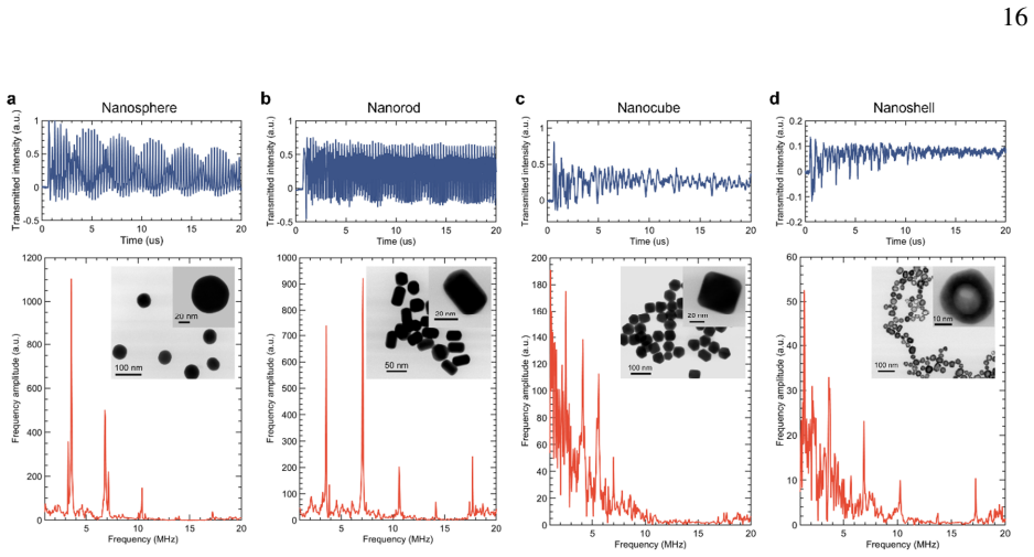
The central discovery is that a photoacoustic event generated by an unbound particle flowing freely in a microfluidic channel can be read out by a whispering-gallery-mode resonator whose optical field never touches the sample, and that the spectral content of that event is rich enough to identify the particle. The authors demonstrate this with four gold-nanoparticle geometries and five species of red blood cells, as well as whole blood from five species, using only 1% dilutions and no purification, labeling, or incubation. Detected signals show SNR exceeding 30 dB, and the excitation laser can be scanned along the capillary so that particles up to 6 mm from the resonator are still measured. Classification is done by transforming PA signals with FFT and feeding the spectra to a 1D CNN with prototype learning, achieving 99.6% accuracy on AuNP geometry and 98.7% accuracy on red-blood-cell species in random 80/20 train/test splits. The paper presents these results as the first demonstration of free-flowing particle detection beyond the reach of the evanescent field of an optical micro-sensor.
Load-bearing premise
The classification results stand only if the frequency-domain differences the model learns are intrinsic to the particle or cell type, and not artifacts of measurement session, laser spot position, flow conditions, concentration, or sample batch; the paper's random split of 1000 signals per class into training and test sets does not test that.
Editorial extensions
If this is right
- Particles and cells can be measured while flowing, without capture, functionalization, or waiting for diffusion to a sensing surface.
- The sensing volume is extended along the capillary: scanning the excitation laser lets the same resonator detect targets far from the optical mode, with arrival delay giving position information.
- Because the optical mode is isolated from the sample, the sensor keeps its Q and signal-to-noise ratio in strongly absorbing or scattering media, enabling direct measurement in whole blood.
- Photoacoustic spectra carry shape-, composition-, and morphology-dependent fingerprints: gold-nanoparticle geometry and red-blood-cell species are classified with high accuracy by a CNN with prototype learning.
- Matching the excitation wavelength to the target's absorption (e.g., 532 nm for hemoglobin) gives selectivity in complex mixtures, and PA amplitude tracks concentration for quantitative detection.
Reading between the lines
- A direct test of whether these fingerprints are intrinsic to the particle would be cross-session or cross-batch generalization: train on one day or donor and test on another; the present random 80/20 split within 1000-signal-per-class datasets does not establish that, so improved generalization would strengthen the 'physical fingerprint' interpretation.
- If the signatures generalize, the same platform could screen for red-blood-cell disorders with altered shape or hemoglobin state—the paper names sickle-cell disease, hemoglobin C, and thalassemia as future targets—without staining or fixing cells.
- Multi-wavelength excitation (for instance via the frequency comb the paper mentions) would add an optical-absorption axis to the acoustic-response axis, likely separating many more particle classes than a single 532 nm wavelength can.
- The acoustic-mediated readout should work for any sufficiently absorbing target, not just gold and hemoglobin; testing non-gold nanoparticles, bacteria, or cultured cells would reveal how broad the fingerprinting mechanism is.
Editorial analysis
A structured set of objections, weighed in public.
Referee Report
Summary. The manuscript proposes and demonstrates an optofluidic whispering-gallery-mode (WGM) microbubble resonator sensor that detects photoacoustic (PA) signals from free-flowing nanoparticles and cells. Absorption of 532 nm pulsed light by analytes in the microfluidic core generates acoustic waves that modulate the WGM readout, allowing detection away from the resonator surface and directly in complex media such as whole blood. The authors report PA signals from four gold nanoparticle geometries and five red blood cell species, and they use a one-dimensional CNN with prototype learning to classify the signals, reporting near-perfect accuracy on held-out subsets of the collected datasets. The central claim is that particles and cells can be identified and classified by an intrinsic photoacoustic fingerprint that captures shape, composition, and morphology.
Significance. If the claims are borne out by stronger validation, this is a potentially significant advance for optical microsensor technology: the acoustic-mediated detection mechanism spatially decouples the optical mode from the analyte, extends the sensing volume beyond the evanescent field, and offers a path to label-free, immobilization-free analysis in complex biological matrices. The design of a thick-walled microbubble resonator that protects the WGM from solution absorption and scattering is an elegant solution to a known limitation of evanescent sensing. However, the paper does not provide public data, code, or machine-checked analyses, and the experimental validation as presented is not sufficient to establish that the machine-learning classification reflects intrinsic physical fingerprints rather than session- or batch-specific systematics.
major comments (4)
- [Supplementary Section 5.1; Methods: Machine learning] The classification evaluation splits 1000 PA signals per class acquired in a single measurement session into a random 80/20 train/test split. This controls only within-session variance; it does not control for measurement session, laser alignment, flow conditions, sample batch, or biological donor. The near-perfect test accuracy reported in Supplementary Table 1 is therefore compatible with the model learning session-specific systematics rather than intrinsic photoacoustic fingerprints. The central claim that the signatures are physical fingerprints requires independent measurement sessions, freshly prepared samples, and biological replicates, with the classifier tested across sessions.
- [Materials and methods: Sample preparation; Supplementary Fig. S4] The four AuNP geometries are measured at different concentrations: spheres, rods, and cubes at 10^10 nps/mL versus shells at 10^12 nps/mL. Since PA amplitude increases with concentration (Supplementary Fig. S4a) and the CNN input includes FFT amplitudes, the classifier could separate the shell class by amplitude or concentration rather than by geometry. The claim that different shapes of the same material produce unique PA signals requires matched-concentration measurements or an amplitude-invariant feature analysis; without this, the geometry-fingerprint claim is confounded.
- [Results: Detection of cells and identification from different species; Materials and methods] Each red blood cell 'species' is represented by a single purchased sample, so species identity is confounded with donor, preparation, and shipment batch. The repeatability shown in Fig. 3f is within a single sample. Independent biological replicates and cross-batch training/test splits are needed before the results support species classification rather than sample-specific discrimination.
- [Results: Nanoparticle sensing; Fig. 1b; Supplementary Fig. S1] No experimental negative control is shown for the photoacoustic detection mechanism: no-analyte or non-absorbing-particle runs are not reported. The schematic in Fig. 1b(ii) and the discussion in Supplementary Fig. S1 state that no PA signal is detected in the absence of the analyte, but this is asserted rather than demonstrated. A control with pure buffer and with a suspension of non-absorbing particles would establish that the recorded transients originate from target absorption rather than from laser-induced or flow-induced artifacts.
minor comments (5)
- [Supplementary Section 5.1] The headings 'Reb blood cells' and 'AU nanoparticles' contain typos and should be corrected to 'Red blood cells' and 'Au nanoparticles'.
- [Methods: Photoacoustic excitation] The phrase 'reputation rate' should be 'repetition rate.'
- [Fig. 3f and Supplementary Fig. S9] Please clarify whether the 10 repeat measurements are repeated acquisitions from one sample or independent sample preparations, and report the statistical spread (for example, standard deviation or confidence intervals) rather than only offset spectra.
- [Supplementary Section 5.3] The accuracy, recall, precision, and F1 scores are reported as point values without confidence intervals; reporting bootstrap or repeated-split intervals would help assess stability.
- [Data availability] The statement that data are not publicly available limits reproducibility; at minimum, processed spectra and the trained-model code should be made available so the classification results can be audited.
Circularity Check
No significant circularity: the machine-learning classification is a genuine held-out prediction, and the physical fingerprint interpretation is an inductive claim with external-validity caveats, not a derivation from the fitted inputs.
full rationale
The paper's central experimental chain is not circular. The sensor readout (acoustic modulation of a WGM) is characterized independently of the classification targets: the extended sensing range and concentration response are measured directly (Supplementary Figs. S3-S4), and the mode-protection claim is tested with black dye (Supplementary Fig. S2). The ML pipeline is supervised: class labels are inputs to training, and the reported accuracies (0.9961 for AuNPs, 0.9870 for RBCs) are evaluated on a held-out 20% of the same datasets (Supplementary Section 5.1), so the test predictions are not on the same data points used to fit the model. No fitted parameter is renamed as a prediction, and no equation reduces to its own input. The claim that PA spectra are 'photoacoustic fingerprints' capturing shape, composition, or morphology is an inductive interpretation of measured differences; possible confounds such as concentration mismatch for nanoshells or single-session data collection are external-validity limitations, not circularity. Self-citations (refs. 18, 37, 38) are background technical references for WGM sensing and microbubble fabrication and are not load-bearing for the classification result. The prototype-learning limitation statement acknowledges that CNNs can learn surface statistical regularities, but that is a robustness caveat, not a circularity admission. Overall, no circular step is identifiable by the paper's own equations or definitions.
Assumptions & free parameters
free parameters (3)
- prototype loss weight lambda =
0.1
- number of prototype embeddings per class =
1
- CNN architecture and training hyperparameters =
2 conv layers, 4 kernels, stride 20, latent dimension 64, dropout 0.3, learning rate 0.001, 100 epochs
assumptions (5)
- domain assumption Photoacoustic effect: absorption of pulsed laser light by a particle causes rapid thermoelastic expansion generating an acoustic wave.
- domain assumption Acoustic waves propagating through the fluid and capillary wall modulate the WGM optical resonance, and the resulting transmission change is the measured signal.
- domain assumption The WGM field is confined within the thick silica wall with negligible overlap with the liquid core, so sample absorption and refractive-index changes do not affect the optical readout.
- domain assumption At 532 nm, hemoglobin absorption dominates the photoacoustic signal of red blood cells, enabling selective detection in whole blood.
- ad hoc to paper The training and test signals for each class are representative of the class, with no systematic batch or session differences that the model could exploit.
Cite this review
Pith. "Pith review of Whispering-Gallery-Mode Resonators for Detection and Classification of Free-Flowing Nanoparticles and Cells through Photoacoustic Signatures." pith.science (2026). https://pith.science/paper/I33RO2C4
@misc{pith2026241115373,
  author       = {Pith},
  title        = {Pith review of: Whispering-Gallery-Mode Resonators for Detection and Classification of Free-Flowing Nanoparticles and Cells through Photoacoustic Signatures},
  year         = {2026},
  howpublished = {\url{https://pith.science/paper/I33RO2C4}},
  note         = {Machine review of arXiv:2411.15373}
}
read the original abstract
Micro and nanoscale particles are crucial in various fields, from biomedical imaging to environmental processes. While conventional spectroscopy and microscopy methods for characterizing these particles often involve bulky equipment and complex sample preparation, optical micro-sensors have emerged as a promising alternative. However, their broad applicability is limited by the need for surface binding and difficulty in differentiating between sensing targets. This study introduces an optofluidic, high-throughput optical microresonator sensor that captures subtle acoustic signals generated by particles absorbing pulsed light energy. This novel approach enables real-time, label-free detection and interrogation of particles and cells in their native environments across an extended sensing volume. By leveraging unique optical absorption properties, our technique selectively detects and classifies flowing particles without surface binding, even in complex matrices like whole blood samples. We demonstrate the measurement of gold nanoparticles with diverse geometries and different species of red blood cells amidst other cellular elements and proteins. These particles are identified and classified based on their photoacoustic fingerprint, which captures shape, composition, and morphology features. This work opens new avenues for rapid, reliable, and high-throughput particle and cell identification in clinical and industrial applications, offering a valuable tool for understanding complex biological and environmental systems.
Figures


Figures from the paper (1 more)

Reference graph
Works this paper leans on
-
[1]
Cho, E. J. et al. Nanoparticle characterization: State of the art, challenges, and emerging technologies. Mol Pharm 10 , 2093–2110 (2013)
work page 2013
-
[2]
Baek, M. J. et al. Tailoring renal-clearable zwitterionic cyclodextrin for colorectal cancer- selective drug delivery. Nat Nanotechnol 18 , 945–956 (2023)
work page 2023
-
[3]
Riley, R. S. & Day, E. S. Gold nanoparticle-medi ated photothermal therapy: applications and opportunities for multimodal cancer treatment. Wiley Interdiscip Rev Nanomed Nanobiotechnol 9, (2017)
work page 2017
-
[4]
Arami, H. et al. Remotely controlled near-infrared-triggered photothermal treatment of brain tumours in freely behaving mice using gold nanostars. Nat Nanotechnol 17 , 1015– 1022 (2022)
work page 2022
-
[5]
Herran, M. et al. Plasmonic bimetallic two-dimensional supercrystals for H2 generation. Nat Catal 6, 1205–1214 (2023)
work page 2023
-
[6]
Wang, F. et al. Tuning upconversion through energy migration in core-shell nanoparticles. Nat Mater 10 , 968–973 (2011)
work page 2011
-
[7]
Shi, Y. Z. et al. Sculpting nanoparticle dynamics for single-bacteria-level screening and direct binding-efficiency measurement. Nat Commun 9, 1–11 (2018)
work page 2018
-
[8]
Stiles, P. L., Dieringer, J. A., Shah, N. C. & V an Duyne, R. P. Surface-enhanced Raman spectroscopy. Annu. Rev. Anal. Chem. 1, 601–626 (2008)
work page 2008
Show all 61 references
-
[9]
& Yang, L
Mao, W., Li, Y., Jiang, X., Liu, Z. & Yang, L. A whispering-gallery scanning microprobe for Raman spectroscopy and imaging. Light Sci Appl 12 , (2023)
2023
-
[10]
Xu, Y. et al. Artificial intelligence: A powerful paradigm for scientific research. Innovation 2, 4 (2021)
2021
-
[11]
Ho, C. S. et al. Rapid identification of pathogenic bacteria using Raman spectroscopy and deep learning. Nat Commun 10 , 4927 (2019)
2019
-
[12]
M., Gorelikov, I., Matsuura, N
Strohm, E. M., Gorelikov, I., Matsuura, N. & Ko lios, M. C. Modeling photoacoustic spectral features of micron-sized particles. Phys Med Biol 59 , 5795–5810 (2014)
2014
-
[13]
Tam, A. C. Applications of photoacoustic sensin g techniques. Rev Mod Phys 58 , 381–431 (1986)
1986
-
[14]
& Wang, L
Xu, M. & Wang, L. V. Photoacoustic imaging in b iomedicine. Review of Scientific Instruments 77 , 4 (2006)
2006
-
[15]
Yao, J. et al. High-speed label-free functional photoacoustic microscopy of mouse brain in action. Nat Methods 12 , 407–410 (2015)
2015
-
[16]
Zharov, V. P. Ultrasharp nonlinear photothermal and photoacoustic resonances and holes beyond the spectral limit. Nat Photonics 5, 110–116 (2011)
2011
-
[17]
Li, C. & V. Wang, L. Photoacoustic tomography a nd sensing in biomedicine. Clin Lymphoma 54 , R59–R97 (2009)
2009
-
[18]
& Yang, L
Loyez, M., Adolphson, M., Liao, J. & Yang, L. F rom Whispering Gallery Mode Resonators to Biochemical Sensors. ACS Sens 8, 2440–2470 (2023)
2023
-
[19]
Yu, D. et al. Whispering-gallery-mode sensors for biological and physical sensing. Nature Reviews Methods Primers 1, 83 (2021)
2021
-
[20]
H., Maier, S
Altug, H., Oh, S. H., Maier, S. A. & Homola, J. Advances and applications of nanophotonic biosensors. Nat Nanotechnol 17 , 5–16 (2022)
2022
-
[21]
Heylman, K. D. et al. Optical Microresonators for Sensing and Transduction: A Materials Perspective. Advanced Materials 29 , 1–29 (2017). 13
2017
-
[22]
Vollmer, F. et al. Protein detection by optical shift of a resonant microcavity. Appl Phys Lett 80 , 4057–4059 (2002)
2002
-
[23]
Baaske, M. D. & Vollmer, F. Optical observation of single atomic ions interacting with plasmonic nanorods in aqueous solution. Nat Photonics 10 , 733–739 (2016)
2016
-
[24]
& Fan, X
Chen, Y.-C., Chen, Q. & Fan, X. Lasing in blood . Optica 3, 809 (2016)
2016
-
[25]
Martino, N. et al. Wavelength-encoded laser particles for massively multiplexed cell tagging. Nat Photonics 13 , 720–727 (2019)
2019
-
[26]
Schubert, M. et al. Monitoring contractility in cardiac tissue with cellular resolution using biointegrated microlasers. Nat Photonics 14 , 452–458 (2020)
2020
-
[27]
Heylman, K. D. et al. Optical microresonators as single-particle absorption spectrometers. Nat Photonics 10 , 788–795 (2016)
2016
-
[28]
P., Madsen, L
Mauranyapin, N. P., Madsen, L. S., Taylor, M. A ., Waleed, M. & Bowen, W. P. Evanescent single-molecule biosensing with quantum-limited precision. Nat Photonics 11 , 477–481 (2017)
2017
-
[29]
Yu, X. C. et al. Single-molecule optofluidic microsensor with interface whispering gallery modes. Proceedings of the National Academy of Sciences 119 , 6 (2022)
2022
-
[30]
Y., Xavier, J., Joshi, L
Watanabe, K., Wu, H. Y., Xavier, J., Joshi, L. T. & Vollmer, F. Single Virus Detection on Silicon Photonic Crystal Random Cavities. Small 18 , 15 (2022)
2022
-
[31]
K., Zhu, J., Kim, W
He, L., Özdemir, Ş. K., Zhu, J., Kim, W. & Yang, L. Detecting single viruses and nanoparticles using whispering gallery microlasers. Nat Nanotechnol 6, 428–432 (2011)
2011
-
[32]
Shao, L. et al. Detection of single nanoparticles and lentiviruses using microcavity resonance broadening. Advanced Materials 25 , 5616–5620 (2013)
2013
-
[33]
Wu, X. et al. Optofluidic laser for dual-mode sensitive biomolecular detection with a large dynamic range. Nat Commun 5, 1–7 (2014)
2014
-
[34]
Tang, S.-J. et al. Single-particle vibrational spectroscopy using optical microresonators. Nat Photonics 17 , 951–956 (2023)
2023
-
[35]
K., Carmon, T
Jing, H., Lü, H., Özdemir, S. K., Carmon, T. & Nori, F. Nanoparticle sensing with a spinning resonator. Optica 5, 1424 (2018)
2018
-
[36]
N., Murugan, G
Zervas, M. N., Murugan, G. S., Petrovich, M. N. & Wilkinson, J. S. Hollow-bottle optical microresonators. Opt Express 19 , 2915–2917 (2011)
2011
-
[37]
& Yang, L
Liao, J. & Yang, L. Optical whispering-gallery mode barcodes for high-precision and wide-range temperature measurements. Light Sci Appl 10 , 32 (2021)
2021
-
[38]
Liao, J., Wu, X., Liu, L. & Xu, L. Fano resonan ce and improved sensing performance in a spectral-simplified optofluidic micro-bubble resonator by introducing selective modal losses. Opt Express 24 , 8574 (2016)
2016
-
[39]
& Liu, C
Yang, H., Zhang, X., Yin, F. & Liu, C. Robust C lassification with Convolutional Prototype Learning. in Proceedings of the IEEE conference on computer vision and pattern recognition 3474–3482 (2018)
2018
-
[40]
Shen, B. et al. Integrated turnkey soliton microcombs. Nature 582 , 365–369 (2020)
2020
-
[41]
Friedlein, J. T. et al. Dual-comb photoacoustic spectroscopy. Nat Commun 11 , (2020)
2020
-
[42]
& Razansky, D
Manohar, S. & Razansky, D. Photoacoustics: a hi storical review. Adv Opt Photonics 8, 586–617 (2016)
2016
-
[43]
Wang, L. V. & Yao, J. A practical guide to phot oacoustic tomography in the life sciences. Nat Methods 13 , 627–638 (2016)
2016
-
[44]
Daniels, D. E. et al. Human cellular model systems of β-thalassemia enable in-depth analysis of disease phenotype. Nat Commun 14 , 1 (2023). 14
2023
-
[45]
For hemophilia and thalassemia, a new era of ‘one-and-done’ gene therapies has arrived
Sheridan, C. For hemophilia and thalassemia, a new era of ‘one-and-done’ gene therapies has arrived. Nat Biotechnol 40 , 1531–1533 (2022)
2022
-
[46]
M., Hunter, I
Fathy, A., Sabry, Y. M., Hunter, I. W., Khalil, D. & Bourouina, T. Direct Absorption and Photoacoustic Spectroscopy for Gas Sensing and Analysis: A Critical Review. Laser Photon Rev 16 , 8 (2022)
2022
-
[47]
Szegedy, C. et al. Intriguing properties of neural networks. 2nd International Conference on Learning Representations, ICLR 2014 - Conference Track Proceedings 1–10 (2014). 15 Figure 1: Label-free all-optical photoacoustic (PA) microresonator-based sensors. a, Principle of lon...
2014
-
[48]
Sensing mechanism of long-range acoustic-mediated sensing Conventional resonance shift sensing is a straightforward method to measure the target of interest by tracking induced changes in the resonance wavele ngth (or frequency) of an optical resonator. Fig. S1 (a) illustrates...
-
[49]
Consequently, when the core filling switches from d eionized (DI) water to black dye, there's negligible change observed in the WGM spectra
Excellent stability for reliable measurement Since the light field is confined within the thick wall of the microbubble resonator (MBR), direct interaction between whispering gallery modes (WGMs) and the sample solution is prevented. Consequently, when the core filling switche...
-
[50]
Extended sensing range By adjusting the beam spot position of the pulsed l aser, we achieve an extended sensing range, enabling the detection of particles even at distanc es far from the microresonator. Fig. S3 (a) illustrates the scanning of the pulse laser along t he transpa...
-
[51]
PA signal amplitude as a function of nanoparticle concentration and input laser power The photoacoustic response observed from the gold nanoparticles varies with the concentration of the nanoparticles in the solution and the input pow er delivered by the pulsed laser. Fig. S4 ...
-
[52]
(a) The PA signals are preprocessed with Fourier Transform as input data
AI analysis for feature learning and particle classification Figure S5: Machine learning pipeline for classifying PA signals. (a) The PA signals are preprocessed with Fourier Transform as input data. The learned prototype embe ddings of the different classes are used for infer...
2000
-
[53]
Consequently, small variations of the spectrum may lead to misclassifications by a well-trained CNN
A highly sensitive optical sensor (e.g., WGM) can detect small environmental c hanges. Consequently, small variations of the spectrum may lead to misclassifications by a well-trained CNN. To enhance the robustness of the model, we implement prototype learning 6 (as described i...
-
[54]
With only the raw features, the data of different species overlap considerably, making it challenging to differentiate the different species
Principal Component Analysis (PCA) without prototype learning We performed PCA directly on the PA signals of red blood cells and whole blood samples, without the aid of AI. With only the raw features, the data of different species overlap considerably, making it challenging to...
-
[55]
These increased deviat ions can be attributed to the complex composition of whole blood
Deviations in signals from whole blood samples The PA signals obtained from whole blood samples ex hibit larger deviations compared to those from red blood cell samples. These increased deviat ions can be attributed to the complex composition of whole blood. In addition to red...
2000
-
[56]
& Haffner, P
Lecun, Y., Bottou, L., Bengio, Y. & Haffner, P. Gradient-based learning applied to document recognition. Proc. IEEE 86 , 2278–2324 (1998)
1998
-
[57]
Kim, U. J. et al. Drug classification with a spectral barcode obtained with a smartphone Raman spectrometer. Nat. Commun. 14 , 1–9 (2023)
2023
-
[58]
Huang, L. et al. Rapid, label-free histopathological diagnosis of liver cancer based on Raman spectroscopy and deep learning. Nat. Commun. 14 , (2023)
2023
-
[59]
Seddiki, K. et al. Cumulative learning enables convolutional neural network representations for small mass spectrometry data classification. Nat. Commun. 11 , (2020)
2020
-
[60]
Szegedy, C. et al. Intriguing properties of neural networks. in 2nd International Conference on Learning Representations, ICLR 2014 (2014)
2014
-
[61]
Robust classification with convolutional prototy pe learning
Yang, Hong-Ming, et al . "Robust classification with convolutional prototy pe learning." Proceedings of the IEEE conference on computer visi on and pattern recognition, CVPR 2018 (2018)
2018
Reviewed August 12, 2026 · model on record in the stance chip above.
Discussion (0). Continue with ORCID to comment.