REVIEW 4 major objections 6 minor 57 references
Accelerated Hydration Site Localization and Thermodynamic Profiling
T0 review · 4 major / 6 minor · reviewed 2026-08-12 · deepseek-v4-flash
Pith's one-line read This paper presents a geometric deep network that predicts hydration site locations and thermodynamic profiles from a static protein structure in one shot, at accuracy close to explicit-water molecular dynamics.
desk verdict Large-scale ML surrogate for MD-based hydration site profiling, with real code and data, but the headline accuracy claim rests on emulating one MD pipeline and the single external x-ray check is far weaker. read the letter →
The pith
A machine-rendered reading of the paper's core claim, the machinery that carries it, and where it could break.
The reading
What carries the argument
A two-stage equivariant graph neural network. The first stage seeds water nodes at every atom with solvent-accessible surface area above 0.1, then applies five SE(3)-equivariant attention layers with distance-based graph updates so that predicted water positions rotate and translate with the protein while protein atoms stay fixed; it is trained with a Gaussian-mixture Kullback-Leibler loss against WATsite hydration sites. The second stage builds a graph connecting protein atoms and hydration sites within 8 Å, applies graph attention layers, and regresses per-site enthalpy and entropy. The equivariant updates are what let the network represent water-water and water-protein interactions without imposing a fixed grid.
What would settle it
Run the trained model on a set of proteins with high-resolution neutron crystallography or multi-force-field consensus hydration sites, and measure the ground-truth recovery rate. If recovery at 1.0 Å falls to the level of the appendix's x-ray prediction hit rate (11.5%) rather than the MD-based 80.2%, the claim that the model achieves near-dynamics-level accuracy on real experimental waters is not supported.
Extended reading notes
Core claim
The central claim is that hydration site localization and thermodynamic profiling, normally requiring lengthy explicit-water molecular dynamics, can be done in one shot by an equivariant graph neural network with near-dynamics-level accuracy. The model first places water nodes at solvent-exposed protein atoms and refines their positions through five equivariant attention layers, learning multi-body water networks that respect the protein surface. A second graph attention network then predicts the enthalpy and entropy of each site, and the predicted water displacement free energies correlate with measured binding affinities in the MUP ligand series (Pearson R = 0.930). The authors argue the model is fast, robust to point mutations and conformational changes, and generalizes to unseen protein sequences.
Load-bearing premise
The entire pipeline inherits whatever bias sits in the MD-based WATsite labels: if the simulated hydration site locations, occupancies, or entropy estimates are wrong, the model's predictions are wrong in the same way, and the appendix's lower x-ray hit rates indicate that this mismatch with experiment is real.
Editorial extensions
If this is right
- Hydration site positions and thermodynamic profiles for a single protein structure are produced on the seconds timescale, making high-throughput scanning across many conformations practical.
- The model can attach physically reasonable water networks to predicted protein structures from structure-prediction models, which normally omit water, opening hydration analysis for uncharacterized proteins.
- Displacing predicted hydration sites yields desolvation free energies that correlate with experimental ligand binding affinities in the MUP case study, suggesting a fast scoring component for lead optimization.
- The model responds to point mutations and conformational rearrangements without retraining, so it can be applied to mutant analysis and to conformational ensembles.
- The architecture could be integrated into deep learning pipelines for co-folding, dynamics, or free-energy estimation that currently ignore explicit water.
Reading between the lines
- The reported accuracy is dominated by first-shell waters: second-shell sites are recovered at only 26.9% within 1 Å, so practical applications should treat second-shell predictions as candidates for refinement rather than final answers.
- Because the training labels come from one MD force field and WATsite's occupancy and entropy approximations, absolute thermodynamic values may shift if labels are regenerated with a different water model, even if rankings are stable.
- The same two-stage architecture could be retrained on hydration data for protein-ligand complexes or protein-protein interfaces, extending the method beyond apo protein surfaces toward biologics and co-solvent mapping.
- The appendix's x-ray comparison shows much lower hit rates than the MD-based evaluation, suggesting that a sharper experimental validation against high-resolution neutron or curated crystallographic waters would be a more demanding test of the model's real-world accuracy.
Editorial analysis
A structured set of objections, weighed in public.
Referee Report
Summary. The manuscript presents a two-stage deep learning pipeline for protein hydration site analysis. In the first stage, an equivariant graph neural network is trained to predict hydration site coordinates from a static protein structure, with a loss based on a Gaussian mixture surrogate for KL divergence. In the second stage, a graph attention network predicts per-site enthalpy and entropy values, conditioned on hydration site coordinates. Training and test labels are generated by a single WATsite/Desmond explicit-water MD protocol applied to 4,148 protein structures, split by sequence similarity at 35% identity. The authors report ground-truth recovery rates and prediction hit rates on the held-out test set, layer- and occupancy-stratified results, thermodynamic correlations against WATsite references, several qualitative case studies (DsbA, TIM, HSP90, Clarin-2, MUP), and a MUP ligand-binding study correlating predicted desolvation free energies with experimental affinities. The central claim is that the model achieves near dynamics-level hydration site localization and thermodynamic profiling in a one-shot, fixed-time manner.
Significance. If the central claims hold, this work would be a substantial practical advance: it would replace expensive, multi-hour explicit-water MD simulations for hydration site localization and thermodynamic profiling with a seconds-scale deep learning model, and it would enable hydration analysis for AlphaFold-predicted structures and large conformational ensembles. The paper has concrete strengths: a large training dataset with a sequence-similarity-based held-out split, publicly available code and data, clearly specified evaluation metrics (GTRR/PHR), and several qualitative case studies probing generalization to mutations, conformational change, and membrane proteins. However, the load-bearing evidence for the 'near dynamics-level accuracy' and 'high fidelity' claims is incomplete. The only external experimental comparison, x-ray crystallographic waters in Appendix B.1, shows markedly lower prediction hit rates than the WATsite-based evaluation, and this discrepancy is not discussed in the main text.
major comments (4)
- [Appendix B.1, Table 7] The x-ray validation reports a prediction hit rate of 6.26% at 0.5 Å and 11.5% at 1.0 Å against crystallographic waters, whereas the WATsite-based test set (Table 1) reports 48.3% and 65.9% at the same cutoffs. The main text (Sections 1 and 4.1, and the Conclusion) claims that the model 'can reproduce results from molecular dynamics and experimentally resolved structures with high fidelity,' but the large gap between the WATsite and x-ray PHR values is not discussed anywhere in the main text. If incompleteness of deposited crystal waters is the explanation, the PHR should be recomputed on a curated set of ordered first-shell waters; if the gap persists, the experimental support for the central claim is much weaker than stated. This needs to be addressed explicitly.
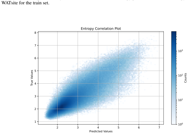
- [Section 4.2 and Section 4.3.5] The thermodynamic profiling evaluation in Table 5 is explicitly performed 'given the coordinates of the true hydration sites' (Section 4.2). In actual use, the thermodynamic model consumes predicted coordinates, so coordinate errors can propagate into enthalpy and entropy errors. The only evaluation on predicted coordinates is the MUP case study (Section 4.3.5), which uses 12 complexes, a displacement tolerance of 2.4 Å selected post hoc, and 'transformed predictions obtained by a linear regression of the experimental values on the model predictions' (footnote to Table 6). The reported Pearson R of 0.930 is therefore not a direct measure of the untransformed model's predictive correlation, and the tolerance choice is not justified. Please report thermodynamic accuracy on the held-out test set using predicted hydration sites with a fixed, pre-specified matching protocol, and report both raw and calibrated correlations for the MUP study.
- [Section 3.3 and Eq. (7)] All training targets and all test references are generated by a single computational protocol: 20 ns Desmond simulations with 50 kcal/mol/Ų heavy-atom restraints, WATsite clustering, and the entropy estimate in Eq. (7) based on an external-mode probability density. Because the model is trained and evaluated on labels from this same protocol, Tables 1–5 establish that the network imitates WATsite/Desmond output on sequence-distant proteins; they do not independently establish agreement with converged explicit-water thermodynamics or with experiment. This is a limitation of the evidence, not a circularity of the held-out split, but the paper should state it plainly and, if possible, include a small convergence or force-field sensitivity check (for example, longer simulations or a second water model on a subset of proteins) to bound the magnitude of protocol-dependent bias.
- [Section 3.1.1, Eq. (5)] The loss L1 in Eq. (5) is described as 'a simplified surrogate for the symmetrized Kullback-Leibler divergence KL(p|q) + KL(q|p),' but as written it evaluates the mixture densities q and p only at the component centers (q(u_j) and p(x_j)) rather than integrating over the mixture components, so it is not a standard symmetrized KL between the two Gaussian mixtures. The paper does not provide a derivation of this surrogate, nor any sensitivity analysis for the Gaussian width σ = 0.5 or the weight penalty α in Eq. (6). These hyperparameters directly shape the predicted spatial distribution, and the reader cannot judge whether the reported accuracies are robust to reasonable variations. Please clarify the relationship to the true symmetrized KL and provide an ablation or sensitivity analysis.
minor comments (6)
- [Throughout] There are several typographical errors, including 'it's' for 'its' in the Abstract and Section 1, and 'signficantly' for 'significantly' in Section 4.1. These should be corrected.
- [Table 6] The PDB ID column lists both '1IO6' and '1I06' for different ligands (SBT, PT, IPT, ET, MT); this appears to be a typographical inconsistency, and the correct identifiers should be verified.
- [Appendix B.2, Table 13] The last row of Table 13 repeats 'r = 1.5' instead of 'r = 2.0', which is presumably a typo. Please correct the cutoff labels.
- [References] Reference [35] contains malformed author names ('Xiaohan Kuang, Zhaoqian Su Su, ... Jesse , Tyler Derr') and should be cleaned up. Reference [44] lacks a title; the full Bowers et al. SC2006 paper title should be provided.
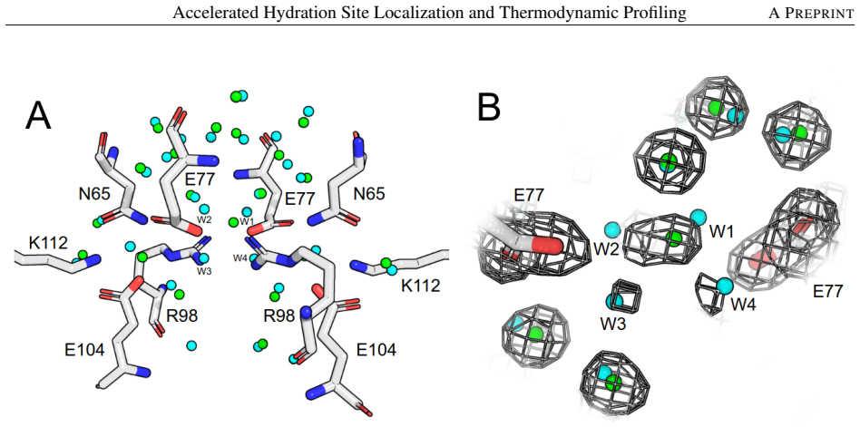
- [Section 4.3.1 and Section 4.3.3] The case study claims that all hydration sites were identified 'within 1.0 Å' (DsbA) or that '20/21 crystal waters predicted within 1.0 Å' (TIM), but the criteria for matching crystal waters to predictions (e.g., any-vs-unique matching, handling of absent waters) are not specified. Please state the matching rule used in these qualitative comparisons.
- [Abstract and Conclusion] The phrase 'near dynamics-level accuracy' is defined only through the WATsite-based metrics; given the x-ray results in Table 7, the wording should be qualified (e.g., 'near WATsite/Desmond-level accuracy') or the experimental validation should be strengthened.
Circularity Check
MUP case-study 'predictions' are post hoc linear regressions on the experimental values; core MD-emulator evaluation is held-out but the experimental validation claim reduces to a fit.
-
fitted input called prediction
[Section 4.3.5 (Major Urinary Protein), Table 6 footnote and Figure 9]
"Shown are the transformed predictions obtained by a linear regression of the experimental values on the model predictions. ... At a water displacement tolerance level of 2.4 Å, we obtain a Pearson R correlation of 0.930."
Table 6's 'Method' column and Figure 9's 'Predictions' are expressly 'transformed predictions obtained by a linear regression of the experimental values on the model predictions.' The reported desolvation free energies are therefore fitted to the very experimental binding free energies they are then said to correlate with (R=0.930). These are not independent predictions: they are the in-sample least-squares fit of experiment on model output, so the agreement statistic is a fit diagnostic rather than a validation. The comparison to MM-GB/SA is also unequal because MM-GB/SA is not given the same post hoc regression. This makes the paper's main experimental thermodynamic validation reduce, by the paper's own footnote, to a fit rather than a prediction.
full rationale
The central localization and thermodynamic models are trained on and evaluated against WATsite/Desmond-derived labels on sequence-distant held-out proteins; that is a standard supervised benchmark and not circular by construction, since the network does not see the WATsite output of the test proteins at inference. The low hit rates against crystallographic waters (Appendix B.1, Table 7) are a serious external-validity concern but are not a circularity. Several citations are to the authors' own WATsite and entropy-estimation work ([28], [39], [46]); these are method citations to the tool that generated the labels, not a self-citation chain that forces the conclusions. The one definite circular step is the MUP case study: the paper explicitly transforms its predictions by linear regression on the experimental values and then presents the resulting R=0.930 as evidence of agreement with experiment. Because this showcased experimental validation reduces to an in-sample fit, the overall circularity score is 6: the central claim has independent held-out content, but one reported 'prediction' is fitted by construction.
Assumptions & free parameters
free parameters (9)
- Gaussian width sigma in loss =
0.5 Å
- Loss weight alpha =
not reported
- Prediction weight cutoff wc =
0.035
- Clustering distance threshold =
2 Å
- Cluster weight cutoff =
0.1
- Graph edge distance cutoffs =
6 Å and 8 Å
- SASA initial placement cutoff =
0.1
- MUP water displacement tolerance =
2.4 Å
- MUP linear calibration coefficients =
not reported
assumptions (4)
- domain assumption WATsite MD-derived locations and thermodynamic values are accurate ground truth for hydration sites
- ad hoc to paper The Gaussian mixture surrogate for KL divergence with sigma=0.5 is a suitable training objective
- domain assumption Representative protein selection by UniProt ID and 35% sequence-similarity split prevents leakage
- domain assumption The force field and explicit water model used in Desmond simulations produce reliable hydration thermodynamics
Cite this review
Pith. "Pith review of Accelerated Hydration Site Localization and Thermodynamic Profiling." pith.science (2026). https://pith.science/paper/UJHFX4TU
@misc{pith2026241115618,
  author       = {Pith},
  title        = {Pith review of: Accelerated Hydration Site Localization and Thermodynamic Profiling},
  year         = {2026},
  howpublished = {\url{https://pith.science/paper/UJHFX4TU}},
  note         = {Machine review of arXiv:2411.15618}
}
read the original abstract
Water plays a fundamental role in the structure and function of proteins and other biomolecules. The thermodynamic profile of water molecules surrounding a protein are critical for ligand binding and recognition. Therefore, identifying the location and thermodynamic behavior of relevant water molecules is important for generating and optimizing lead compounds for affinity and selectivity to a given target. Computational methods have been developed to identify these hydration sites, but are largely limited to simplified models that fail to capture multi-body interactions, or dynamics-based methods that rely on extensive sampling. Here we present a method for fast and accurate localization and thermodynamic profiling of hydration sites for protein structures. The method is based on a geometric deep neural network trained on a large, novel dataset of explicit water molecular dynamics simulations. We confirm the accuracy and robustness of our model on experimental data and demonstrate it's utility on several case studies.
Figures


Figures from the paper (9 more)




Reference graph
Works this paper leans on
-
[1]
Hugh Savage and Alexander Wlodawer. [11] determination of water structure around biomolecules using x-ray and neutron diffraction methods. In Methods in enzymology, volume 127, pages 162–183. Elsevier, 1986
work page 1986
-
[2]
Protein hydration dynamics in solution: a critical survey
Bertil Halle. Protein hydration dynamics in solution: a critical survey. Philosophical Transactions of the Royal Society of London. Series B: Biological Sciences, 359(1448):1207–1224, 2004
work page 2004
-
[3]
Three-dimensional model of purple membrane obtained by electron microscopy
Richard Henderson and P Nigel T Unwin. Three-dimensional model of purple membrane obtained by electron microscopy. Nature, 257(5521):28–32, 1975
work page 1975
-
[4]
The way to nmr structures of proteins
Kurt Wüthrich. The way to nmr structures of proteins. Nature structural biology, 8(11):923–925, 2001
work page 2001
-
[5]
Balázs Zoltán Zsidó, Bayartsetseg Bayarsaikhan, Rita Börzsei, Viktor Szél, Violetta Mohos, and Csaba Hetényi. The advances and limitations of the determination and applications of water structure in molecular engineering. International Journal of Molecular Sciences, 24(14):11784, 2023
work page 2023
-
[6]
Andriy Kovalenko and Fumio Hirata. Three-dimensional density profiles of water in contact with a solute of arbitrary shape: a rism approach. Chemical Physics Letters, 290(1-3):237–244, 1998
work page 1998
-
[7]
Andriy Kovalenko and Fumio Hirata. Self-consistent description of a metal–water interface by the kohn–sham density functional theory and the three-dimensional reference interaction site model. The Journal of chemical physics, 110(20):10095–10112, 1999
work page 1999
-
[8]
Water molecules in protein–ligand interfaces
Eva Nittinger, Paul Gibbons, Charles Eigenbrot, Doug R Davies, Brigitte Maurer, Christine L Yu, James R Kiefer, Andreas Kuglstatter, Jeremy Murray, Daniel F Ortwine, et al. Water molecules in protein–ligand interfaces. evaluation of software tools and sar comparison. Journal of computer-aided molecular design, 33:307–330, 2019
work page 2019
Show all 57 references
-
[9]
Modelling of solvent positions around polar groups in proteins
William R Pitt and Julia M Goodfellow. Modelling of solvent positions around polar groups in proteins. Protein Engineering, Design and Selection, 4(5):531–537, 1991
1991
-
[10]
Evaluating free energies of binding and conservation of crystallographic waters using szmap
Alexander S Bayden, Demetri T Moustakas, Diane Joseph-McCarthy, and Michelle L Lamb. Evaluating free energies of binding and conservation of crystallographic waters using szmap. Journal of chemical information and modeling, 55(8):1552–1565, 2015
2015
-
[11]
High end gpcr design: crafted ligand design and druggability analysis using protein structure, lipophilic hotspots and explicit water networks
Jonathan S Mason, Andrea Bortolato, Dahlia R Weiss, Francesca Deflorian, Benjamin Tehan, and Fiona H Marshall. High end gpcr design: crafted ligand design and druggability analysis using protein structure, lipophilic hotspots and explicit water networks. In Silico Pharmacology...
2013
-
[12]
A common reference framework for analyzing/comparing proteins and ligands
Massimo Baroni, Gabriele Cruciani, Simone Sciabola, Francesca Perruccio, and Jonathan S Mason. A common reference framework for analyzing/comparing proteins and ligands. fingerprints for ligands and proteins (flap): theory and application. Journal of chemical information and m...
2007
-
[13]
Acquaalta: a directional approach to the solvation of ligand–protein complexes
Gianluca Rossato, Beat Ernst, Angelo Vedani, and Martin Smiesko. Acquaalta: a directional approach to the solvation of ligand–protein complexes. Journal of chemical information and modeling, 51(8):1867–1881, 2011
2011
-
[14]
Algorithm for the systematic solvation of proteins based on the directionality of hydrogen bonds
Angelo Vedani and David W Huhta. Algorithm for the systematic solvation of proteins based on the directionality of hydrogen bonds. Journal of the American Chemical Society, 113(15):5860–5862, 1991
1991
-
[15]
Solvated docking: introducing water into the modelling of biomolecular complexes
Aalt DJ Van Dijk and Alexandre MJJ Bonvin. Solvated docking: introducing water into the modelling of biomolecular complexes. Bioinformatics, 22(19):2340–2347, 2006
2006
-
[16]
Placement of water molecules in protein structures: from large-scale evaluations to single-case examples
Eva Nittinger, Florian Flachsenberg, Stefan Bietz, Gudrun Lange, Robert Klein, and Matthias Rarey. Placement of water molecules in protein structures: from large-scale evaluations to single-case examples. Journal of Chemical Information and Modeling, 58(8):1625–1637, 2018
2018
-
[17]
Watgen: an algorithm for modeling water networks at protein–protein interfaces
Huynh-Hoa Bui, Alexandra J Schiewe, and Ian S Haworth. Watgen: an algorithm for modeling water networks at protein–protein interfaces. Journal of computational chemistry, 28(14):2241–2251, 2007
2007
-
[18]
Prediction of water and metal binding sites and their affinities by using the fold-x force field.Proceedings of the National Academy of Sciences, 102(29):10147–10152, 2005
Joost WH Schymkowitz, Frederic Rousseau, Ivo C Martins, Jesper Ferkinghoff-Borg, Francois Stricher, and Luis Serrano. Prediction of water and metal binding sites and their affinities by using the fold-x force field.Proceedings of the National Academy of Sciences, 102(29):10147...
2005
-
[19]
Waterdock 2.0: Water placement prediction for holo- structures with a pymol plugin
Akshay Sridhar, Gregory A Ross, and Philip C Biggin. Waterdock 2.0: Water placement prediction for holo- structures with a pymol plugin. PloS one, 12(2):e0172743, 2017
2017
-
[20]
Protein pharmacophore selection using hydration-site analysis.Journal of chemical information and modeling, 52(4):1046–1060, 2012
Bingjie Hu and Markus A Lill. Protein pharmacophore selection using hydration-site analysis.Journal of chemical information and modeling, 52(4):1046–1060, 2012
2012
-
[21]
Analysis of factors influencing hydration site prediction based on molecular dynamics simulations
Ying Yang, Bingjie Hu, and Markus A Lill. Analysis of factors influencing hydration site prediction based on molecular dynamics simulations. Journal of Chemical Information and Modeling, 54(10):2987–2995, 2014. 14 Accelerated Hydration Site Localization and Thermodynamic Profi...
2014
-
[22]
Dissecting the influence of protein flexibility on the location and thermodynamic profile of explicit water molecules in protein–ligand binding
Ying Yang and Markus A Lill. Dissecting the influence of protein flexibility on the location and thermodynamic profile of explicit water molecules in protein–ligand binding. Journal of chemical theory and computation , 12(9):4578–4592, 2016
2016
-
[23]
Prediction of the favorable hydration sites in a protein binding pocket and its application to scoring function formulation
Yan Li, Yingduo Gao, M Katharine Holloway, and Renxiao Wang. Prediction of the favorable hydration sites in a protein binding pocket and its application to scoring function formulation. Journal of Chemical Information and Modeling, 60(9):4359–4375, 2020
2020
-
[24]
Modeling the hydration layer around proteins: Hypred
Jouko J Virtanen, Lee Makowski, Tobin R Sosnick, and Karl F Freed. Modeling the hydration layer around proteins: Hypred. Biophysical journal, 99(5):1611–1619, 2010
2010
-
[25]
Exploration of interfacial hydration networks of target–ligand complexes
Norbert Jeszenoi, Mónika Bálint, István Horváth, David van der Spoel, and Csaba Hetényi. Exploration of interfacial hydration networks of target–ligand complexes. Journal of chemical information and modeling , 56(1):148–158, 2016
2016
-
[26]
Mobility-based prediction of hydration structures of protein surfaces
Norbert Jeszen ˝oi, István Horváth, Mónika Bálint, David van der Spoel, and Csaba Hetényi. Mobility-based prediction of hydration structures of protein surfaces. Bioinformatics, 31(12):1959–1965, 2015
1959
-
[27]
Role of the active-site solvent in the thermodynamics of factor xa ligand binding
Robert Abel, Tom Young, Ramy Farid, Bruce J Berne, and Richard A Friesner. Role of the active-site solvent in the thermodynamics of factor xa ligand binding. Journal of the American Chemical Society, 130(9):2817–2831, 2008
2008
-
[28]
Watsite: Hydration site prediction program with pymol interface, 2014
Bingjie Hu and Markus A Lill. Watsite: Hydration site prediction program with pymol interface, 2014
2014
-
[29]
Watsite2
Ying Yang, Bingjie Hu, and Markus A Lill. Watsite2. 0 with pymol plugin: Hydration site prediction and visualization. Protein Function Prediction: Methods and Protocols, pages 123–134, 2017
2017
-
[30]
Ying Yang, Matthew R Masters, Amr H Mahmoud, Bingjie Hu, and Markus A Lill. Watsite. user guide a gpu-accelerated hydration site prediction program with pymol plugin. 2018
2018
-
[31]
Galaxywater-cnn: Prediction of water positions on the protein structure by a 3d-convolutional neural network
Sangwoo Park and Chaok Seok. Galaxywater-cnn: Prediction of water positions on the protein structure by a 3d-convolutional neural network. Journal of Chemical Information and Modeling, 62(13):3157–3168, 2022
2022
-
[32]
Hydraprot: A new deep learning tool for fast and accurate prediction of water molecule positions for protein structures
Andreas Zamanos, George Ioannakis, and Ioannis Z Emiris. Hydraprot: A new deep learning tool for fast and accurate prediction of water molecule positions for protein structures. Journal of Chemical Information and Modeling, 64(7):2594–2611, 2024
2024
-
[33]
Instantaneous generation of protein hydration properties from static structures
Ahmadreza Ghanbarpour, Amr H Mahmoud, and Markus A Lill. Instantaneous generation of protein hydration properties from static structures. Communications Chemistry, 3(1):188, 2020
2020
-
[34]
Water position prediction with se (3)-graph neural network
Sangwoo Park. Water position prediction with se (3)-graph neural network. bioRxiv, pages 2024–03, 2024
2024
-
[35]
Superwater: Predicting water molecule positions on protein structures by generative ai
Xiaohan Kuang, Zhaoqian Su Su, Yunchao Liu, Xiaobo Lin, Jesse , Tyler Derr, Yinghao Wu, and Jens Meiler. Superwater: Predicting water molecule positions on protein structures by generative ai. bioRxiv, pages 2024–11, 2024
2024
-
[36]
Prediction of molecular field points using se(3)-transformer model
Florian B Hinz, Amr H Mahmoud, and Markus A Lill. Prediction of molecular field points using se(3)-transformer model. Machine Learning: Science and Technology, 4(3):035016, aug 2023
2023
-
[37]
Pedregosa, G
F. Pedregosa, G. Varoquaux, A. Gramfort, V . Michel, B. Thirion, O. Grisel, M. Blondel, P. Prettenhofer, R. Weiss, V . Dubourg, J. Vanderplas, A. Passos, D. Cournapeau, M. Brucher, M. Perrot, and E. Duchesnay. Scikit-learn: Machine learning in Python. Journal of Machine Learni...
2011
-
[38]
Graph attention networks, 2018
Petar Veliˇckovi´c, Guillem Cucurull, Arantxa Casanova, Adriana Romero, Pietro Liò, and Yoshua Bengio. Graph attention networks, 2018
2018
-
[39]
Efficient and accurate hydration site profiling for enclosed binding sites
Matthew R Masters, Amr H Mahmoud, Ying Yang, and Markus A Lill. Efficient and accurate hydration site profiling for enclosed binding sites. Journal of chemical information and modeling, 58(11):2183–2188, 2018
2018
-
[40]
The pdbbind database: method- ologies and updates
Renxiao Wang, Xueliang Fang, Yipin Lu, Chao-Yie Yang, and Shaomeng Wang. The pdbbind database: method- ologies and updates. Journal of medicinal chemistry, 48(12):4111–4119, 2005
2005
-
[41]
Comparative assessment of scoring functions: the casf-2016 update
Minyi Su, Qifan Yang, Yu Du, Guoqin Feng, Zhihai Liu, Yan Li, and Renxiao Wang. Comparative assessment of scoring functions: the casf-2016 update. Journal of chemical information and modeling, 59(2):895–913, 2018
2016
-
[42]
Mmseqs2 enables sensitive protein sequence searching for the analysis of massive data sets
Martin Steinegger and Johannes Söding. Mmseqs2 enables sensitive protein sequence searching for the analysis of massive data sets. Nature biotechnology, 35(11):1026–1028, 2017
2017
-
[43]
Protein and ligand preparation: parameters, protocols, and influence on virtual screening enrichments
G Madhavi Sastry, Matvey Adzhigirey, Tyler Day, Ramakrishna Annabhimoju, and Woody Sherman. Protein and ligand preparation: parameters, protocols, and influence on virtual screening enrichments. Journal of computer-aided molecular design, 27:221–234, 2013. 15 Accelerated Hydra...
2013
-
[44]
Scalable algorithms for molecular dynam- ics simulations on commodity clusters
Kevin J Bowers, Edmond Chow, Huafeng Xu, Ron O Dror, Michael P Eastwood, Brent A Gregersen, John L Klepeis, Istvan Kolossvary, Mark A Moraes, Federico D Sacerdoti, et al. Scalable algorithms for molecular dynam- ics simulations on commodity clusters. In Proceedings of the 2006...
2006
-
[45]
Vmd: visual molecular dynamics
William Humphrey, Andrew Dalke, and Klaus Schulten. Vmd: visual molecular dynamics. Journal of molecular graphics, 14(1):33–38, 1996
1996
-
[46]
Calculation of thermodynamic properties of bound water molecules
Ying Yang, Amr HA Abdallah, and Markus A Lill. Calculation of thermodynamic properties of bound water molecules. Computational Drug Discovery and Design, pages 389–402, 2018
2018
-
[47]
Mechanisms of disulfide bond formation in nascent polypeptides entering the secretory pathway
Philip J Robinson and Neil J Bulleid. Mechanisms of disulfide bond formation in nascent polypeptides entering the secretory pathway. Cells, 9(9):1994, 2020
1994
-
[48]
A buried water network modulates the activity of the escherichia coli disulphide catalyst dsba
Geqing Wang, Jilong Qin, Anthony D Verderosa, Lilian Hor, Carlos Santos-Martin, Jason J Paxman, Jennifer L Martin, Makrina Totsika, and Begoña Heras. A buried water network modulates the activity of the escherichia coli disulphide catalyst dsba. Antioxidants, 12(2):380, 2023
2023
-
[49]
Towards out-of- distribution generalization: A survey
Jiashuo Liu, Zheyan Shen, Yue He, Xingxuan Zhang, Renzhe Xu, Han Yu, and Peng Cui. Towards out-of- distribution generalization: A survey. arXiv preprint arXiv:2108.13624, 2021
2021 arXiv
-
[50]
The critical role of the loops of triosephosphate isomerase for its oligomerization, dynamics, and functionality
Ataur R Katebi and Robert L Jernigan. The critical role of the loops of triosephosphate isomerase for its oligomerization, dynamics, and functionality. Protein Sci., 23(2):213–228, 2014
2014
-
[51]
Accurate structure prediction of biomolecular interactions with alphafold 3
Josh Abramson, Jonas Adler, Jack Dunger, Richard Evans, Tim Green, Alexander Pritzel, Olaf Ronneberger, Lindsay Willmore, Andrew J Ballard, Joshua Bambrick, et al. Accurate structure prediction of biomolecular interactions with alphafold 3. Nature, pages 1–3, 2024
2024
-
[52]
Generalized biomolecular modeling and design with rosettafold all-atom
Rohith Krishna, Jue Wang, Woody Ahern, Pascal Sturmfels, Preetham Venkatesh, Indrek Kalvet, Gyu Rie Lee, Felix S Morey-Burrows, Ivan Anishchenko, Ian R Humphreys, et al. Generalized biomolecular modeling and design with rosettafold all-atom. Science, 384(6693):eadl2528, 2024
2024
-
[53]
Overcoming the challenges of membrane protein crystallography
Elisabeth P Carpenter, Konstantinos Beis, Alexander D Cameron, and So Iwata. Overcoming the challenges of membrane protein crystallography. Current opinion in structural biology, 18(5):581–586, 2008
2008
-
[54]
A guide to membrane protein x-ray crystallography
Ali A Kermani. A guide to membrane protein x-ray crystallography. The FEBS journal, 288(20):5788–5804, 2021
2021
-
[55]
Clarin-2 is essential for hearing by maintaining stereocilia integrity and function
Lucy A Dunbar, Pranav Patni, Carlos Aguilar, Philomena Mburu, Laura Corns, Helena RR Wells, Sedigheh Delmaghani, Andrew Parker, Stuart Johnson, Debbie Williams, et al. Clarin-2 is essential for hearing by maintaining stereocilia integrity and function. EMBO Molecular Medicine,...
2019
-
[56]
Clarin-2 gene supplementation durably preserves hearing in a model of progressive hearing loss
Clara Mendia, Thibault Peineau, Mina Zamani, Chloé Felgerolle, Nawal Yahiaoui, Nele Christophersen, Samantha Papal, Audrey Maudoux, Reza Maroofian, Pranav Patni, et al. Clarin-2 gene supplementation durably preserves hearing in a model of progressive hearing loss. Molecular Th...
2024
-
[57]
ground truth
Avital Adato, Sarah Vreugde, Tarja Joensuu, Nili Avidan, Riikka Hamalainen, Olga Belenkiy, Tsviya Olender, Batsheva Bonne-Tamir, Edna Ben-Asher, Carmen Espinos, et al. Ush3a transcripts encode clarin-1, a four- transmembrane-domain protein with a possible role in sensory synap...
2002
Reviewed August 12, 2026 · model on record in the stance chip above.
Discussion (0). Continue with ORCID to comment.