REVIEW 4 major objections 6 minor 1 cited by
Glo-In-One-v2: Holistic Identification of Glomerular Cells, Tissues, and Lesions in Human and Mouse Histopathology
T0 review · 4 major / 6 minor · reviewed 2026-08-12 · deepseek-v4-flash
Pith's one-line read One model maps 14 glomerular classes in mice and humans
desk verdict Large useful glomerular dataset and a working containerized tool, but the headline numbers rest on inconsistent test-set definitions that need fixing. read the letter →
The pith
A machine-rendered reading of the paper's core claim, the machinery that carries it, and where it could break.
The reading
What carries the argument
The central mechanism is the class-aware dynamic head: a residual U-Net backbone extracts image features, a global-average-pooled representation is concatenated with a one-hot task encoding for the target class, and a small convolutional controller generates the three convolution kernels of a lightweight head that predicts that class's mask from the decoder features. This lets a single model serve all 14 classes under partial labeling, and is what the paper claims lets it handle hierarchical relationships such as Bowman's capsule, tuft, and mesangium being nested regions.
What would settle it
Re-run the released Glo-In-One-v2 model on the test set while reassigning the 227 mesangial-expansion annotations from human to rodent (or vice versa, per Table 1) and recompute the average Dice; if the rodent average drops below 76.5% or the human transfer gain vanishes, the central claims are not reproducible.
Extended reading notes
Core claim
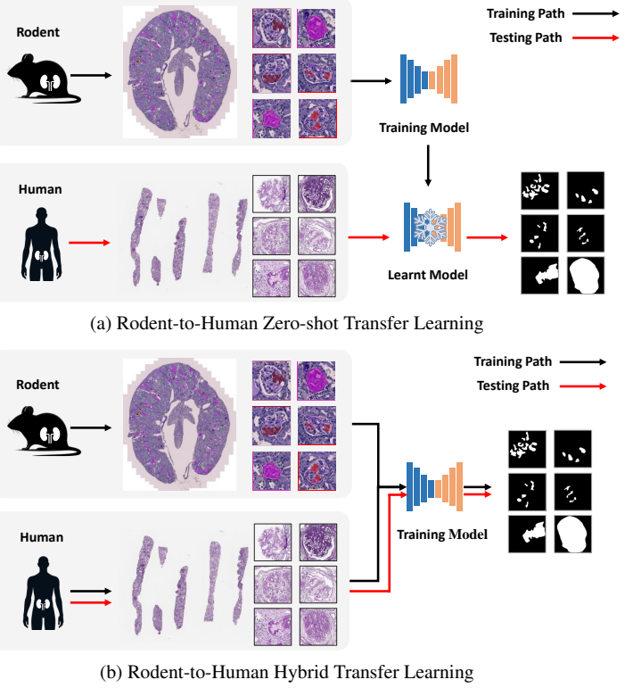
On its own terms, the paper demonstrates that a single residual U-Net with a dynamic, class-aware head can jointly handle 14 segmentation classes even when each training image is annotated for only one class (partially labeled data). The task is encoded as a one-hot vector that is concatenated with the global-pooled image features; a small controller produces the convolution kernels for the head, so one network can output a mask for any requested class. Across the rodent test set the model achieves a 76.5% average Dice, with per-class scores from 57.1% (mesangial lysis) to 96.3% (Bowman's capsule). In the rodent-to-human transfer experiments, the hybrid training strategy R&H2H (rodent + human lesion data) yields 70.4% average human lesion Dice, and R&H2H+T (adding all tissue data) yields 71.2%, compared with 67.9% for human-only H2H training. The paper attributes the gains to the dynamic head's ability to reason about overlapping and subset/superset classes, and releases the toolkit as a Docker container.
Load-bearing premise
The headline Dice averages assume the data tables correctly assign every annotation to human or rodent; if mesangial-expansion annotations are placed in the wrong species column, the 76.5% and 3.3-point figures would not describe the claimed 14-class model.
Editorial extensions
If this is right
- Clinicians can run the released Docker toolkit on raw whole-slide images and get per-class masks for all 14 glomerular structures and lesions in a single command.
- Rodent histopathology can serve as auxiliary training data to improve human lesion segmentation when human annotations are scarce, with a reported 3.3-point Dice gain.
- The dynamic-head design outperforms multi-head baselines on classes with anatomical overlap, such as global sclerosis covering the Bowman's capsule region.
- The curated dataset of 23,529 partially labeled glomeruli, split at patient level, provides a shared benchmark for cross-species intraglomerular segmentation.
Reading between the lines
- A natural extension would be to check whether adding rodent tissue data helps human tissue segmentation as much as it helps lesion segmentation, a comparison the paper does not report.
- The transfer gain is reported only as an average; disaggregating by lesion class would show whether the +3.3 points is driven by one class (e.g., nodular or segmental sclerosis) or is uniform.
- The same dynamic-head scheme could be applied to other partially labeled multi-class pathology problems, such as tubular, vascular, and interstitial compartments in the same kidney sections.
- Because the paper releases the model and weights, the reproducibility of the headline numbers can be tested directly against the curated test sets, including the mesangial-expansion annotations whose species assignment is unclear from the tables.
Editorial analysis
A structured set of objections, weighed in public.
Referee Report
Summary. The paper introduces Glo-In-One-v2, a dynamic-head segmentation network and Dockerized toolkit for holistic intraglomerular analysis. The model is trained on partially labeled human and rodent histopathology patches to segment 14 classes: five tissue/cell classes (Bowman's capsule, tuft, mesangium, mesangial cells, podocytes) and nine lesion classes (adhesion, capsular drop, global sclerosis, hyalinosis, mesangial lysis, microaneurysm, nodular sclerosis, mesangial expansion, segmental sclerosis). The authors report a 76.5% average Dice on rodent glomeruli, outperforming several CNN and transformer baselines, and report that adding rodent data plus tissue data to human lesion training (R&H2H+T) improves human lesion Dice by 3.3 points over human-only training (H2H). The paper also describes a web-mined unannotated image pool and a companion detection module. The central architecture and dataset contributions are plausible, but the headline numeric claims rest on inconsistent test-set definitions that need to be fixed.
Significance. If the reported results survive a corrected evaluation, the paper would make a useful contribution: it tackles a genuinely hard partially labeled, multi-class, cross-species segmentation setting; it provides a large annotated glomerular dataset (23,529 patches) and a public codebase; and its transfer-learning comparison is a genuine held-out evaluation rather than a circular one. The dynamic-head formulation is a reasonable way to handle overlapping and hierarchical classes such as Bowman's capsule containing tuft and mesangium. The manuscript is also honest about data-sharing restrictions. However, the current inconsistencies in which test set produced the reported averages prevent the paper from being accepted as is; the claims need to be re-anchored to a single, coherent class/test-set definition.
major comments (4)
- [§4.1, Table 1; §4.3.1, Table 3] Table 1 reports no rodent mesangial-expansion (ME) annotations (rodent ME is listed as '—', while human ME is 227), yet Table 3, introduced as results 'over the entire rodent dataset,' reports an ME Dice of 66.2 and includes it in the 76.5% average. This is internally inconsistent: either a rodent ME test set exists and Table 1 is incomplete, or ME was evaluated on human patches inside a rodent-only table. The authors must state exactly which test patches produce the ME column and recompute the average (and the headline 76.5%) under that definition.
- [§4.3.2, Table 4] The paragraph introducing Table 4 says it presents 'performance metrics for all lesion classes in the complete human dataset,' but the table has only five lesion columns (GS, HS, MA, NS, SS) and omits ME, the second-largest human lesion class by annotation count (227, per Table 1). Consequently, the abstract's 'more than 3%' improvement from 67.9 (H2H) to 71.2 (R&H2H+T) is measured on five classes only, not on all nine human lesion classes, and the effect of transfer on ME is unmeasured. Please add ME with a clear test-set definition, or revise the claim to state explicitly that it applies to the five-class subset.
- [§4.2, §4.3.2] The R&H2H+T condition differs from H2H by adding both rodent lesion data and all tissue-segmentation data, so the +3.3 Dice gain cannot be attributed specifically to rodent-to-human transfer; the paper's own numbers show that R&H2H (lesion data only) gives +2.5 over H2H, implying the tissue data adds roughly 0.8 Dice points. This is a confounded comparison for the stated transfer-learning conclusion. Please add a controlled ablation and report uncertainty (multiple seeds with means and confidence intervals) before claiming a transfer-driven improvement.
- [§4.3.1, Table 3] Several baseline entries are at or near 50% Dice for lesion classes (e.g., U-Nets AH 50.2 and CD 50.8; Multi-class ME 49.1, MA 49.3, and ML 49.6; Segmenter MA 55.5 and SS 50.2), a pattern consistent with degenerate predictions that always predict background or a single class. With many classes near that level, the statement that the dynamic-head model 'outperforms all baselines' is not yet a meaningful comparison. Please report per-class precision/recall or the proportion of non-empty predictions, and characterize or exclude degenerate baseline runs.
minor comments (6)
- [§3.1, Eq. (1)] The task vector is called m-dimensional and the text refers to 'the i th class of lesion,' but the ordering of the 14 classes is never defined; please specify the class ordering used for the one-hot encoding.
- [§4.2] The image-pool and training descriptions say the pool size matches the number of classes and that the best model is chosen by average Dice over 200 epochs, but no hyperparameters (learning rate, optimizer, batch size, augmentation, epochs) are reported; a reproducibility appendix with these values is needed.
- [Data Availability] The abstract emphasizes the large dataset, but the Data Availability section states that a portion of the data cannot be made public; please clarify how much of the 23,529-patch dataset is publicly accessible and under what terms.
- [Table 5] In the Swinunetr row, '73.3-' appears instead of a Dice value for HS; also, Table 5 is referenced in §4.3.2 as being in the appendix but it appears after §6, so the numbering/placement should be adjusted.
- [Throughout] There are multiple typos and grammatical errors that should be corrected: 'glomeular' in the keywords, 'reprensents' in §3.1, 'Adhension' in Fig. 4, 'hybird' and 'stragety' in Fig. 6, and 'of nine require' in §2.
- [§3.2] The web-mining subsection describes collecting 10,000 compound figures and obtaining over 30,000 unannotated glomerular images, but the paper never reports an experiment using these images; please state whether they were used in pretraining or contrastive learning, or remove the subsection if they are not part of the pipeline.
Circularity Check
No material circularity: the reported Dice values are held-out test measurements, and self-citations to prior architecture/tool work are not load-bearing.
full rationale
The paper's central claims are empirical: the 76.5% average Dice (Table 3) and the more-than-3% transfer gain (Table 4) are computed on test splits described in Table 2 (6:1:3 patient-level split) and are not fitted to the test set. The architecture is adopted from the authors' prior dynamic-head paper (ref 25) and from external DoDNet-style dynamic filter generation (ref 27), but those citations supply implementation components, not evidence for the transfer-learning claim; the comparisons against U-Net, DeepLabV3, Swin-UNETR, and Segmenter are independent baselines. The paper's self-citations (refs 3, 25, 29) refer to the previous toolkit, network design, and compound-figure separation tool; none is a uniqueness theorem or a definition of the metric, and none forces the reported Dice values by construction. The internal inconsistency between Tables 1, 3, and 4 regarding mesangial-expansion annotations is a data-reporting/correctness concern, not a circular reduction: no equation or fitted parameter equates the reported prediction to the input. At most there is a minor pattern of self-citation, but no circular step is present.
Assumptions & free parameters
free parameters (2)
- training hyperparameters (learning rate, optimizer, batch size, augmentation, epochs) =
not reported
- image pool size =
14 (number of classes)
assumptions (3)
- domain assumption Manual annotations by expert nephropathologists are treated as the ground-truth reference standard without measuring inter-observer variability.
- domain assumption Overlapping/hierarchical classes (e.g., global sclerosis covering Bowman's capsule) can be learned as independent per-pixel labels from partially labeled data.
- domain assumption The patient-level stratified 6:1:3 split prevents data leakage.
Cite this review
Pith. "Pith review of Glo-In-One-v2: Holistic Identification of Glomerular Cells, Tissues, and Lesions in Human and Mouse Histopathology." pith.science (2026). https://pith.science/paper/UJACKHMH
@misc{pith2026241116961,
  author       = {Pith},
  title        = {Pith review of: Glo-In-One-v2: Holistic Identification of Glomerular Cells, Tissues, and Lesions in Human and Mouse Histopathology},
  year         = {2026},
  howpublished = {\url{https://pith.science/paper/UJACKHMH}},
  note         = {Machine review of arXiv:2411.16961}
}
read the original abstract
Segmenting glomerular intraglomerular tissue and lesions traditionally depends on detailed morphological evaluations by expert nephropathologists, a labor-intensive process susceptible to interobserver variability. Our group previously developed the Glo-In-One toolkit for integrated detection and segmentation of glomeruli. In this study, we leverage the Glo-In-One toolkit to version 2 with fine-grained segmentation capabilities, curating 14 distinct labels for tissue regions, cells, and lesions across a dataset of 23,529 annotated glomeruli across human and mouse histopathology data. To our knowledge, this dataset is among the largest of its kind to date.In this study, we present a single dynamic head deep learning architecture designed to segment 14 classes within partially labeled images of human and mouse pathology data. Our model was trained using a training set derived from 368 annotated kidney whole-slide images (WSIs) to identify 5 key intraglomerular tissues covering Bowman's capsule, glomerular tuft, mesangium, mesangial cells, and podocytes. Additionally, the network segments 9 glomerular lesion classes including adhesion, capsular drop, global sclerosis, hyalinosis, mesangial lysis, microaneurysm, nodular sclerosis, mesangial expansion, and segmental sclerosis. The glomerulus segmentation model achieved a decent performance compared with baselines, and achieved a 76.5 % average Dice Similarity Coefficient (DSC). Additional, transfer learning from rodent to human for glomerular lesion segmentation model has enhanced the average segmentation accuracy across different types of lesions by more than 3 %, as measured by Dice scores. The Glo-In-One-v2 model and trained weight have been made publicly available at https: //github.com/hrlblab/Glo-In-One_v2.
Figures


Figures from the paper (3 more)


Forward citations
Cited by 1 Pith paper
-
KPIs 2024 Challenge: Advancing Glomerular Segmentation from Patch- to Slide-Level
The KPIs 2024 challenge created a benchmark for glomerular segmentation in PAS-stained mouse kidney slides from four CKD models, and top models achieved Dice scores near 94 percent.
Reference graph
Works this paper leans on
-
[1]
Y. Huo, R. Deng, Q. Liu, et al. , ``Ai applications in renal pathology,'' Kidney international (6), 1309--1320 (2021)
work page 2021
- [2]
-
[3]
T. Yao, Y. Lu, J. Long, et al. , ``Glo-in-one: holistic glomerular detection, segmentation, and lesion characterization with large-scale web image mining,'' Journal of Medical Imaging (5), 052408--052408 (2022)
work page 2022
-
[4]
A. Janowczyk and A. Madabhushi, ``Deep learning for digital pathology image analysis: A comprehensive tutorial with selected use cases,'' Journal of pathology informatics (1), 29 (2016)
work page 2016
-
[5]
D. Komura and S. Ishikawa, ``Machine learning approaches for pathologic diagnosis,'' Virchows Archiv , 131--138 (2019)
work page 2019
-
[6]
M. Gadermayr, A.-K. Dombrowski, B. M. Klinkhammer, et al. , ``Cnn cascades for segmenting sparse objects in gigapixel whole slide images,'' Computerized Medical Imaging and Graphics , 40--48 (2019)
work page 2019
- [7]
-
[8]
S. Wang, D. M. Yang, R. Rong, et al. , ``Pathology image analysis using segmentation deep learning algorithms,'' The American journal of pathology (9), 1686--1698 (2019)
work page 2019
Show all 37 references
-
[9]
F. N. Saikia, Y. Iwahori, T. Suzuki, et al. , ``Mlp-unet: Glomerulus segmentation,'' IEEE Access (2023)
2023
-
[10]
Kim, S.-W
H.-R. Kim, S.-W. Park, H.-J. Cho, et al. , ``Comparative gene expression profiles of intestinal transporters in mice, rats and humans,'' Pharmacological research (3), 224--236 (2007)
2007
-
[11]
A. L. Smith and D. J. Corrow, ``Modifications to husbandry and housing conditions of laboratory rodents for improved well-being,'' ILAR journal (2), 140--147 (2005)
2005
-
[12]
D. K. Brubaker and D. A. Lauffenburger, ``Translating preclinical models to humans,'' Science (6479), 742--743 (2020)
2020
-
[13]
C. H. Leenaars, C. Kouwenaar, F. R. Stafleu, et al. , ``Animal to human translation: A systematic scoping review of reported concordance rates,'' Journal of translational medicine , 1--22 (2019)
2019
-
[14]
Ritskes-Hoitinga, C
M. Ritskes-Hoitinga, C. Leenaars, W. Beumer, et al. , ``Improving translation by identifying evidence for more human-relevant preclinical strategies,'' Animals (7), 1170 (2020)
2020
-
[15]
Hermsen, T
M. Hermsen, T. de Bel, M. Den Boer, et al. , ``Deep learning--based histopathologic assessment of kidney tissue,'' Journal of the American Society of Nephrology (10), 1968--1979 (2019)
2019
-
[16]
Bouteldja, B
N. Bouteldja, B. M. Klinkhammer, R. D. B \"u low, et al. , ``Deep learning--based segmentation and quantification in experimental kidney histopathology,'' Journal of the American Society of Nephrology (1), 52--68 (2021)
2021
-
[17]
Ginley, B
B. Ginley, B. Lutnick, K.-Y. Jen, et al. , ``Computational segmentation and classification of diabetic glomerulosclerosis,'' Journal of the American Society of Nephrology: JASN (10), 1953 (2019)
2019
-
[18]
C. Zeng, Y. Nan, F. Xu, et al. , ``Identification of glomerular lesions and intrinsic glomerular cell types in kidney diseases via deep learning,'' The Journal of pathology (1), 53--64 (2020)
2020
-
[19]
Kawazoe, K
Y. Kawazoe, K. Shimamoto, R. Yamaguchi, et al. , ``Computational pipeline for glomerular segmentation and association of the quantified regions with prognosis of kidney function in iga nephropathy,'' Diagnostics (12), 2955 (2022)
2022
-
[20]
J. D. Bukowy, A. Dayton, D. Cloutier, et al. , ``Region-based convolutional neural nets for localization of glomeruli in trichrome-stained whole kidney sections,'' Journal of the American Society of Nephrology: JASN (8), 2081 (2018)
2018
-
[21]
H. Yang, R. Deng, Y. Lu, et al. , ``Circlenet: Anchor-free glomerulus detection with circle representation,'' in Medical Image Computing and Computer Assisted Intervention--MICCAI 2020: 23rd International Conference, Lima, Peru, October 4--8, 2020, Proceedings, Part IV 23 , 35...
2020
-
[22]
Sheehan, S
S. Sheehan, S. Mawe, R. E. Cianciolo, et al. , ``Detection and classification of novel renal histologic phenotypes using deep neural networks,'' The American Journal of Pathology (9), 1786--1796 (2019)
2019
-
[23]
Y. Nan, F. Li, P. Tang, et al. , ``Automatic fine-grained glomerular lesion recognition in kidney pathology,'' Pattern Recognition , 108648 (2022)
2022
-
[24]
Akatsuka and Y
A. Akatsuka and Y. Horai, ``Automated recognition of glomerular lesions in the kidneys of mice by using deep learning,'' Journal of Pathology Informatics , 100129 (2022)
2022
-
[25]
R. Deng, Q. Liu, C. Cui, et al. , ``Single dynamic network for multi-label renal pathology image segmentation,'' in International Conference on Medical Imaging with Deep Learning , 304--314, PMLR (2022)
2022
-
[26]
Q. Chen, J. Xu, and V. Koltun, ``Fast image processing with fully-convolutional networks,'' in Proceedings of the IEEE International Conference on Computer Vision , 2497--2506 (2017)
2017
-
[27]
Zhang, Y
J. Zhang, Y. Xie, Y. Xia, et al. , ``Dodnet: Learning to segment multi-organ and tumors from multiple partially labeled datasets,'' in Proceedings of the IEEE/CVF conference on computer vision and pattern recognition , 1195--1204 (2021)
2021
-
[28]
Demner-Fushman, S
D. Demner-Fushman, S. Antani, M. Simpson, et al. , ``Design and development of a multimodal biomedical information retrieval system,'' Journal of Computing Science and Engineering (2), 168--177 (2012)
2012
-
[29]
T. Yao, C. Qu, Q. Liu, et al. , ``Compound figure separation of biomedical images with side loss,'' (2021)
2021
-
[30]
The results here are in whole or part based upon data generated by the Kidney Precision Medicine Project
``Kidney precision medicine project data.'' https://www.kpmp.org. The results here are in whole or part based upon data generated by the Kidney Precision Medicine Project. Accessed August 30, 2024. Funded by the National Institute of Diabetes and Digestive and Kidney Diseases ...
2024
-
[31]
J.-Y. Zhu, T. Park, P. Isola, et al. , ``Unpaired image-to-image translation using cycle-consistent adversarial networks,'' in Proceedings of the IEEE international conference on computer vision , 2223--2232 (2017)
2017
-
[32]
Ronneberger, P
O. Ronneberger, P. Fischer, and T. Brox, ``U-net: Convolutional networks for biomedical image segmentation,'' in Medical Image Computing and Computer-Assisted Intervention--MICCAI 2015: 18th International Conference, Munich, Germany, October 5-9, 2015, Proceedings, Part III 18...
2015
-
[33]
Lutnick, B
B. Lutnick, B. Ginley, D. Govind, et al. , ``An integrated iterative annotation technique for easing neural network training in medical image analysis,'' Nature machine intelligence (2), 112--119 (2019)
2019
-
[34]
Gonz \'a lez, G
G. Gonz \'a lez, G. R. Washko, and R. San Jos \'e Est \'e par, ``Multi-structure segmentation from partially labeled datasets. application to body composition measurements on ct scans,'' in International Workshop on Reconstruction and Analysis of Moving Body Organs , 215--224,...
2018
-
[35]
Hatamizadeh, V
A. Hatamizadeh, V. Nath, Y. Tang, et al. , ``Swin unetr: Swin transformers for semantic segmentation of brain tumors in mri images,'' in International MICCAI Brainlesion Workshop , 272--284, Springer (2021)
2021
-
[36]
Strudel, R
R. Strudel, R. Garcia, I. Laptev, et al. , ``Segmenter: Transformer for semantic segmentation,'' in Proceedings of the IEEE/CVF international conference on computer vision , 7262--7272 (2021)
2021
-
[37]
write newline
" write newline "" before.all 'output.state := FUNCTION blank.sep after.quote 'output.state := FUNCTION fin.entry output.state after.quoted.block = 'skip 'add.period if write newline FUNCTION new.block output.state before.all = 'skip output.state after.quote = after.quoted.blo...
Reviewed August 12, 2026 · model on record in the stance chip above.
Discussion (0). Continue with ORCID to comment.