REVIEW 4 major objections 8 minor 56 references
MedAutoCorrect: Image-Conditioned Autocorrection in Medical Reporting
T0 review · 4 major / 8 minor · reviewed 2026-08-11 · deepseek-v4-flash
Pith's one-line read This paper claims that a two-stage, image-conditioned system can find and fix factual errors in chest X-ray reports, whether written by radiologists or by AI, and can lift a basic retrieval generator to state-of-the-art quality.
desk verdict Useful task framing and a solid synthetic pipeline, but the central claim that images help correction is never actually tested. read the letter →
The pith
A machine-rendered reading of the paper's core claim, the machinery that carries it, and where it could break.
The reading
What carries the argument
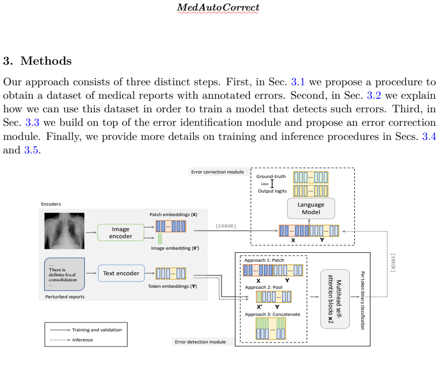
The load-bearing object is the [ERROR] mask. Error detection is framed as per-token binary classification over sequences that combine a fine-tuned Vision Transformer's patch embeddings (or pooled embedding) with GatorTron token embeddings, processed by two multihead self-attention blocks and trained with focal loss to handle class imbalance. Error correction is a fine-tuned GPT-2 Medium conditioned on image patch embeddings concatenated with the report tokens, trained with cross-entropy computed only at the [ERROR] positions, so the model learns to rewrite exactly the flagged spans. The synthetic corruption process—large-language-model-generated and manual edits following four error categories defined by prior work—makes supervised detection and correction possible.
What would settle it
Run the trained detector and corrector on a corpus of naturally occurring erroneous radiology reports whose corrections have been adjudicated by expert radiologists, and compare detection recall and post-correction clinical accuracy against the synthetic-error test set. A substantial drop—especially on errors of omission or on compounding errors—would show that the measured gains are tied to the injection distribution rather than to real reporting errors.
Extended reading notes
Core claim
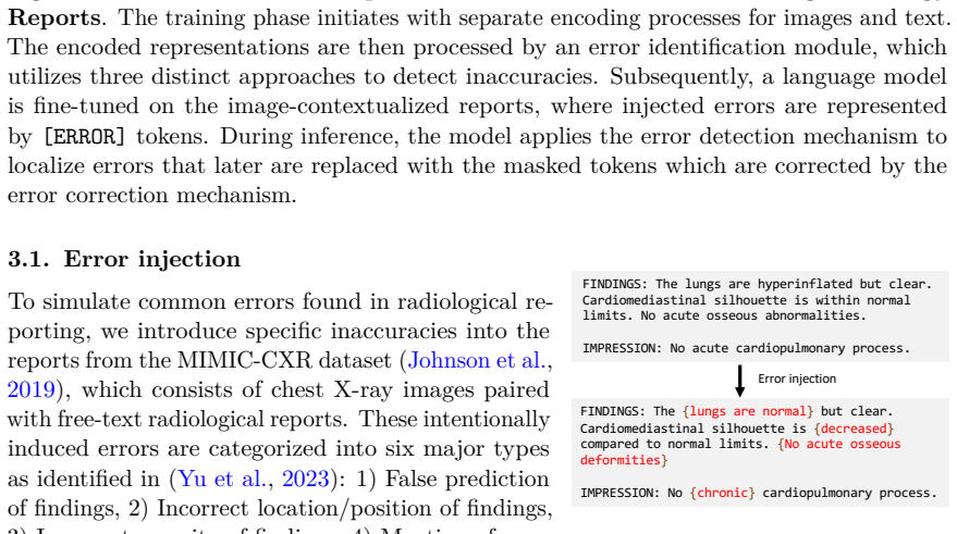
The paper's central claim is that a report's factual errors can be pinpointed and corrected by conditioning on the chest X-ray itself, not just on the text. The authors build a synthetic error-correction dataset by having a large language model and manual edits introduce errors of four clinically defined types into MIMIC-CXR reports, then train two modules: a per-token detector that labels each word as correct or erroneous, and a correction module that, after erroneous spans are replaced with a special [ERROR] token, generates replacements. They report that conditioning the detector on image patch embeddings outperforms a pooled image embedding or per-token concatenation, and that the full pipeline nearly doubles the clinical efficacy F1 of a retrieval-based report generator while improving all NLG metrics, reaching levels comparable to or above established generators. The authors position the framework as a guardrail that does not require retraining the underlying generator, while acknowledging that errors must be flagged before they can be corrected and that errors requiring full clinical inference fall outside the correction scope.
Load-bearing premise
The load-bearing premise is that the errors injected by a large language model and by manual edits are representative of the errors that radiologists and automated generators actually make in practice, and that the original MIMIC-CXR reports are correct references.
Editorial extensions
If this is right
- Autocorrection can be layered on top of existing report generators without retraining them for generation.
- A retrieval model that is not state-of-the-art can be elevated to state-of-the-art report quality, with BLEU-1 rising from 0.216 to 0.370 and CheXpert F1 from 0.183 to 0.330.
- Patch-level image conditioning is the best of the three tested ways to bring visual information into error detection.
- Errors the detector fails to flag are never corrected, so the ceiling of the whole system is set by detection recall.
- The framework is intended as a decision-support guardrail, with clinicians remaining in the loop, rather than as an autonomous reporter.
Reading between the lines
- If real-world errors differ from the injected ones—for instance, compounding errors in poorly written reports—the measured gains may not transfer; a direct test would compare detection and correction on an expert-annotated corpus of naturally occurring report errors.
- The same inject-detect-correct recipe could be adapted to other imaging modalities and clinical documents, with error categories tailored to each setting, as the paper itself suggests for hospital-specific deployment.
- Because correction quality depends on detection, a deployment version could expose detection confidence to clinicians, letting them decide which flagged spans to accept rather than applying corrections automatically.
- Pairing the detector with structured clinical entity extraction could separate style from content and reduce corrections that change wording without changing clinical meaning.
Signed reviews
Editorial analysis
A structured set of objections, weighed in public.
Referee Report
Summary. MedAutoCorrect proposes a two-stage framework for detecting and correcting errors in radiology reports conditioned on chest X-ray images. Errors are synthetically injected into MIMIC-CXR reports using GPT-4 prompts and manual edits across four categories (false prediction, wrong location, wrong severity, spurious comparison). A per-token error detection module is trained under three image-conditioning strategies (Patch, Pool, Concatenate), and a fine-tuned GPT-2 Medium is used to replace detected erroneous spans, represented by [ERROR] tokens, with corrected text. Experiments report detection F1 around 0.63, correction gains over the uncorrected input on NLG and CheXpert metrics (Table 2), and application to a retrieval-based report generator with improved metrics (Table 3). The paper also includes qualitative attention analyses and a brief discussion of limitations.
Significance. The task is timely and practically motivated, and the synthetic error-injection procedure is a reasonable response to the lack of paired erroneous/correct radiology reports. Concrete strengths include the two-stage detection-plus-correction formulation, the release of the synthetic dataset, and the reporting of confidence intervals for the correction ablation in Table 2. The central claim, however, is that the framework uses visual information for autocorrection, and that claim is not yet supported: no text-only correction baseline is evaluated, so the observed gains could in principle come entirely from the language model's masked-infilling behavior. In addition, the main quantitative evaluation is on held-out errors from the same GPT-4/manual injection pipeline used for training, which limits the support for the paper's broader generalization claims. If the missing ablation is added and the claims are appropriately tempered, the framework could serve as a useful guardrail; as it stands, the evidence is stronger for a text-autocorrection system than for an image-grounded one.
major comments (4)
- [Sec. 4.3, Table 3; Sec. 1] The claim that the method 'uses visual information to detect and auto-correct errors' is not tested, because no ablation removes the image. The Baseline row in Table 2 is the uncorrected input, not a text-only correction baseline, and all three conditioning approaches in Table 2 include image embeddings. A text-only variant of the same DETECT+CORRECT pipeline, for example with image embeddings dropped or replaced by a single learned vector, is needed to attribute the observed gains (e.g., BLEU-1 0.216 to 0.370 in Table 3) to image grounding rather than to the language model's prior plus the [ERROR] masking mechanism.
- [Sec. 3.1–3.2] The procedure for obtaining token-level error labels y_ji for GPT-4-injected errors is not described. Since GPT-4 can insert, delete, or rephrase spans, the mapping from altered tokens back to original tokens is nontrivial. The paper should specify the alignment or annotation method used to create the per-token supervision for detection training and the [ERROR] masking for correction training; otherwise the supervision signal for the detector is underspecified.
- [Sec. 7; Sec. 4.3] The held-out test set for Tables 1 and 2 is generated by the same GPT-4/manual error-injection pipeline used for training, so the reported detection and correction numbers measure reversal of the injection distribution. The paper acknowledges this in Sec. 7 but still claims the framework can fix errors from human radiologists and automated generators (Sec. 1). To support that generalization, the evaluation should include errors from an independent source, or the conclusions should be explicitly limited to the synthetic-error setting.
- [Table 3; Sec. 5.3] The statement that autocorrection 'elevates their outputs to SOTA levels' is based on comparing a retrieval-plus-autocorrection pipeline with published numbers from other papers, without a common evaluation protocol or significance tests. Since the retrieval baseline is intentionally not optimized and the comparison is cross-paper, the claim is stronger than the evidence supports. Please report matched-set comparisons or clearly state the protocol differences as a caveat in the text and abstract.
minor comments (8)
- [Sec. 4.2] The description of the error sensitivity threshold appears reversed: a threshold set closer to 1.0 should identify fewer, not more, potential errors; please correct the sentence.
- [Sec. 3.2, Eq. (4)] The Focal Loss reference is an unresolved citation '(?)' in the text; also clarify whether alpha is a scalar or per-token and whether gamma is fixed at 2, as stated in the surrounding text.
- [Sec. 3.3] The statement that GPT-2 Medium was 'fine-tuned on a corpus of PubMed abstracts' lacks a citation; the reference list includes Papanikolaou and Pierleoni (2020), but it is not cited in the text.
- [Sec. 5.1] The sentence 'conditioning the error detection module on token embeddings' should read 'image patch embeddings' to match the Patch approach defined in Sec. 3.2.
- [Sec. 5.2] There is a duplicated phrase: 'misidentification of anatomical location misidentification and severity of findings'; please revise.
- [Table 1] The detection results in Table 1 lack confidence intervals; adding them would be consistent with the presentation in Table 2.
- [Sec. 4.2] The phrase 'the number of erroneous tokens is much less than the correct tokens' should read 'much smaller than the number of correct tokens'.
- [Fig. 8 caption] The caption says 'Lengths greater than 250 are not included'; please clarify whether long reports are excluded from the figure only or from training as well.
Circularity Check
Core detection/correction numbers are measured on held-out errors generated by the same GPT-4/manual injection pipeline that defines the training labels, so the headline claims largely quantify inversion of that synthetic corruption process rather than correction of independently observed clinical errors.
-
fitted input called prediction
[Sec. 3.1 error injection; Sec. 3.3 Eq. (7); Sec. 4.1 dataset and evaluation]
"The error injection process involves the intentional systematic introduction of errors into the MIMIC-CXR radiological reports... We use the X-rays and the altered reports from the MIMIC-CXR dataset to train and evaluate our the error detection module, while the original, unaltered reports are reserved for error correction training and evaluation purposes."
Error labels and correction targets are defined by the injection procedure: GPT-4/manual edits corrupt the original report, Eq. (7) trains only at those injected positions, and the held-out 6,000 'image/erroneous text reports' are made by the same pipeline. The corrected-report labels are exactly the pre-injection originals used to create the erroneous inputs, so Tables 1-2 measure inversion of that synthetic corruption distribution, not correction of independently observed clinical errors. The model is fitted to reverse this distribution, yet Sec. 1 claims it can 'identify and rectify errors whether they originate from machine learning models or human radiologists.' Sec.
full rationale
The only defensible circularity is the self-contained synthetic error loop: errors are generated by GPT-4/manual edits, the model is trained to reverse them at masked positions, and the test errors are produced by the same pipeline. This makes the central detection/correction results (Tables 1-2) partly a measure of inverting the injection distribution, and the paper's own Sec. 7 limitation confirms the generalization gap. I did not score higher because the Table 3 comparison against externally published radiology generators and the retrieval experiment provide some independent content, and because the Rajpurkar self-citations (Endo et al., CheXpert, MIMIC-CXR) are data/baseline citations rather than a load-bearing uniqueness argument. The missing image-free correction baseline is a serious experimental gap for the 'image-conditioned' claim, but an omitted control is not itself a circular reduction; it is a correctness risk. On balance the core benchmark is partially circular (score 6), not fully forced (8-10).
Assumptions & free parameters
free parameters (3)
- Error sensitivity threshold =
0.7
- Focal loss alpha and gamma =
alpha=0.85, gamma=2
- Token truncation length =
200 tokens
assumptions (5)
- domain assumption Original MIMIC-CXR reports are correct ground truth
- ad hoc to paper GPT-4 and manual injections produce errors representative of real radiology errors
- domain assumption Per-token binary labels for LLM-injected errors are obtainable and correct
- domain assumption NLG metrics plus CheXpert disease classification are valid proxies for clinical correctness
- domain assumption Image encoder fine-tuned on CheXpert provides clinically relevant visual features
Cite this review
Pith. "Pith review of MedAutoCorrect: Image-Conditioned Autocorrection in Medical Reporting." pith.science (2026). https://pith.science/paper/PJEQR5Y5
@misc{pith2026241202971,
  author       = {Pith},
  title        = {Pith review of: MedAutoCorrect: Image-Conditioned Autocorrection in Medical Reporting},
  year         = {2026},
  howpublished = {\url{https://pith.science/paper/PJEQR5Y5}},
  note         = {Machine review of arXiv:2412.02971}
}
read the original abstract
In medical reporting, the accuracy of radiological reports, whether generated by humans or machine learning algorithms, is critical. We tackle a new task in this paper: image-conditioned autocorrection of inaccuracies within these reports. Using the MIMIC-CXR dataset, we first intentionally introduce a diverse range of errors into reports. Subsequently, we propose a two-stage framework capable of pinpointing these errors and then making corrections, simulating an \textit{autocorrection} process. This method aims to address the shortcomings of existing automated medical reporting systems, like factual errors and incorrect conclusions, enhancing report reliability in vital healthcare applications. Importantly, our approach could serve as a guardrail, ensuring the accuracy and trustworthiness of automated report generation. Experiments on established datasets and state of the art report generation models validate this method's potential in correcting medical reporting errors.
Figures

Figures from the paper (7 more)


Reference graph
Works this paper leans on
-
[1]
Nguyen, H., Nie, D., Badamdorj, T., Liu, Y., Zhu, Y., Truong, J., and Cheng, L. (2021). Automated generation of accurate & fluent medical X-ray reports. In Moens, M.-F., Huang, X., Specia, L., and Yih, S. W., editors,Proceedings of the 2021 Conference on Empirical Methods in Natural Language Processing, pages 3552–3569, Online and Punta Cana, Dominican Re...
work page 2021
-
[2]
Jing, B., Xie, P., and Xing, E. (2018). On the automatic generation of medical imaging reports. In
work page 2018
-
[3]
Sanchez, M., Alford, K., Krishna, V., Huynh, T. M., Nguyen, C. D., Lungren, M. P., Truong, S. Q., and Rajpurkar, P. (2023). AI-clinician collaboration via disagreement prediction: A decision pipeline and retrospective analysis of real-world radiologist-AI interactions.Cell Reports Medicine, 4(10)
work page 2023
-
[4]
D., Harned, Z., Banerjee, O., Abràmoff, M
Saenz, A. D., Harned, Z., Banerjee, O., Abràmoff, M. D., and Rajpurkar, P. (2023). Autonomous AI systems in the face of liability, regulations and costs.NPJ digital medicine, 6(1):185
work page 2023
-
[5]
Agarwal, N., Moehring, A., Rajpurkar, P., and Salz, T. (2023). Combining human expertise with artificial intelligence: Experimental evidence from radiology. Technical report, National Bureau of Economic Research
work page 2023
-
[6]
Jeong, J., Tian, K., Li, A., Hartung, S., Behzadi, F., Calle, J., Osayande, D., Pohlen, M., Adithan, S., and Rajpurkar, P. (2023). Multimodal image-text matching improves retrieval-based chest X-ray report generation.arXiv preprint arXiv:2303.17579
arXiv 2023
-
[7]
Saenz, A., and Rajpurkar, P. (2023). Style-aware radiology report generation with radgraph and few-shot prompting. arXiv preprint arXiv:2310.17811
arXiv 2023
- [8]
Show all 56 references
-
[9]
H., Duong, H., Saenz, A., and Rajpurkar, P
Khanna, S., Dejl, A., Yoon, K., Truong, Q. H., Duong, H., Saenz, A., and Rajpurkar, P. (2023). Rad- graph2: Modeling disease progression in radiology reports via hierarchical information extraction. arXiv preprint arXiv:2308.05046
2023 arXiv
-
[10]
Miura, Y., Zhang, Y., Tsai, E., Langlotz, C., and Jurafsky, D. (2021). Improving factual completeness and consistency of image-to-text radiology report generation. In Toutanova, K., Rumshisky, A., Zettlemoyer, L., Hakkani-Tur, D., Beltagy, I., Bethard, S., Cotterell, R., Chakr...
2021
-
[11]
Y., et al
Ng, A. Y., et al. (2023). Evaluating progress in automatic chest X-ray radiology report generation. Patterns, 4(9):100802
2023
-
[12]
Johnson, A., Pollard, T., Mark, R., Berkowitz, S., and Horng, S. (2019). MIMIC-CXR database. PhysioNet
2019
-
[13]
Brady, A. P. (2018). Radiology reporting—from Hemingway to HAL? Insights into Imaging, 9:237–246. European Society of Radiology (2011). Good practice for radiological reporting. Guidelines from the European Society of Radiology (ESR).Insights into Imaging, 2(2):93–96
2018
-
[14]
Brady, A. P. (2017). Error and discrepancy in radiology: inevitable or avoidable?Insights into Imaging, 8(1):171–182
2017
-
[15]
Chen, S., Jin, Q., Wang, P., and Wu, Q. (2020). Say as you wish: Fine-grained control of image caption generation with abstract scene graphs.arXiv preprint arXiv:2003.00387
2020 arXiv
-
[16]
Y., Liang, X., Hu, Z., and Xing, E
Li, C. Y., Liang, X., Hu, Z., and Xing, E. P. (2018). Hybrid retrieval-generation reinforced agent for medical image report generation. InAdvances in Neural Information Processing Systems, pages 1537–1547, Montréal, Canada. Curran Associates Inc
2018
-
[17]
Wang, X., Peng, Y., Lu, L., Lu, Z., and Summers, R. M. (2018). TieNet: Text-image embedding network for common thorax disease classification and reporting in chest X-rays.arXiv preprint arXiv:1801.04334
2018 arXiv
-
[18]
Y., and Rajpurkar, P
Endo, M., Krishnan, R., Krishna, V., Ng, A. Y., and Rajpurkar, P. (2021). Retrieval-based chest X-ray report generation using a pre-trained contrastive language-image model. In Roy, S., Pfohl, S., Rocheteau, E., Tadesse, G. A., Oala, L., Falck, F., Zhou, Y., Shen, L., Zamzmi, ...
2021
-
[19]
Polosukhin, I. (2017). Attention is all you need. In Guyon, I., Luxburg, U. V., Bengio, S., Wallach, H., Fergus, R., Vishwanathan, S., and Garnett, R., editors,Advances in Neural Information Processing Systems, volume 30, pages 5998–6008. Curran Associates, Inc
2017
-
[20]
Yuan, J., Liao, H., Luo, R., and Luo, J. (2019). Automatic radiology report generation based on multi-view image fusion and medical concept enrichment.arXiv preprint arXiv:1907.09085
2019 arXiv
-
[21]
Chen, Z., Shen, Y., Song, Y., and Wan, X. (2022). Cross-modal memory networks for radiology report generation. arXiv preprint arXiv:2204.13258
2022 arXiv
-
[22]
Wang, L., Ning, M., Lu, D., Wei, D., Zheng, Y., and Chen, J. (2022). An inclusive task-aware framework for radiology report generation. InMedical Image Computing and Computer Assisted Intervention – MICCAI 2022: 25th International Conference, Singapore, September 18–22, 2022,
2022
-
[23]
Li, X., Jiang, S., andHan, J. (2019). Learningobject contextfor dense captioning. InProceedings of the Thirty-Third AAAI Conference on Artificial Intelligence and Thirty-First Innovative Applications of Artificial Intelligence Conference and Ninth AAAI Symposium on Educational...
2019
-
[24]
Alfarghaly, O., Khaled, R., Elkorany, A., Helal, M., and Fahmy, A. (2021). Automated radiology report generation using conditioned transformers.Informatics in Medicine Unlocked, 24:100557
2021
-
[25]
Shao, Z., Han, J., Marnerides, D., and Debattista, K. (2022). Region-object relation-aware dense captioning via transformer.IEEE Transactions on Neural Networks and Learning Systems
2022
-
[26]
Cornia, M., Stefanini, M., Baraldi, L., and Cucchiara, R. (2020). Meshed-memory transformer for image captioning. arXiv preprint arXiv:1912.08226
2020 arXiv
-
[27]
Sanh, V., Debut, L., Chaumond, J., and Wolf, T. (2020). DistilBERT, a distilled version of BERT: smaller, faster, cheaper and lighter.arXiv preprint arXiv:1910.01108
2020 arXiv
-
[28]
L., and Parikh, D
Vedantam, R., Zitnick, C. L., and Parikh, D. (2015). CIDEr: Consensus-based image description evaluation. arXiv preprint arXiv:1411.5726
2015 arXiv
-
[29]
Yang, X., Chen, A., PourNejatian, N., et al. (2022). A large language model for electronic health records. npj Digital Medicine, 5:194
2022
-
[30]
Heigold, G., Gelly, S., Uszkoreit, J., and Houlsby, N. (2021). An image is worth 16x16 words: Transformers for image recognition at scale.arXiv preprint arXiv:2010.11929
2021 arXiv
-
[31]
Radford, A., Wu, J., Child, R., Luan, D., Amodei, D., and Sutskever, I. (2019). Language models are unsupervised multitask learners
2019
-
[32]
and Pierleoni, A
Papanikolaou, Y. and Pierleoni, A. (2020). DARE: Data augmented relation extraction with GPT-2. arXiv preprint arXiv:2004.13845
2020 arXiv
-
[33]
and Hutter, F
Loshchilov, I. and Hutter, F. (2019). Decoupled weight decay regularization. arXiv preprint arXiv:1711.05101
2019 arXiv
-
[34]
and Hutter, F
Loshchilov, I. and Hutter, F. (2017). SGDR: Stochastic gradient descent with warm restarts.arXiv preprint arXiv:1608.03983
2017 arXiv
-
[35]
and Al-Onaizan, Y
Freitag, M. and Al-Onaizan, Y. (2017). Beam search strategies for neural machine translation. In
2017
-
[36]
Holtzman, A., Buys, J., Du, L., Forbes, M., and Choi, Y. (2020). The curious case of neural text degeneration. arXiv preprint arXiv:1904.09751
2020 arXiv
-
[37]
Maskell, G. (2019). Error in radiology-where are we now? British Journal of Radiology, 92(1095):20180845. OpenAI (2023). GPT-4 technical report.arXiv preprint arXiv:2303.08774
2019 arXiv
-
[38]
Lin, C.-Y. (2004). ROUGE: A package for automatic evaluation of summaries. InText Summarization Branches Out, pages 74–81, Barcelona, Spain. Association for Computational Linguistics
2004
-
[39]
and Lavie, A
Banerjee, S. and Lavie, A. (2005). METEOR: An automatic metric for MT evaluation with improved correlation with human judgments. In Goldstein, J., Lavie, A., Lin, C.-Y., and Voss, C., editors,Proceedings of the ACL Workshop on Intrinsic and Extrinsic Evaluation Measures for Ma...
2005
-
[40]
Nooralahzadeh, F., Perez Gonzalez, N., Frauenfelder, T., Fujimoto, K., and Krauthammer, M. (2021). Progressive transformer-based generation of radiology reports. In Moens, M.-F., Huang, X., Specia, L., and Yih, S. W., editors,Findings of the Association for Computational Lingu...
2021
-
[41]
Liu, F., Yin, C., Wu, X., Ge, S., Zhang, P., and Sun, X. (2021). Contrastive attention for automatic chest X-ray report generation. In Zong, C., Xia, F., Li, W., and Navigli, R., editors,Findings of the Association for Computational Linguistics: ACL-IJCNLP 2021, pages 269–280,...
2021
-
[42]
You, D., Liu, F., Ge, S., Xie, X., Zhang, J., and Wu, X. (2022). AlignTransformer: Hierarchi- cal alignment of visual regions and disease tags for medical report generation.arXiv preprint arXiv:2203.10095
2022 arXiv
-
[43]
Cornia, M., Stefanini, M., Baraldi, L., and Cucchiara, R. (2020). Meshed-memory transformer for image captioning. InProceedings of the IEEE/CVF Conference on Computer Vision and Pattern Recognition
2020
-
[44]
Schaffer, J., O’Donovan, J., Michaelis, J., Raglin, A., and Höllerer, T. (2019). I can do better than your AI: Expertise and explanations. InProceedings of the 24th International Conference on Intelligent User Interfaces, pages 240–251, Marina del Ray, California. Association ...
2019
-
[45]
H., McDermott, M., Boag, W., Weng, W.-H., Szolovits, P., and Ghassemi, M
Liu, G., Hsu, T.-M. H., McDermott, M., Boag, W., Weng, W.-H., Szolovits, P., and Ghassemi, M. (2019). Clinically accurate chest X-ray report generation.arXiv preprint arXiv:1904.02633
2019 arXiv
-
[46]
Tanida, T., Müller, P., Kaissis, G., and Rueckert, D. (2023). Interactive and explainable region-guided radiology report generation. In Proceedings of the 2023 IEEE/CVF Conference on Computer Vision and Pattern Recognition (CVPR), pages 718–727. IEEE
2023
-
[47]
Abad, Z. S. H., Ng, A. Y., et al. (2023). Evaluating progress in automatic chest X-ray radiology report generation. Patterns, 4(9):100802
2023
-
[48]
Chen, Z., Song, Y., Chang, T.-H., and Wan, X. (2022). Generating radiology reports via memory- driven transformer. arXiv preprint arXiv:2010.16056. Available at: https://arxiv.org/abs/ 2010.16056
2022 arXiv
-
[49]
You, D., Liu, F., Ge, S., Xie, X., Zhang, J., and Wu, X. (2021). AlignTransformer: Hierarchical alignment of visual regions and disease tags for medical report generation. In de Bruijne, M.,
2021
-
[50]
C., Cotin, S., Padoy, N., Speidel, S., Zheng, Y., and Essert, C., editors,Medical Image Computing and Computer Assisted Intervention – MICCAI 2021, pages 72–82, Cham
Cattin, P. C., Cotin, S., Padoy, N., Speidel, S., Zheng, Y., and Essert, C., editors,Medical Image Computing and Computer Assisted Intervention – MICCAI 2021, pages 72–82, Cham. Springer International Publishing
2021
-
[51]
Liu, F., Wu, X., Ge, S., Fan, W., and Zou, Y. (2021). Exploring and distilling posterior and prior knowledge for radiology report generation. In2021 IEEE/CVF Conference on Computer Vision and Pattern Recognition (CVPR), pages 13748–13757. URL: https://api.semanticscholar. org/...
2021
-
[52]
W., Hallacy, C., Ramesh, A., Goh, G., Agarwal, S., Sastry, G., Askell, A., Mishkin, P., Clark, J., Krueger, G., and Sutskever, I
Radford, A., Kim, J. W., Hallacy, C., Ramesh, A., Goh, G., Agarwal, S., Sastry, G., Askell, A., Mishkin, P., Clark, J., Krueger, G., and Sutskever, I. (2021). Learning transferable visual models from natural language supervision. arXiv preprint arXiv:2103.00020. URL: https: //...
2021 arXiv
-
[53]
Y., and Rajpurkar, P
Endo, M., Krishnan, R., Krishna, V., Ng, A. Y., and Rajpurkar, P. (2021). Retrieval-based chest X-ray report generation using a pre-trained contrastive language-image model. In Roy, S., Pfohl, S., Rocheteau, E., Tadesse, G. A., Oala, L., Falck, F., Zhou, Y., Shen, L., Zamzmi, ...
2021
-
[54]
Johnson, J., Karpathy, A., and Fei-Fei, L. (2015). DenseCap: Fully convolutional localization networks for dense captioning.arXiv preprint arXiv:1511.07571. URL: https://arxiv.org/abs/ 1511.07571
2015 arXiv
-
[55]
Yin, G., Sheng, L., Liu, B., Yu, N., Wang, X., and Shao, J. (2019). Context and attribute grounded dense captioning. arXiv preprint arXiv:1904.01410. URL: https://arxiv.org/abs/1904.01410
2019 arXiv
-
[56]
B., Langlotz, C
Larson, D. B., Langlotz, C. P., Patel, B. N., Lungren, M. P., and Ng, A. Y. (2019). CheXpert: A large chest radiograph dataset with uncertainty labels and expert comparison.arXiv preprint arXiv:1901.07031. 20
2019 arXiv
Reviewed August 11, 2026 · model on record in the stance chip above.
Discussion (0). Continue with ORCID to comment.