REVIEW 4 major objections 6 minor 1 cited by
4DRGS: 4D Radiative Gaussian Splatting for Efficient 3D Vessel Reconstruction from Sparse-View Dynamic DSA Images
T0 review · 4 major / 6 minor · reviewed 2026-08-11 · deepseek-v4-flash
Pith's one-line read This paper claims that 3D vessel reconstruction from sparse dynamic DSA images can be done in minutes with 4D radiative Gaussian splatting, a decoupling of static vessel geometry from time-varying contrast attenuation, at quality matching…
desk verdict Genuinely new and fast Gaussian-splatting pipeline for dynamic DSA reconstruction, but the 3D SOTA claim hinges on an asymmetric-threshold metric with margins inside the noise. read the letter →
The pith
A machine-rendered reading of the paper's core claim, the machinery that carries it, and where it could break.
The reading
What carries the argument
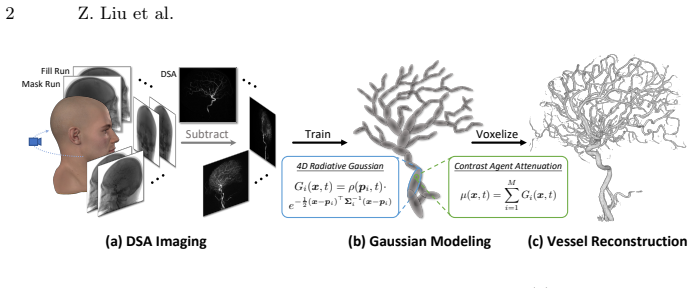
The load-bearing object is the 4D radiative Gaussian kernel $G_i(x,t) = \rho(p_i,t)\exp\left(-\frac{1}{2}(x-p_i)^\top \Sigma_i^{-1}(x-p_i)\right)$, where $\Sigma_i = R_i S_i S_i^\top R_i^\top$ encodes fixed orientation and size and $\rho(p_i,t)$ is the time-varying central attenuation predicted by a compact neural field. This single identity does two jobs: it constrains the reconstruction to static vessel geometry, which regularizes the severely ill-posed sparse-view problem, and it lets contrast-agent dynamics be represented as a scalar function of position and time rather than as a full 4D volume. X-ray rasterization from radiative Gaussian splatting integrates these kernels along rays to synthesize DSA images, and GPU voxelization converts the trained kernels into an attenuation volume. Accumulated attenuation pruning and bounded scaling activation are the two supporting mechanisms that keep the kernel set clean and compact during optimization.
What would settle it
Run 4DRGS and the previous method on a synthetic or physical phantom with known vessel centerlines, generate sparse-view dynamic DSA projections from a known ground-truth attenuation volume, and measure Chamfer and Hausdorff distances against the true geometry at identical mesh thresholds; if the gap between methods shrinks or reverses, or if the 0.025-versus-0.008 threshold choice explains the reported margin, then the paper's accuracy claim is a measurement artifact.
Extended reading notes
Core claim
The authors claim that 4DRGS is the first Gaussian-splatting framework for 3D vessel reconstruction from sparse-view dynamic DSA images. The central discovery is that time-varying contrast-agent flow and static vessel anatomy can be decoupled in a single explicit representation: vessels are static in space, so only attenuation needs to change over time. Each 4D radiative Gaussian kernel carries time-invariant position, rotation, and scale, and a neural attenuation field predicts the kernel's central attenuation from its position and timestamp. The kernels are optimized with an L1 plus D-SSIM loss against real DSA frames under temporal perturbation, and the trained kernels are voxelized and averaged across timestamps to produce the final vessel volume. The paper further introduces accumulated attenuation pruning, which removes kernels whose attenuation averaged over training is consistently small, and bounded scaling activation, which keeps kernels within a fixed size range to avoid needle artifacts. With these components, the method reports state-of-the-art or comparable Chamfer and Hausdorff distances for 3D reconstruction and PSNR/SSIM for 2D DSA synthesis across 30, 40, 50, and 60 input views, while training in minutes.
Load-bearing premise
The reported 3D accuracy rests on the scanner's inbuilt FDK volumes being accurate enough to serve as reference geometry, even though the paper says they are 'not entirely accurate,' and on comparing surfaces that were meshed at different attenuation thresholds for reference versus reconstruction.
Editorial extensions
If this is right
- A full 3D vessel reconstruction from 30 to 60 DSA views converges in about 13 minutes on a single GPU, and a fast 10k-iteration version reaches the previous method's quality in about 5 minutes, a 12x to 32x speedup.
- The method reports the best or second-best Chamfer and Hausdorff distances against scanner-volume references and the best PSNR/SSIM on held-out DSA frames in most tested view counts.
- Because kernels only cover vascular structures rather than the whole scan volume, training and rendering time scale with vessel sparsity instead of scene size.
- Accumulated attenuation pruning preserves vessels that are not yet opacified at a given timestamp, which random or instantaneous-threshold pruning removes.
- Bounded scaling activation removes the needle-like elongated Gaussians that unbounded exponential scaling produces in DSA reconstruction.
Reading between the lines
- Editorial inference: the reported 3D accuracy is measured against scanner-inbuilt FDK volumes that the paper itself calls 'not entirely accurate,' and reference meshes are extracted at a higher attenuation threshold (0.025) than reconstructed meshes (0.008); an independent ground-truth phantom study would determine how much of the Chamfer/Hausdorff margin is real geometry fidelity.
- Editorial inference: the static-geometry assumption means the approach would likely degrade with patient motion or cardiac-driven vessel displacement; extending the kernels with per-kernel temporal displacement or motion compensation is the natural next test.
- Editorial inference: the same 'static structure, time-varying attenuation' decomposition should transfer to other dynamic tomographic problems, such as contrast-enhanced cone-beam CT or 4D CT, where a compact Gaussian-plus-MLP representation could replace full 4D volume optimization.
- Editorial inference: because the neural attenuation field is compact and rasterization is differentiable, the trained 4DRGS model could be used for near-real-time re-rendering of DSA frames at arbitrary angles, which would help interventional navigation; the paper does not demonstrate this but the representation supports it.
Signed reviews
Editorial analysis
A structured set of objections, weighed in public.
Referee Report
Summary. The paper introduces 4DRGS, a 4D radiative Gaussian splatting framework for reconstructing 3D vessel structures from sparse-view dynamic DSA images. Vessels are represented by Gaussian kernels with static geometry (position, rotation, scale) and a time-varying central attenuation predicted by a compact hash-encoded MLP (DNAF). The model is trained by differentiable X-ray rasterization against captured DSA images, with an accumulated-attenuation pruning criterion and a bounded scaling activation to improve quality. The final volume is obtained by voxelizing and averaging the trained kernels over time. Experiments on 15 real patient cases at 30, 40, 50, and 60 views compare against FDK, R2-Gaussian, TOGS, and VPAL using Chamfer/Hausdorff distances, PSNR/SSIM on held-out views, and training time. The paper claims state-of-the-art or comparable reconstruction quality in most scenarios with a 12x-32x speedup over VPAL. The 2D synthesis results are based on held-out views and appear internally consistent, but the 3D comparison relies on an admittedly inaccurate FDK reference meshed at a different attenuation threshold than the reconstructed volumes, which undermines the central 3D-quality claim as currently presented.
Significance. If the 3D-quality claim were substantiated, this would be a practically important contribution: it is the first Gaussian-splatting method for sparse-view dynamic DSA reconstruction, and the reported training times (about 13 minutes for the full version and 5 minutes for the fast version) are dramatically lower than the roughly 2.6 hours reported for VPAL. The core representation is well motivated: static vessel geometry with time-varying attenuation is a natural fit for DSA, and the two proposed components, accumulated attenuation pruning and bounded scaling activation, are clearly explained and ablated. The use of 15 real clinical cases and multiple view settings is a strength relative to many medical-imaging papers. However, the paper does not release code or data, and the main quantitative evidence for 3D superiority depends on a threshold-sensitive comparison against a reference that the paper itself describes as inaccurate. Because the 3D metric is load-bearing for the headline claim, the current evidence does not yet support the stated level of certainty.
major comments (4)
- [Sec. 4.1] The 3D evaluation against the FDK reference is not yet convincing. The paper states that "the provided volumes are not entirely accurate" and then uses them as reference geometry; it also meshes the reference at attenuation threshold 0.025 while meshing all reconstructed volumes at 0.008. Since the iso-surface of a thresholded Gaussian attenuation field depends strongly on the chosen threshold, a 3.1x asymmetry can systematically favor methods whose reconstructed attenuation scale is lower than the reference. CD and HD may therefore be measuring attenuation calibration rather than geometric accuracy. I ask for a threshold-sweep analysis (e.g., varying both thresholds and reporting CD/HD curves), a calibration procedure for attenuation scales across methods, or validation against a trustworthy ground-truth geometry (e.g., a phantom or a high-quality 3D-DSA volume) before the 3D superiority claim can be accepted.
- [Sec. 4.2, Table 2] The claim of "SOTA or comparable" performance in most scenarios is not statistically supported by the reported numbers. At 30 views the Chamfer distance is 1.72 +/- 0.29 for 4DRGS versus 1.79 +/- 0.51 for VPAL; at 50 views VPAL wins CD (1.58 +/- 0.19 versus 1.67 +/- 0.29); and most PSNR and HD differences are within one standard deviation over 15 cases. The paper should report paired significance tests (e.g., Wilcoxon signed-rank) and effect sizes, and should identify explicitly at which view settings the differences are statistically significant. Without this, the abstract's "SOTA" phrasing overstates what the data show.
- [Sec. 3.1 and Sec. 3.2] The method depends on several manually set quantities, including the FDK initialization threshold delta = 0.016, the scale bounds smin = 0.1 and smax = 10 times voxel spacing, and the pruning threshold epsilon = 1e-6, yet no sensitivity analysis is provided. Since the central claim is that the method works robustly for sparse-view clinical data, the paper should show how CD, HD, PSNR, and SSIM vary with these parameters over reasonable ranges, or otherwise justify that the reported results are not a tuned operating point.
- [Sec. 4.1, competing methods] The efficiency comparison would be more complete if the paper reported convergence criteria and loss curves for VPAL and for 4DRGS. The current table reports final iteration counts for 4DRGS (10k and 30k) but does not state whether VPAL was run to its own convergence or was stopped at a fixed schedule. If VPAL was stopped early or was not tuned, the "12x-32x speedup" could partly reflect implementation choices. Reporting training loss over time for all methods would make the efficiency claim more robust.
minor comments (6)
- [Sec. 4.1, Table 1] The row labeled "Others" is ambiguous; please list the cases it covers or give per-case configuration details in supplementary material.
- [Eq. (7)] The summation notation in Eq. (7) is typeset as "P iter" without a subscript; it should be a sum over iterations between neighboring pruning operations.
- [Eq. (6)] Please clarify whether the temporal perturbation tau is sampled once per image or per pixel/ray, since this affects the interpretation of the loss and the temporal-consistency claim.
- [Sec. 3.1] In Eq. (3), s_i denotes the activated scale vector while earlier s_i is used as a scale parameter; the notation should be made consistent, for example by writing s_i as a vector and sigma as elementwise sigmoid.
- [Sec. 4.1, TOGS baseline] The TOGS volume baseline is reconstructed by rendering 720 views in a full circle and applying FDK; please justify the choice of a full circle rather than the original 198-degree arc, since this may affect the comparison.
- [General] The paper does not state whether code or data will be made available; given the overlap of the authors with the VPAL and R2-Gaussian baselines, releasing source code or detailed baseline configurations would substantially improve reproducibility.
Circularity Check
No circular derivation: optimization is image-driven and evaluated on held-out views against an external (if imperfect) FDK reference; overlapping baselines are code-reproduced and not load-bearing.
full rationale
4DRGS's derivation is self-contained. Geometry and attenuation parameters are optimized exclusively against real DSA images via the L1 + D-SSIM loss in Eq. (6); 2D evaluation is performed on held-out frames not used in training, and the 3D volume is obtained by voxelizing the trained Gaussian kernels (Sec. 3.3) rather than by copying the FDK initialization or the Siemens reference. The CD/HD comparison uses the system FDK volumes as an external reference, and the paper explicitly discloses their imperfection ('the provided volumes are not entirely accurate'), which is a metric-validity limitation rather than a circular step. Self-citations to VPAL [15], R2-Gaussian [30], and TOGS [32] are used as baselines or as borrowed software modules; these are code-reproduced comparisons, not unverified premises that force the result. The meshing-threshold asymmetry (0.025 vs 0.008) and FDK-based initialization are potential experimental confounds, but no equation in the paper makes the reported prediction equal to an input by construction. Therefore no circular step is identified.
Assumptions & free parameters
free parameters (6)
- Scale bounds s_min, s_max =
0.1 x voxel size, 10 x voxel size
- FDK initialization threshold delta =
0.016
- Accumulated attenuation pruning threshold epsilon =
1e-6
- Loss weight lambda_ssim =
0.2
- Hash encoding capacity =
2^19 entries, 2D features, 12 levels (3D), similar 4D configuration
- Number of kernels M =
30,000
assumptions (5)
- standard math X-ray attenuation follows the Beer-Lambert law, and DSA images are log-subtracted line integrals of attenuation
- domain assumption Vessels remain static during scanning while only attenuation changes over time
- domain assumption Contrast-agent attenuation can be approximated by a sum of Gaussian kernel responses
- domain assumption The FDK reconstructed volume from the system, though 'not entirely accurate', is a valid reference for ranking methods
- ad hoc to paper The sparse-view FDK volume provides a good-enough initialization for kernel positions
invented entities (3)
-
4D radiative Gaussian kernel
independent evidence
-
Dynamic neural attenuation field (DNAF)
independent evidence
-
Accumulated attenuation A_i
Cite this review
Pith. "Pith review of 4DRGS: 4D Radiative Gaussian Splatting for Efficient 3D Vessel Reconstruction from Sparse-View Dynamic DSA Images." pith.science (2026). https://pith.science/paper/DAXAETCF
@misc{pith2026241212919,
  author       = {Pith},
  title        = {Pith review of: 4DRGS: 4D Radiative Gaussian Splatting for Efficient 3D Vessel Reconstruction from Sparse-View Dynamic DSA Images},
  year         = {2026},
  howpublished = {\url{https://pith.science/paper/DAXAETCF}},
  note         = {Machine review of arXiv:2412.12919}
}
read the original abstract
Reconstructing 3D vessel structures from sparse-view dynamic digital subtraction angiography (DSA) images enables accurate medical assessment while reducing radiation exposure. Existing methods often produce suboptimal results or require excessive computation time. In this work, we propose 4D radiative Gaussian splatting (4DRGS) to achieve high-quality reconstruction efficiently. In detail, we represent the vessels with 4D radiative Gaussian kernels. Each kernel has time-invariant geometry parameters, including position, rotation, and scale, to model static vessel structures. The time-dependent central attenuation of each kernel is predicted from a compact neural network to capture the temporal varying response of contrast agent flow. We splat these Gaussian kernels to synthesize DSA images via X-ray rasterization and optimize the model with real captured ones. The final 3D vessel volume is voxelized from the well-trained kernels. Moreover, we introduce accumulated attenuation pruning and bounded scaling activation to improve reconstruction quality. Extensive experiments on real-world patient data demonstrate that 4DRGS achieves impressive results in 5 minutes training, which is 32x faster than the state-of-the-art method. This underscores the potential of 4DRGS for real-world clinics.
Figures


Figures from the paper (2 more)


Forward citations
Cited by 1 Pith paper
-
3D Gaussian Adaptive Reconstruction for Fourier Light-Field Microscopy
3DGAT uses 3D Gaussian kernels plus the FLFM optical model to self-supervisedly reconstruct volumetric fluorescence, beating Richardson-Lucy deconvolution in resolution on simulated and real data.
Reference graph
Works this paper leans on
-
[1]
Ultrasonic imaging6(1), 81–94 (1984)
Andersen,A.H.,Kak,A.C.:Simultaneousalgebraicreconstructiontechnique(sart): a superior implementation of the art algorithm. Ultrasonic imaging6(1), 81–94 (1984)
work page 1984
-
[2]
IEEE Transactions onPatternAnalysisandMachineIntelligence 14(2),239–256(1992)
Besl, P., McKay, N.D.: A method for registration of 3-d shapes. IEEE Transactions onPatternAnalysisandMachineIntelligence 14(2),239–256(1992). https://doi. org/10.1109/34.121791
-
[3]
Biomedical Physics & Engineering Express2(5), 055010 (2016)
Biguri, A., Dosanjh, M., Hancock, S., Soleimani, M.: Tigre: a matlab-gpu toolbox for cbct image reconstruction. Biomedical Physics & Engineering Express2(5), 055010 (2016)
work page 2016
-
[4]
In: European Conference on Computer Vision
Cai, Y., Liang, Y., Wang, J., Wang, A., Zhang, Y., Yang, X., Zhou, Z., Yuille, A.: Radiative gaussian splatting for efficient x-ray novel view synthesis. In: European Conference on Computer Vision. pp. 283–299. Springer (2025)
work page 2025
-
[5]
Cai, Y., Wang, J., Yuille, A., Zhou, Z., Wang, A.: Structure-aware sparse-view x-ray 3d reconstruction. In: CVPR (2024)
work page 2024
-
[6]
American journal of neuroradiology18(8), 1507–1514 (1997)
Fahrig, R., Fox, A., Lownie, S., Holdsworth, D.: Use of a c-arm system to generate true three-dimensional computed rotational angiograms: preliminary in vitro and in vivo results. American journal of neuroradiology18(8), 1507–1514 (1997)
work page 1997
-
[7]
arXiv preprint arXiv:2211.17048 (2022)
Fang, Y., Mei, L., Li, C., Liu, Y., Wang, W., Cui, Z., Shen, D.: Snaf: Sparse-view cbct reconstruction with neural attenuation fields. arXiv preprint arXiv:2211.17048 (2022)
arXiv 2022
-
[8]
Feldkamp, L.A., Davis, L.C., Kress, J.W.: Practical cone-beam algorithm. Josa a 1(6), 612–619 (1984)
work page 1984
Show all 33 references
-
[9]
arXiv preprint arXiv:2406.02518 (2024)
Gao, Z., Planche, B., Zheng, M., Chen, X., Chen, T., Wu, Z.: Ddgs-ct: Direction- disentangled gaussian splatting for realistic volume rendering. arXiv preprint arXiv:2406.02518 (2024)
2024 arXiv
-
[10]
SIAM (2001)
Kak, A.C., Slaney, M.: Principles of computerized tomographic imaging. SIAM (2001)
2001
-
[11]
ACM Trans
Kerbl, B., Kopanas, G., Leimkühler, T., Drettakis, G.: 3d gaussian splatting for real-time radiance field rendering. ACM Trans. Graph.42(4), 139–1 (2023)
2023
-
[12]
In: Interna- tional Conference on Learning Representations (ICLR) (2015)
Kingma, D.P., Ba, J.: Adam: A method for stochastic optimization. In: Interna- tional Conference on Learning Representations (ICLR) (2015)
2015
-
[13]
American Journal of Neuroradiology38(6), 1169– 1176 (2017)
Lang, S., Gölitz, P., Struffert, T., Rösch, J., Rössler, K., Kowarschik, M., Strother, C., Doerfler, A.: 4d dsa for dynamic visualization of cerebral vasculature: a single- center experience in 26 cases. American Journal of Neuroradiology38(6), 1169– 1176 (2017)
2017
-
[14]
arXiv preprint arXiv:2312.15676 (2023)
Li, Y., Fu, X., Zhao, S., Jin, R., Zhou, S.K.: Sparse-view ct reconstruction with 3d gaussian volumetric representation. arXiv preprint arXiv:2312.15676 (2023)
2023 arXiv
-
[15]
arXiv preprint arXiv:2405.10705 (2024)
Liu, Z., Zhao, H., Qin, W., Zhou, Z., Wang, X., Wang, W., Lai, X., Zheng, C., Shen, D., Cui, Z.: 3d vessel reconstruction from sparse-view dynamic dsa images via vessel probability guided attenuation learning. arXiv preprint arXiv:2405.10705 (2024)
2024 arXiv
-
[16]
In: Seminal graphics: pioneering efforts that shaped the field, pp
Lorensen, W.E., Cline, H.E.: Marching cubes: A high resolution 3d surface con- struction algorithm. In: Seminal graphics: pioneering efforts that shaped the field, pp. 347–353 (1998) 14 Z. Liu et al
1998
-
[17]
Commu- nications of the ACM65(1), 99–106 (2021)
Mildenhall, B., Srinivasan, P.P., Tancik, M., Barron, J.T., Ramamoorthi, R., Ng, R.: Nerf: Representing scenes as neural radiance fields for view synthesis. Commu- nications of the ACM65(1), 99–106 (2021)
2021
-
[18]
ACM Transactions on Graphics (ToG)41(4), 1– 15 (2022)
Müller,T.,Evans,A.,Schied,C.,Keller,A.:Instantneuralgraphicsprimitiveswith a multiresolution hash encoding. ACM Transactions on Graphics (ToG)41(4), 1– 15 (2022)
2022
-
[19]
In: Proceedings of the 27th international conference on machine learning (ICML-10)
Nair, V., Hinton, G.E.: Rectified linear units improve restricted boltzmann ma- chines. In: Proceedings of the 27th international conference on machine learning (ICML-10). pp. 807–814 (2010)
2010
-
[20]
Park,S.,Son,M.,Jang,S.,Ahn,Y.C.,Kim,J.Y.,Kang,N.:Temporalinterpolation isallyouneedfordynamicneuralradiancefields.In:ProceedingsoftheIEEE/CVF Conference on Computer Vision and Pattern Recognition. pp. 4212–4221 (2023)
2023
-
[21]
ACM Transactions on Graphics (TOG)41(4), 1–13 (2022)
Rückert, D., Wang, Y., Li, R., Idoughi, R., Heidrich, W.: Neat: Neural adaptive tomography. ACM Transactions on Graphics (TOG)41(4), 1–13 (2022)
2022
-
[22]
American Journal of Neuroradiology 42(2), 214–220 (2021)
Ruedinger, K., Schafer, S., Speidel, M., Strother, C.: 4d-dsa: development and current neurovascular applications. American Journal of Neuroradiology 42(2), 214–220 (2021)
2021
-
[23]
American Journal of Neuroradiology36(10), 1959–1963 (2015)
Sandoval-Garcia, C., Royalty, K., Aagaard-Kienitz, B., Schafer, S., Yang, P., Strother, C.: A comparison of 4d dsa with 2d and 3d dsa in the analysis of normal vascular structures in a canine model. American Journal of Neuroradiology36(10), 1959–1963 (2015)
2015
-
[24]
Journal of neurointer- ventional surgery (2015)
Sandoval-Garcia, C., Royalty, K., Yang, P., Niemann, D., Ahmed, A., Aagaard- Kienitz, B., Başkaya, M.K., Schafer, S., Strother, C.: 4d dsa a new technique for arteriovenous malformation evaluation: a feasibility study. Journal of neurointer- ventional surgery (2015)
2015
-
[25]
Physics in Medicine & Biology 53(17), 4777 (2008)
Sidky, E.Y., Pan, X.: Image reconstruction in circular cone-beam computed tomog- raphy by constrained, total-variation minimization. Physics in Medicine & Biology 53(17), 4777 (2008)
2008
-
[26]
IEEE transactions on image processing 13(4), 600–612 (2004)
Wang, Z., Bovik, A.C., Sheikh, H.R., Simoncelli, E.P.: Image quality assessment: from error visibility to structural similarity. IEEE transactions on image processing 13(4), 600–612 (2004)
2004
-
[27]
IEEE Transactions on Computa- tional Imaging (2023)
Wu, Q., Feng, R., Wei, H., Yu, J., Zhang, Y.: Self-supervised coordinate projection network for sparse-view computed tomography. IEEE Transactions on Computa- tional Imaging (2023)
2023
-
[28]
arXiv preprint arXiv:2403.14621 (2024)
Xu, Y., Shi, Z., Yifan, W., Chen, H., Yang, C., Peng, S., Shen, Y., Wetzstein, G.: Grm: Large gaussian reconstruction model for efficient 3d reconstruction and generation. arXiv preprint arXiv:2403.14621 (2024)
2024 arXiv
-
[29]
In: Proceedings of the IEEE/CVF International Conference on Computer Vision
Zang, G., Idoughi, R., Li, R., Wonka, P., Heidrich, W.: Intratomo: self-supervised learning-based tomography via sinogram synthesis and prediction. In: Proceedings of the IEEE/CVF International Conference on Computer Vision. pp. 1960–1970 (2021)
2021
-
[30]
arXiv preprint arXiv:2405.20693 (2024)
Zha, R., Lin, T.J., Cai, Y., Cao, J., Zhang, Y., Li, H.: R 2-gaussian: Rectify- ing radiative gaussian splatting for tomographic reconstruction. arXiv preprint arXiv:2405.20693 (2024)
2024 arXiv
-
[31]
In: International Conference on Medical Image Computing and Computer-Assisted Intervention
Zha, R., Zhang, Y., Li, H.: Naf: neural attenuation fields for sparse-view cbct reconstruction. In: International Conference on Medical Image Computing and Computer-Assisted Intervention. pp. 442–452. Springer (2022)
2022
-
[32]
arXiv preprint arXiv:2403.19586 (2024) 4DRGS: 4D Radiative Gaussian Splatting 15
Zhang, S., Zhao, H., Zhou, Z., Wu, G., Zheng, C., Wang, X., Liu, W.: Togs: Gaus- sian splatting with temporal opacity offset for real-time 4d dsa rendering. arXiv preprint arXiv:2403.19586 (2024) 4DRGS: 4D Radiative Gaussian Splatting 15
2024 arXiv
-
[33]
Cell Reports Medicine 3(10) (2022)
Zhao,H.,Zhou,Z.,Wu,F.,Xiang,D.,Zhao,H.,Zhang,W.,Li,L.,Li,Z.,Huang,J., Hu, H., et al.: Self-supervised learning enables 3d digital subtraction angiography reconstruction from ultra-sparse 2d projection views: a multicenter study. Cell Reports Medicine 3(10) (2022)
2022
Reviewed August 11, 2026 · model on record in the stance chip above.
Discussion (0). Continue with ORCID to comment.