REVIEW 3 major objections 6 minor 1 cited by
HCMA-UNet: A Hybrid CNN-Mamba UNet with Axial Self-Attention for Efficient Breast Cancer Segmentation
T0 review · 3 major / 6 minor · reviewed 2026-08-10 · deepseek-v4-flash
Pith's one-line read A 3D hybrid CNN-Mamba UNet with axial self-attention reaches state-of-the-art Dice on three breast DCE-MRI datasets while using a fraction of the compute of prior Mamba models.
desk verdict A coherent hybrid CNN-Mamba UNet with a clean internal ablation story, but the state-of-the-art claim rests on a fixed-protocol baseline comparison without error bars or significance tests. read the letter →
The pith
A machine-rendered reading of the paper's core claim, the machinery that carries it, and where it could break.
The reading
What carries the argument
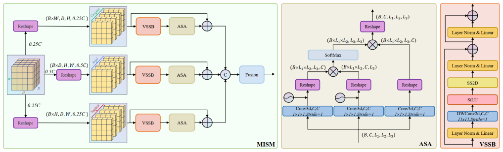
The load-bearing object is the Multi-view Axial Self-Attention Mamba (MISM) module, a hybrid block that replaces direct 3D Mamba scanning with tri-planar processing: the volume is cut into axial, coronal, and sagittal slices, each slice runs through VMamba's Visual State Space Block (VSSB) with bidirectional cross-scanning for intra-slice features, and a linear-complexity 1D Axial Self-Attention (ASA) mechanism then models inter-slice correlations along the orthogonal direction. The Asymmetric Split Channel (ASC) strategy feeds 50% of the channels through the axial plane and 25% through each of the other planes, which the paper argues cuts redundancy because the axial plane already shows the complete structure. The second carrier is the Feature-guided Region-aware Loss (FRLoss), computed on feature maps before the output block: it averages foreground features into a foreground center, pulls positive pixels toward that center (positive compactness), penalizes negative pixels near the boundary that resemble the center (boundary aware), and mines the top-N most similar negative pixels (hard negative mining).
What would settle it
Retrain the nine baselines on the same three datasets using each method's own recommended or individually tuned settings and several random seeds, then compare Dice distributions. If a tuned baseline matches or exceeds HCMA-UNet's Dice, or if HCMA-UNet's lead falls inside run-to-run variation, the state-of-the-art claim is refuted; a second check is to remove FRLoss and confirm the paper's reported 1.01-point Dice improvement disappears.
Extended reading notes
Core claim
On the paper's own terms, HCMA-UNet is a 3D encoder-decoder whose encoder combines a MobileNetV2-style Res Block and a Hybrid Block, and whose decoder uses transpose convolutions with HCMA-Up blocks. The novel part is the MISM module, which reslices each 3D volume along the axial, coronal, and sagittal planes instead of flattening the whole volume into one sequence; each plane is processed by VMamba's Visual State Space Block for intra-slice context, and an Axial Self-Attention mechanism runs along the orthogonal direction to model inter-slice dependencies. An Asymmetric Split Channel strategy allocates 50% of the channels to the axial view and 25% to each of the other two, reducing redundant computation. With this design the paper reports state-of-the-art Dice on all three datasets, outperforming nine baselines including nnU-Net, SwinUNETR, UNETR++, U-Mamba, LKM-UNet, and MambaClinix, and the FRLoss experiments show consistent gains when its three terms are added one by one.
Load-bearing premise
The comparison assumes every baseline model performs near its best under the single shared training recipe (patch size $128^3$, batch size 2, 500 epochs, AdamW at $10^{-4}$), with no method-specific tuning or multi-seed averaging, so the reported gaps could be configuration artifacts rather than real architectural differences.
Editorial extensions
If this is right
- If the reported results hold, 3D breast lesion segmentation no longer requires multi-hundred-million-parameter Mamba stacks: HCMA-UNet uses roughly 4% of U-Mamba's parameters and about 1% of its FLOPs while scoring higher Dice on all three datasets.
- The tri-planar MISM recipe gives a general way to adapt sequence-native Mamba blocks to volumetric medical images without flattening the volume into an unnatural 1D order, so the approach should transfer to other 3D segmentation tasks.
- FRLoss behaves as a plug-in loss: the comparison tables show that most of the nine baselines improve when FRLoss is added, which means existing DCE-MRI segmentation models can likely be upgraded without changing their architectures.
- On the private and public datasets evaluated, the complete HCMA-UNet with FRLoss sets a concrete numeric benchmark (Dice 80.28 / 76.14 / 78.05) for future breast cancer segmentation work to compare against.
Reading between the lines
- A testable extension the paper leaves implicit: the 50/25/25 axial-first channel split is motivated by the axial plane showing the complete breast structure, so a learnable or dataset-adaptive split could be benchmarked in organs without a dominant axis, such as liver or brain.
- Because FRLoss attacks a generic failure mode—negative pixels whose features resemble the foreground center—it plausibly transfers to other modalities and lesion types, but the paper demonstrates it only on DCE-MRI breast data, so that transfer is my inference rather than a paper claim.
- The shared training protocol makes the headline comparison a configuration-equality test rather than a best-configuration test; an external benchmark with per-method tuned settings and multi-seed confidence intervals would separate genuine architectural gains from configuration effects.
- If FRLoss's cross-architecture gains are real, a lighter design point follows that the paper does not explore: keep a fixed backbone and add FRLoss, since the loss terms cost nothing at inference time.
Signed reviews
Editorial analysis
A structured set of objections, weighed in public.
Referee Report
Summary. The paper proposes HCMA-UNet, a hybrid CNN-Mamba UNet for 3D breast cancer lesion segmentation in DCE-MRI. The architecture combines a lightweight residual CNN backbone with a Multi-view Axial Self-Attention Mamba (MISM) module, which uses Visual State Space Blocks and axial self-attention under an asymmetric channel-splitting scheme to process the three anatomical views. The paper also introduces a Feature-guided Region-aware Loss (FRLoss) composed of positive compactness, boundary-aware, and hard-negative-mining terms. The method is evaluated on one private and two public datasets against nine baselines, reporting state-of-the-art Dice scores of 80.28%, 76.14%, and 78.05% with 2.87M parameters and 126.44 GFLOPs. Ablations in Tables II and III support the individual contributions of the MISM components and the FRLoss terms. Source code is made available.
Significance. If the reported results hold, the paper makes a useful practical contribution: a compact architecture that reportedly outperforms much larger models while using substantially fewer parameters and FLOPs, together with a loss function that improves several baseline architectures. The availability of code, the internal consistency of the reported numbers, and the component-wise ablations are strengths. However, the central state-of-the-art claim currently rests on a baseline comparison that is not sufficiently controlled, and the lack of uncertainty quantification leaves the size of the reported gains unverified. The architectural idea and the proposed loss are coherent and worth pursuing, but the evidence presented does not yet support the headline claim as stated.
major comments (3)
- [Section III-C, Table I] The state-of-the-art claim rests on a comparison in which all nine baselines were trained under a single fixed protocol (patch size 128^3, batch size 2, 500 epochs, AdamW with learning rate 1e-4), with no per-method hyperparameter tuning, no multiple-seed averaging, and no statistical tests. Published recipes for several baselines (e.g., SwinUNETR, UMamba, LKM-UNet, MedNeXt) differ substantially in patch size, learning-rate schedule, and training length, and some are sensitive to batch size and regularization. The reported Dice gaps over the best baseline are 1.85, 2.30, and 2.52 points on Datasets I, II, and III respectively, which are within the range that configuration artifacts can produce. The authors should either tune each baseline under the same budget, report results from the baselines' own recommended settings, or explicitly restrict the claim to the fixed protocol and add significance testing.
- [Section III-A/B, Table I] Statistical robustness is not established. Dataset III has only 25 test samples from a random split, Dataset I's random split is not described as seeded, and no error bars, confidence intervals, or significance tests are reported anywhere. Run-to-run variability from weight initialization and data ordering can be comparable to the reported improvements in medical segmentation benchmarks, so the cross-dataset gains in Table I and the component gains in Tables II and III need to be supported by repeated runs, multiple random splits, or paired statistical tests. Without this, the claims of consistent and significant improvement are not quantitatively supported.
- [Section II-B2, Eqs. (1)-(2)] The paper states that the Axial Self-Attention mechanism 'achieves linear computational complexity.' The scaled dot-product attention in Eq. (2) computes pairwise similarities over the L3 dimension, which is O(H*W*L3^2*C) operations per volume, i.e., quadratic in the slice dimension and super-linear in the total volume H*W*L3. This is not linear complexity. If the intended meaning is linear in the slice length per query, that should be stated explicitly; otherwise the efficiency claim as written is inaccurate and should be corrected. The reported GFLOPs may still be favorable, but the complexity statement needs revision.
minor comments (6)
- [Introduction, first paragraph] There is a typo: 'surrocunding tissues' should be 'surrounding tissues.'
- [Section II-B3] The 50/25/25 channel split in ASC is justified by a heuristic about the axial plane displaying complete and symmetrical features, but no ablation with alternative split ratios is provided. A sensitivity analysis of this ratio would make the design choice more convincing.
- [Section II-D4, Eq. (7)] The symbol N is used both for the number of top hard-negative pixels and for the dilated hard-negative set obtained after the dilation operation. Rename one of the two to avoid notational confusion.
- [Section III-C] The FRLoss hyperparameters (lambda=5, T1=10, T2=10, N=250) are fixed across all experiments without sensitivity analysis. Please report whether the results are stable around these values, particularly across the three datasets.
- [Section III-D, Fig. 4] The single-slice Dice comparison in Fig. 4 (a 10.81% improvement over UMamba) is anecdotal. A quantitative summary over the test set or several representative slices would be more informative than one selected case.
- [Table I] Please specify the input size and the exact definition of GFLOPs used for the complexity numbers, for example whether they correspond to a forward pass on a patch of size 128^3. This is needed for a fair comparison with the baseline complexity figures.
Circularity Check
No significant circularity: architecture and loss claims are evaluated empirically on external and private datasets against standard metrics; no fitted input is renamed as a prediction.
full rationale
The paper's central claims are empirical and self-contained. HCMA-UNet is a concrete architecture whose components (VSSB, ASA, ASC, FRLoss) are defined by explicit formulas and then tested on one private and two public datasets using standard metrics (Dice, IoU, Precision, Recall, VS). The FRLoss terms (Eqs. 3-9) are explicit loss objectives, not fitted predictions: the foreground center is computed from current features, and the loss pulls features toward that center, which is a feature-regularization scheme rather than a hidden reuse of the evaluation metric. The reported parameter counts, GFLOPs, and Dice scores are measurements from training runs, not derivations from assumptions. The MISM module adapts published blocks (VMamba's VSSB and RDNet's Dense Block) by citation, but the combination and the axial self-attention mechanism are new design choices tested via ablations. The paper invokes no uniqueness theorem, and no load-bearing argument reduces to a self-citation chain. The uniform training protocol for all baselines in Section III-C could raise a benchmarking-fairness concern, but that is a question of experimental control and external validity, not circularity. No fitted input is renamed as a prediction, and no equation is equivalent to its input by construction. Therefore, the circularity score is 0.
Assumptions & free parameters
free parameters (6)
- lambda (FRLoss weight) =
5
- T1 (boundary dilation iterations) =
10
- T2 (hard-negative dilation iterations) =
10
- N (top hard negative pixels) =
250
- ASC channel split ratio =
50/25/25
- Patch size, batch size, epochs, learning rate =
128^3, 2, 500, 1e-4
assumptions (4)
- domain assumption Mamba's selective state space model provides effective long-range dependency modeling for 2D visual slices.
- ad hoc to paper The axial plane displays the complete structure and symmetrical features of breast lesions, justifying 50% channel allocation.
- standard math Standard deep learning components (scaled dot-product attention, residual blocks, dense connectivity) behave as described in the cited works.
- domain assumption The reported baseline performance reflects near-optimal configurations under the shared nnUNet-based training protocol.
Cite this review
Pith. "Pith review of HCMA-UNet: A Hybrid CNN-Mamba UNet with Axial Self-Attention for Efficient Breast Cancer Segmentation." pith.science (2026). https://pith.science/paper/QSUX7LBG
@misc{pith2026250100751,
  author       = {Pith},
  title        = {Pith review of: HCMA-UNet: A Hybrid CNN-Mamba UNet with Axial Self-Attention for Efficient Breast Cancer Segmentation},
  year         = {2026},
  howpublished = {\url{https://pith.science/paper/QSUX7LBG}},
  note         = {Machine review of arXiv:2501.00751}
}
read the original abstract
Breast cancer lesion segmentation in DCE-MRI remains challenging due to heterogeneous tumor morphology and indistinct boundaries. To address these challenges, this study proposes a novel hybrid segmentation network, HCMA-UNet, for lesion segmentation of breast cancer. Our network consists of a lightweight CNN backbone and a Multi-view Axial Self-Attention Mamba (MISM) module. The MISM module integrates Visual State Space Block (VSSB) and Axial Self-Attention (ASA) mechanism, effectively reducing parameters through Asymmetric Split Channel (ASC) strategy to achieve efficient tri-directional feature extraction. Our lightweight model achieves superior performance with 2.87M parameters and 126.44 GFLOPs. A Feature-guided Region-aware loss function (FRLoss) is proposed to enhance segmentation accuracy. Extensive experiments on one private and two public DCE-MRI breast cancer datasets demonstrate that our approach achieves state-of-the-art performance while maintaining computational efficiency. FRLoss also exhibits good cross-architecture generalization capabilities. The source code is available at https://github.com/Haoxuanli-Thu/HCMA-UNet.
Figures


Figures from the paper (1 more)

Forward citations
Cited by 1 Pith paper
-
Johnson-Lindenstrauss Lemma Guided Network for Efficient 3D Medical Segmentation
VeloxSeg pairs multi-scale window attention with JL-lemma-motivated grouped convolutions and Gram-matrix distillation to achieve efficient 3D medical segmentation at 1.66M parameters.
Reference graph
Works this paper leans on
-
[1]
Key steps for effective breast cancer prevention,
Kara L Britt, Jack Cuzick, and Kelly-Anne Phillips, “Key steps for effective breast cancer prevention,” Nature Reviews Cancer, vol. 20, no. 8, pp. 417–436, 2020
work page 2020
-
[2]
Early diagnosis and detection of breast cancer,
Marina Milosevic, Dragan Jankovic, Aleksandar Milenkovic, and Dra- gan Stojanov, “Early diagnosis and detection of breast cancer,” Tech- nology and Health Care , vol. 26, no. 4, pp. 729–759, 2018
work page 2018
-
[3]
U-net: Con- volutional networks for biomedical image segmentation,
Olaf Ronneberger, Philipp Fischer, and Thomas Brox, “U-net: Con- volutional networks for biomedical image segmentation,” in Medical image computing and computer-assisted intervention–MICCAI 2015: 18th international conference, Munich, Germany, October 5-9, 2015, proceedings, part III 18 . Springer, 2015, pp. 234–241
2015
-
[4]
Swin unetr: Swin transformers for semantic segmentation of brain tumors in mri images,
Ali Hatamizadeh, Vishwesh Nath, Yucheng Tang, Dong Yang, Holger R Roth, and Daguang Xu, “Swin unetr: Swin transformers for semantic segmentation of brain tumors in mri images,” in International MICCAI brainlesion workshop . Springer, 2021, pp. 272–284
work page 2021
-
[5]
Unetr++: delving into efficient and accurate 3d medical image segmentation,
Abdelrahman M Shaker, Muhammad Maaz, Hanoona Rasheed, Salman Khan, Ming-Hsuan Yang, and Fahad Shahbaz Khan, “Unetr++: delving into efficient and accurate 3d medical image segmentation,” IEEE Transactions on Medical Imaging , 2024
work page 2024
-
[6]
Swinhr: Hemodynamic-powered hierarchical vision transformer for breast tumor segmentation,
Zhihe Zhao, Siyao Du, Zeyan Xu, Zhi Yin, Xiaomei Huang, Xin Huang, Chinting Wong, Yanting Liang, Jing Shen, Jianlin Wu, et al., “Swinhr: Hemodynamic-powered hierarchical vision transformer for breast tumor segmentation,” Computers in biology and medicine , vol. 169, pp. 107939, 2024
work page 2024
-
[7]
Msa-vnet: Multi- scale attention-based v-net for dce-mri lesion segmentation,
Chang Yan Xu, Zi Jiang Sang, and Ye Qin Shao, “Msa-vnet: Multi- scale attention-based v-net for dce-mri lesion segmentation,” in 2022 International Conference on Image Processing, Computer Vision and Machine Learning (ICICML) . IEEE, 2022, pp. 309–312
work page 2022
-
[8]
Prototype learning guided hybrid network for breast tumor segmenta- tion in dce-mri,
Lei Zhou, Yuzhong Zhang, Jiadong Zhang, Xuejun Qian, Chen Gong, Kun Sun, Zhongxiang Ding, Xing Wang, Zhenhui Li, Zaiyi Liu, et al., “Prototype learning guided hybrid network for breast tumor segmenta- tion in dce-mri,” IEEE Transactions on Medical Imaging , 2024
work page 2024
Show all 22 references
-
[9]
Mamba: Linear-time sequence modeling with selective state spaces,
Albert Gu and Tri Dao, “Mamba: Linear-time sequence modeling with selective state spaces,” arXiv preprint arXiv:2312.00752 , 2023
2023 arXiv
-
[10]
U-mamba: Enhancing long-range depen- dency for biomedical image segmentation. arxiv 2024,
J Ma, F Li, and B Wang, “U-mamba: Enhancing long-range depen- dency for biomedical image segmentation. arxiv 2024,” arXiv preprint arXiv:2401.04722
2024 arXiv
-
[11]
Lkm-unet: Large kernel vision mamba unet for medical image segmentation,
Jinhong Wang, Jintai Chen, Danny Chen, and Jian Wu, “Lkm-unet: Large kernel vision mamba unet for medical image segmentation,” in International Conference on Medical Image Computing and Computer- Assisted Intervention . Springer, 2024, pp. 360–370
2024
-
[12]
Mambaclinix: Hierarchical gated convolution and mamba-based u-net for enhanced 3d medical image segmentation,
Chenyuan Bian, Nan Xia, Xia Yang, Feifei Wang, Fengjiao Wang, Bin Wei, and Qian Dong, “Mambaclinix: Hierarchical gated convolution and mamba-based u-net for enhanced 3d medical image segmentation,” arXiv preprint arXiv:2409.12533 , 2024
2024 arXiv
-
[13]
Vmamba: Visual state space model,
Yue Liu, Yunjie Tian, Yuzhong Zhao, Hongtian Yu, Lingxi Xie, Yaowei Wang, Qixiang Ye, Jianbin Jiao, and Yunfan Liu, “Vmamba: Visual state space model,” in The Thirty-eighth Annual Conference on Neural Information Processing Systems , 2024
2024
-
[14]
Mobilenetv2: Inverted residuals and linear bottlenecks,
Mark Sandler, Andrew Howard, Menglong Zhu, Andrey Zhmoginov, and Liang-Chieh Chen, “Mobilenetv2: Inverted residuals and linear bottlenecks,” in Proceedings of the IEEE conference on computer vision and pattern recognition , 2018, pp. 4510–4520
2018
-
[15]
Bag of tricks for image classification with convolutional neural networks,
Tong He, Zhi Zhang, Hang Zhang, Zhongyue Zhang, Junyuan Xie, and Mu Li, “Bag of tricks for image classification with convolutional neural networks,” in Proceedings of the IEEE/CVF conference on computer vision and pattern recognition , 2019, pp. 558–567
2019
-
[16]
Densenets reloaded: paradigm shift beyond resnets and vits,
Donghyun Kim, Byeongho Heo, and Dongyoon Han, “Densenets reloaded: paradigm shift beyond resnets and vits,” in European Confer- ence on Computer Vision . Springer, 2025, pp. 395–415
2025
-
[17]
Attention gated networks: Learning to leverage salient regions in medical images,
Jo Schlemper, Ozan Oktay, Michiel Schaap, Mattias Heinrich, Bernhard Kainz, Ben Glocker, and Daniel Rueckert, “Attention gated networks: Learning to leverage salient regions in medical images,” Medical Image Analysis, vol. 53, pp. 197–207, 2019
2019
-
[18]
nnu-net: a self-configuring method for deep learning-based biomedical image segmentation,
Fabian Isensee, Paul F Jaeger, Simon AA Kohl, Jens Petersen, and Klaus H Maier-Hein, “nnu-net: a self-configuring method for deep learning-based biomedical image segmentation,” Nature methods , vol. 18, no. 2, pp. 203–211, 2021
2021
-
[19]
Mednext: transformer-driven scaling of convnets for medical image segmentation,
Saikat Roy, Gregor Koehler, Constantin Ulrich, Michael Baumgartner, Jens Petersen, Fabian Isensee, Paul F Jaeger, and Klaus H Maier- Hein, “Mednext: transformer-driven scaling of convnets for medical image segmentation,” in International Conference on Medical Image Computing a...
2023
-
[20]
3d ux-net: A large kernel volumetric convnet modernizing hierar- chical transformer for medical image segmentation,
Ho Hin Lee, Shunxing Bao, Yuankai Huo, and Bennett A Landman, “3d ux-net: A large kernel volumetric convnet modernizing hierar- chical transformer for medical image segmentation,” arXiv preprint arXiv:2209.15076, 2022
2022 arXiv
-
[21]
Mama- mia: A large-scale multi-center breast cancer dce-mri benchmark dataset with expert segmentations,
Lidia Garrucho, Claire-Anne Reidel, Kaisar Kushibar, Smriti Joshi, Richard Osuala, Apostolia Tsirikoglou, Maciej Bobowicz, Javier del Riego, Alessandro Catanese, Katarzyna Gwo ´zdziewicz, et al., “Mama- mia: A large-scale multi-center breast cancer dce-mri benchmark dataset wi...
2024 arXiv
-
[22]
A robust and efficient ai assistant for breast tumor segmentation from dce-mri via a spatial-temporal framework,
Jiadong Zhang, Zhiming Cui, Zhenwei Shi, Yingjia Jiang, Zhiliang Zhang, Xiaoting Dai, Zhenlu Yang, Yuning Gu, Lei Zhou, Chu Han, et al., “A robust and efficient ai assistant for breast tumor segmentation from dce-mri via a spatial-temporal framework,” Patterns, vol. 4, no. 9, 2023
2023
Reviewed August 10, 2026 · model on record in the stance chip above.
Discussion (0). Continue with ORCID to comment.