REVIEW 4 major objections 5 minor 2 cited by
Segment Anything for Histopathology
T0 review · 4 major / 5 minor · reviewed 2026-08-09 · deepseek-v4-flash
Pith's one-line read PathoSAM, a vision foundation model built by finetuning the Segment Anything Model on six H&E-stained nucleus datasets, claims state-of-the-art automatic and interactive nucleus instance segmentation in histopathology.
desk verdict A useful, well-engineered SAM-for-histopathology model with a broad evaluation; the SOTA claim is plausible but lacks error bars, and the Lizard/CoNSeP overlap needs checking before the OOD generalization claim can be trusted. read the letter →
The pith
A machine-rendered reading of the paper's core claim, the machinery that carries it, and where it could break.
The reading
What carries the argument
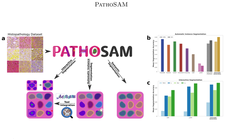
The load-bearing mechanism is joint training of SAM for both interaction and automation. PathoSAM retains SAM's image encoder, prompt encoder, and mask decoder for point, box, and mask prompting, and adds a convolutional decoder that outputs foreground probability, distance to the nearest object center, and distance to the nearest object boundary; seeded watershed converts those maps into instance segmentations. Training alternates between sampling prompts from ground-truth masks and correcting the model's predictions over seven iterations, while the same backbone is simultaneously optimized to reproduce the automatic-segmentation decoder's targets. At inference, the same weights answer a single click or box, refine a mask through further prompts, and run unattended over whole-slide tiles. A separate decoder added on top of the generalist performs semantic nucleus classification, trained with cross-entropy over PanNuke's five classes.
What would settle it
Evaluate PathoSAM on a new H&E dataset whose ground truth was drawn with a deliberately different nucleus convention, such as labeling cytoplasmic extensions or excluding small or overlapping nuclei, and compare automatic mean segmentation accuracy against a specialist model retrained on that dataset. If the generalist does not match or beat the specialist, or if errors concentrate wherever the annotation convention differs, the central generalization claim is falsified. A complementary check is to re-annotate CryoNuSeg to match PathoSAM's nucleus-region convention and see whether the reported quality gap disappears.
Extended reading notes
Core claim
On the paper's own terms, PathoSAM is the new state-of-the-art for automatic and interactive nucleus instance segmentation in histopathology. The model keeps SAM's prompt-driven mask prediction and adds a second decoder that predicts foreground probability and distance maps, which a seeded watershed turns into instance masks; the whole network is trained jointly with an objective that simulates interactive corrections and automatic segmentation on a diverse set of H&E nucleus datasets. In evaluation across 12 datasets, the ViT-L generalist achieves the highest average mean segmentation accuracy among automatic methods, even though for some competing methods the best available model version per dataset was used. For interactive segmentation, PathoSAM outperforms other SAM variants on single point and box prompts and reaches near-perfect accuracy after seven iterative corrections. The paper also reports a semantic segmentation decoder trained on PanNuke that ranks second behind CellViT, with the main shortfall on a rare 'dead cells' class, and a finetuning recipe that yields improved specialist models for lymphocyte and gland segmentation.
Load-bearing premise
The load-bearing premise is that the six H&E training datasets share consistent enough nucleus annotation conventions and enough imaging diversity for one model to generalize; the paper itself shows that when annotations disagree with the visible nucleus region, as in CryoNuSeg, automatic segmentation quality collapses.
Editorial extensions
If this is right
- A single set of PathoSAM weights can replace dataset-specific retraining for nucleus instance segmentation across most H&E histopathology datasets.
- Interactive annotation in tools such as QuPath and napari requires fewer prompts per nucleus, because point and box prompts already capture nuclei accurately.
- Whole-slide images can be segmented automatically with tile-and-stitch scripts in under an hour on a GPU, enabling large-scale nucleus analysis.
- The same finetuning recipe transfers to non-nucleus structures such as lymphocytes and glands when trained on task-specific data.
- Semantic nucleus segmentation is possible from the same generalist backbone, though on PanNuke it trails CellViT mainly on a minority 'dead cells' class.
Reading between the lines
- If annotation protocols were harmonized across datasets, the out-of-domain failures such as CryoNuSeg could shrink; the paper's own analysis attributes that failure to annotation inconsistency rather than image content alone.
- The same joint interactive-plus-automatic training recipe may extend to multi-scale segmentation of glands and tissue regions if the model is given scale-aware prompts or multi-scale training, a step the paper identifies as future work.
- Because PathoSAM's interactive segmentation is near-perfect after a few corrections, it could serve as a pre-annotation engine where human annotators confirm and fix masks instead of drawing nuclei from scratch.
- A direct test of the semantic-segmentation gap would be to train with minority-class oversampling and joint instance-semantic objectives, which the paper proposes; if the gap to CellViT disappears, the current ranking is a training-strategy artifact rather than an architectural limit.
Editorial analysis
A structured set of objections, weighed in public.
Referee Report
Summary. The paper introduces PathoSAM, a finetuned variant of the Segment Anything Model (SAM) for nucleus segmentation in histopathology. It supports interactive segmentation from point/box prompts and automatic instance segmentation via an additional decoder with seeded watershed post-processing (AIS), following the microSAM recipe. The authors train generalist ViT-B/L/H models on a combination of six H&E datasets and evaluate on 12 datasets (6 in-domain test splits and 6 out-of-domain), comparing against HoVerNet, StarDist, CellViT, HoVerNeXt, InstanSeg, SAM, and microSAM. They report that PathoSAM is the new state-of-the-art for automatic and interactive nucleus instance segmentation, while for semantic segmentation on PanNuke it ranks second to CellViT. The paper also presents a user study with QuPath and napari integration, whole-slide-image inference scripts, and open-source code and model weights.
Significance. If the results hold, PathoSAM is a practically valuable contribution: a single open-source model that performs automatic and interactive nucleus segmentation across diverse histopathology datasets without per-dataset retraining, with integration into popular annotation tools. The study's strengths include held-out test splits, best-available-version reporting for multi-version baselines, and public code and models. However, the strength of the state-of-the-art claim is currently weakened by the absence of uncertainty estimates or significance tests, and by a potential overlap between the Lizard training data and the CoNSeP out-of-domain evaluation set, which directly bears on the 'unseen dataset' generalization claim.
major comments (4)
- [Section 2.5 and Table 1] The out-of-domain evaluation may be contaminated by training-data overlap. The Lizard dataset (Graham et al., 2021) is one of the six training datasets, and Lizard is known to be constructed by merging and re-annotating images from existing colorectal datasets, including CoNSeP (Graham et al., 2019), which is later listed among the 'remaining 8 datasets' used for out-of-domain evaluation. The manuscript does not report an image-level or slide-level de-duplication check between the Lizard training split and the CoNSeP test set. If any CoNSeP images or tiles appear in the Lizard training data, then CoNSeP is not 'not directly represented in the training set' as claimed, and the generalization result on CoNSeP is not valid evidence for the 'unseen dataset' claim. The authors must provide a de-duplication analysis (e.g., image hashing or patch matching) and re-run the out-of-domain evaluation excluding any overlapping images, or explicitly state which out-of-domain datasets remain genuinely unseen.
- [Section 3.1, Fig. 2a, Fig. 8, App. D.1] All quantitative results are presented as figures with mean scores only; no error bars, standard deviations, or significance tests are reported. The claim that PathoSAM is 'the new state-of-the-art' for automatic nucleus instance segmentation rests on average scores over test sets, and for some comparisons the differences between variants (e.g., ViT-B vs. ViT-L vs. ViT-H) are described as small. Without uncertainty estimates or paired statistical tests on the same test images, the reader cannot assess whether the observed improvements over baselines are meaningful. Please provide numeric tables with per-dataset means and standard deviations (at least across test images), and perform significance tests for the headline comparisons that support the state-of-the-art claim.
- [Fig. 10 and App. C] The watershed seed thresholds used in AIS appear to be selected by a grid search on 15 validation images of LyNSeC, an out-of-domain dataset. The manuscript should clarify whether these thresholds were fixed before evaluation on all datasets and whether the LyNSeC validation images are disjoint from the LyNSeC test images. If the thresholds were selected on this out-of-domain validation split and then applied to the same dataset, the reported LyNSeC results are optimistically biased; if the thresholds were applied to other out-of-domain datasets, a sensitivity analysis should be reported to show that the main conclusions do not depend on the exact threshold choice.
- [Section 3.2] The interactive segmentation comparison is limited to the original SAM and microSAM. The claim in Section 1 that 'PathoSAM outperforms other SAM variants for interactive segmentation' is broader than the evidence. Please either compare with additional SAM-based methods for histopathology (for example SAM-Path or MedicoSAM) or restrict the claim to the baselines actually evaluated, so that the stated claim matches the experimental scope.
minor comments (5)
- [Appendix C] The training iteration counts state '100,00 iterations' for the PanNuke and specialist models; this should read '100,000 iterations'.
- [Section 2.2] The sentence 'We evaluate interactive segmentation (for SAM or variants) with AMG and, if available, AIS' is confusing because AMG and AIS are automatic segmentation methods, not interactive ones; it should be rephrased to refer to automatic segmentation.
- [Fig. 2 caption] The notation I_P and I_B is used in the caption without a definition; it should be defined in the caption or explained at first use in the main text.
- [Section 3.4] The phrase 'ca. 5 GB of VRAM per tile' should be clarified to mean peak VRAM usage during inference on a tile, not persistent per-tile allocation.
- [Appendix D.1] The text refers to 'D.1 displays results' without identifying whether D.1 is a table or figure; the cross-reference should be made explicit.
Circularity Check
No material circularity: PathoSAM's central claims are empirically evaluated on separate test splits; self-citations to microSAM are independent and non-load-bearing.
full rationale
PathoSAM is an empirical finetuning paper. The central claim, being the new state-of-the-art for automatic and interactive nucleus instance segmentation, is supported by evaluations on held-out test splits of 12 datasets against external methods such as HoVerNet, StarDist, CellViT, HoVerNeXt and InstanSeg. These results are measured, not derived by construction from PathoSAM's training objective or from fitted parameters. The main self-referential component is the adoption of the microSAM training implementation, the additional AIS decoder design, and the interactive evaluation protocol (Sec. 2.1, 2.2, 2.3, App. C). These are citations to an independently published Nature Methods paper by overlapping authors (Archit et al., 2025a); they supply a method and an evaluation scheme, but the state-of-the-art comparison is against external baselines and does not reduce to the cited work. The manuscript also contains honest limitations, stating that PathoSAM does not improve gland or lymphocyte segmentation and that semantic segmentation was only evaluated on a single dataset, which weakens scope claims but does not create circularity. A possible data-leakage concern, that the Lizard training set contains images also appearing in the CoNSeP out-of-domain evaluation, is not established by the manuscript text and would be an empirical correctness issue rather than a circular derivation. Likewise, the watershed-threshold grid search on 15 validation images of LyNSeC (Fig. 10) is a post-processing selection on an OOD validation subset; the paper does not state that the reported test numbers were produced with these tuned thresholds, and even if they were, this is a tuning effect, not a parameter renamed as a prediction. Overall, the derivation chain is self-contained and externally benchmarked, so the circularity score is low.
Assumptions & free parameters
free parameters (2)
- AIS watershed seed thresholds (center and boundary) =
grid-searched on 15 LyNSeC H&E validation images
- Training hyperparameters (iterations, LR, batch size, objects per image) =
250k iterations, LR 1e-5, batch size 2, 40/30/25 objects per image
assumptions (4)
- domain assumption The six H&E-stained training datasets are representative of histopathology nuclei and have sufficiently consistent annotation protocols for joint training.
- domain assumption SAM's pretrained weights and the microSAM training objective transfer to histopathology such that joint interactive and automatic training is beneficial.
- standard math The mean segmentation accuracy metric (averaged over IoU thresholds) is a valid measure of instance segmentation quality for comparing methods.
- domain assumption The out-of-domain datasets are genuinely out-of-domain, i.e., not represented in the training set.
Cite this review
Pith. "Pith review of Segment Anything for Histopathology." pith.science (2026). https://pith.science/paper/PXRRT76B
@misc{pith2026250200408,
  author       = {Pith},
  title        = {Pith review of: Segment Anything for Histopathology},
  year         = {2026},
  howpublished = {\url{https://pith.science/paper/PXRRT76B}},
  note         = {Machine review of arXiv:2502.00408}
}
read the original abstract
Nucleus segmentation is an important analysis task in digital pathology. However, methods for automatic segmentation often struggle with new data from a different distribution, requiring users to manually annotate nuclei and retrain data-specific models. Vision foundation models (VFMs), such as the Segment Anything Model (SAM), offer a more robust alternative for automatic and interactive segmentation. Despite their success in natural images, a foundation model for nucleus segmentation in histopathology is still missing. Initial efforts to adapt SAM have shown some success, but did not yet introduce a comprehensive model for diverse segmentation tasks. To close this gap, we introduce PathoSAM, a VFM for nucleus segmentation, based on training SAM on a diverse dataset. Our extensive experiments show that it is the new state-of-the-art model for automatic and interactive nucleus instance segmentation in histopathology. We also demonstrate how it can be adapted for other segmentation tasks, including semantic nucleus segmentation. For this task, we show that it yields results better than popular methods, while not yet beating the state-of-the-art, CellViT. Our models are open-source and compatible with popular tools for data annotation. We also provide scripts for whole-slide image segmentation. Our code and models are publicly available at https://github.com/computational-cell-analytics/patho-sam.
Figures


Figures from the paper (13 more)


Forward citations
Cited by 2 Pith papers
-
Towards Autonomous and Auditable Medical Imaging Model Development
AMID, a verification-guided multi-agent MLE system for medical imaging, outperforms general MLE agents on 20 ReX-MLE challenges and approaches human challenge solutions on several tasks.
-
Evaluating Vision Foundation Models for Pixel and Object Classification in Microscopy
Vision foundation model features improve pixel and object classification in microscopy over hand-crafted features, and object-guided attentive probing (ObAP) can match or beat supervised baselines with very few labels.
Reference graph
Works this paper leans on
-
[1]
Nuclick: A deep learning framework for interactive segmentation of microscopic images
Navid Alemi Koohbanani, Mostafa Jahanifar, Neda Zamani Tajadin, and Nasir Rajpoot. Nuclick: A deep learning framework for interactive segmentation of microscopic images. Medical Image Analysis, 65: 0 101771, October 2020. ISSN 1361-8415. doi:10.1016/j.media.2020.101771. URL http://doi.org/10.1016/j.media.2020.101771
arXiv 2020
-
[2]
Segment anything for microscopy
Anwai Archit, Luca Freckmann, Sushmita Nair, Nabeel Khalid, Paul Hilt, Vikas Rajashekar, Marei Freitag, Carolin Teuber, Genevieve Buckley, Sebastian von Haaren, Sagnik Gupta, Andreas Dengel, Sheraz Ahmed, and Constantin Pape. Segment anything for microscopy. Nature Methods, 22 0 (3): 0 579–591, February 2025 a . ISSN 1548-7105. doi:10.1038/s41592-024-0258...
-
[3]
Medicosam: Towards foundation models for medical image segmentation, 2025 b
Anwai Archit, Luca Freckmann, and Constantin Pape. Medicosam: Towards foundation models for medical image segmentation, 2025 b . URL https://arxiv.org/abs/2501.11734
arXiv 2025
-
[4]
Peter Bankhead, Maurice B. Loughrey, José A. Fernández, Yvonne Dombrowski, Darragh G. McArt, Philip D. Dunne, Stephen McQuaid, Ronan T. Gray, Liam J. Murray, Helen G. Coleman, Jacqueline A. James, Manuel Salto-Tellez, and Peter W. Hamilton. Qupath: Open source software for digital pathology image analysis. Scientific Reports, 7 0 (1), December 2017. ISSN ...
-
[5]
Nagtegaal, Maria Rodriguez Martinez, and Inti Zlobec
Elias Baumann, Bastian Dislich, Josef Lorenz Rumberger, Iris D. Nagtegaal, Maria Rodriguez Martinez, and Inti Zlobec. Hover-next: A fast nuclei segmentation and classification pipeline for next generation histopathology. In Medical Imaging with Deep Learning, 2024. URL https://openreview.net/forum?id=3vmB43oqIO
work page 2024
-
[6]
Caicedo, Allen Goodman, Kyle W
Juan C. Caicedo, Allen Goodman, Kyle W. Karhohs, Beth A. Cimini, Jeanelle Ackerman, Marzieh Haghighi, CherKeng Heng, Tim Becker, Minh Doan, Claire McQuin, Mohammad Rohban, Shantanu Singh, and Anne E. Carpenter. Nucleus segmentation across imaging experiments: the 2018 data science bowl. Nature Methods, 16 0 (12): 0 1247–1253, October 2019. ISSN 1548-7105....
-
[7]
Junlong Cheng, Jin Ye, Zhongying Deng, Jianpin Chen, Tianbin Li, Haoyu Wang, Yanzhou Su, Ziyan Huang, Jilong Chen, Lei Jiang, Hui Sun, Junjun He, Shaoting Zhang, Min Zhu, and Yu Qiao. Sam-med2d, 2023. URL https://arxiv.org/abs/2308.16184
arXiv 2023
-
[8]
Ruining Deng, Can Cui, Quan Liu, Tianyuan Yao, Lucas Walker Remedios, Shunxing Bao, Bennett A. Landman, Yucheng Tang, Lee E. Wheless, Lori A. Coburn, Keith T. Wilson, Yaohong Wang, Agnes B. Fogo, Haichun Yang, and Yuankai Huo. Segment anything model ( SAM ) for digital pathology: Assess zero-shot segmentation on whole slide imaging. In Medical Imaging wit...
work page 2023
Show all 40 references
-
[9]
An image is worth 16x16 words: Transformers for image recognition at scale
Alexey Dosovitskiy, Lucas Beyer, Alexander Kolesnikov, Dirk Weissenborn, Xiaohua Zhai, Thomas Unterthiner, Mostafa Dehghani, Matthias Minderer, Georg Heigold, Sylvain Gelly, Jakob Uszkoreit, and Neil Houlsby. An image is worth 16x16 words: Transformers for image recognition at...
2021
-
[10]
Pannuke: an open pan-cancer histology dataset for nuclei instance segmentation and classification
Jevgenij Gamper, Navid Alemi Koohbanani, Ksenija Benes, Ali Khuram, and Nasir Rajpoot. Pannuke: an open pan-cancer histology dataset for nuclei instance segmentation and classification. In European Congress on Digital Pathology, pages 11--19. Springer, 2019. URL https://doi.or...
2019 doi
-
[11]
Instanseg: an embedding-based instance segmentation algorithm optimized for accurate, efficient and portable cell segmentation, 2024
Thibaut Goldsborough, Ben Philps, Alan O'Callaghan, Fiona Inglis, Leo Leplat, Andrew Filby, Hakan Bilen, and Peter Bankhead. Instanseg: an embedding-based instance segmentation algorithm optimized for accurate, efficient and portable cell segmentation, 2024. URL https://arxiv....
2024 arXiv
-
[12]
Hover-net: Simultaneous segmentation and classification of nuclei in multi-tissue histology images
Simon Graham, Quoc Dang Vu, Shan E Ahmed Raza, Ayesha Azam, Yee Wah Tsang, Jin Tae Kwak, and Nasir Rajpoot. Hover-net: Simultaneous segmentation and classification of nuclei in multi-tissue histology images. Medical Image Analysis, 58: 0 101563, December 2019. ISSN 1361-8415. ...
2019
-
[13]
Ahmed Raza, Hesham El Daly, Kishore Gopalakrishnan, David Snead, and Nasir M
Simon Graham, Mostafa Jahanifar, Ayesha Azam, Mohammed Nimir, Yee-Wah Tsang, Katherine Dodd, Emily Hero, Harvir Sahota, Atisha Tank, Ksenija Benes, Noorul Wahab, Fayyaz Minhas, Shan E. Ahmed Raza, Hesham El Daly, Kishore Gopalakrishnan, David Snead, and Nasir M. Rajpoot. Lizar...
2021
-
[14]
Roth, and Daguang Xu
Ali Hatamizadeh, Yucheng Tang, Vishwesh Nath, Dong Yang, Andriy Myronenko, Bennett Landman, Holger R. Roth, and Daguang Xu. Unetr: Transformers for 3d medical image segmentation. In Proceedings of the IEEE/CVF Winter Conference on Applications of Computer Vision (WACV), pages ...
2022
-
[15]
o rst, Moritz Rempe, Lukas Heine, Constantin Seibold, Julius Keyl, Giulia Baldini, Selma Ugurel, Jens Siveke, Barbara Gr\
Fabian H\" o rst, Moritz Rempe, Lukas Heine, Constantin Seibold, Julius Keyl, Giulia Baldini, Selma Ugurel, Jens Siveke, Barbara Gr\" u nwald, Jan Egger, and Jens Kleesiek. Cellvit: Vision transformers for precise cell segmentation and classification. Medical Image Analysis, 9...
2024
-
[16]
Deep learning for digital pathology image analysis: A comprehensive tutorial with selected use cases
Andrew Janowczyk and Anant Madabhushi. Deep learning for digital pathology image analysis: A comprehensive tutorial with selected use cases. Journal of Pathology Informatics, 7 0 (1): 0 29, January 2016. ISSN 2153-3539. doi:10.4103/2153-3539.186902. URL http://doi.org/10.4103/...
2016
-
[17]
Berg, Wan-Yen Lo, Piotr Dollar, and Ross Girshick
Alexander Kirillov, Eric Mintun, Nikhila Ravi, Hanzi Mao, Chloe Rolland, Laura Gustafson, Tete Xiao, Spencer Whitehead, Alexander C. Berg, Wan-Yen Lo, Piotr Dollar, and Ross Girshick. Segment anything. In Proceedings of the IEEE/CVF International Conference on Computer Vision ...
2023
-
[18]
A dataset and a technique for generalized nuclear segmentation for computational pathology
Neeraj Kumar, Ruchika Verma, Sanuj Sharma, Surabhi Bhargava, Abhishek Vahadane, and Amit Sethi. A dataset and a technique for generalized nuclear segmentation for computational pathology. IEEE Transactions on Medical Imaging, 36 0 (7): 0 1550–1560, July 2017. ISSN 1558-254X. d...
2017
-
[19]
Cryonuseg: A dataset for nuclei instance segmentation of cryosectioned h&e-stained histological images
Amirreza Mahbod, Gerald Schaefer, Benjamin Bancher, Christine L\" o w, Georg Dorffner, Rupert Ecker, and Isabella Ellinger. Cryonuseg: A dataset for nuclei instance segmentation of cryosectioned h&e-stained histological images. Computers in Biology and Medicine, 132: 0 104349,...
2021
-
[20]
Nuinsseg: A fully annotated dataset for nuclei instance segmentation in h&e-stained histological images
Amirreza Mahbod, Christine Polak, Katharina Feldmann, Rumsha Khan, Katharina Gelles, Georg Dorffner, Ramona Woitek, Sepideh Hatamikia, and Isabella Ellinger. Nuinsseg: A fully annotated dataset for nuclei instance segmentation in h&e-stained histological images. Scientific Dat...
2024 doi
-
[21]
Holy-net: Segmentation of histological images of diffuse large b-cell lymphoma
Hussein Naji, Lucas Sancere, Adrian Simon, Reinhard B\" u ttner, Marie-Lisa Eich, Philipp Lohneis, and Katarzyna Bożek. Holy-net: Segmentation of histological images of diffuse large b-cell lymphoma. Computers in Biology and Medicine, 170: 0 107978, March 2024. ISSN 0010-4825....
2024
-
[22]
Segmentation of nuclei in histopathology images by deep regression of the distance map
Peter Naylor, Marick Laé, Fabien Reyal, and Thomas Walter. Segmentation of nuclei in histopathology images by deep regression of the distance map. IEEE Transactions on Medical Imaging, 38 0 (2): 0 448–459, February 2019. ISSN 1558-254X. doi:10.1109/tmi.2018.2865709. URL http:/...
2019
-
[23]
SAM 2: Segment anything in images and videos
Nikhila Ravi, Valentin Gabeur, Yuan-Ting Hu, Ronghang Hu, Chaitanya Ryali, Tengyu Ma, Haitham Khedr, Roman R \"a dle, Chloe Rolland, Laura Gustafson, Eric Mintun, Junting Pan, Kalyan Vasudev Alwala, Nicolas Carion, Chao-Yuan Wu, Ross Girshick, Piotr Dollar, and Christoph Feich...
2025
-
[24]
U-Net: Convolutional Networks for Biomedical Image Segmentation, page 234–241
Olaf Ronneberger, Philipp Fischer, and Thomas Brox. U-Net: Convolutional Networks for Biomedical Image Segmentation, page 234–241. Springer International Publishing, 2015. ISBN 9783319245744. doi:10.1007/978-3-319-24574-4_28. URL http://doi.org/10.1007/978-3-319-24574-4_28
2015 doi
-
[25]
A novel dataset for nuclei and tissue segmentation in melanoma with baseline nuclei segmentation and tissue segmentation benchmarks
Mark Schuiveling, Hong Liu, Daniel Eek, Gerben E Breimer, Karijn P M Suijkerbuijk, Willeke A M Blokx, and Mitko Veta. A novel dataset for nuclei and tissue segmentation in melanoma with baseline nuclei segmentation and tissue segmentation benchmarks. GigaScience, 14, 2025. ISS...
2025 doi
-
[26]
Pluim, Hao Chen, Xiaojuan Qi, Pheng-Ann Heng, Yun Bo Guo, Li Yang Wang, Bogdan J
Korsuk Sirinukunwattana, Josien P.W. Pluim, Hao Chen, Xiaojuan Qi, Pheng-Ann Heng, Yun Bo Guo, Li Yang Wang, Bogdan J. Matuszewski, Elia Bruni, Urko Sanchez, Anton B\" o hm, Olaf Ronneberger, Bassem Ben Cheikh, Daniel Racoceanu, Philipp Kainz, Michael Pfeiffer, Martin Urschler...
2017 doi
-
[27]
Nicholas Sofroniew, Talley Lambert, Grzegorz Bokota, Juan Nunez-Iglesias, Peter Sobolewski, Andrew Sweet, Lorenzo Gaifas, Kira Evans, Alister Burt, Draga Doncila Pop, Kevin Yamauchi, Melissa Weber Mendon c a, Genevieve Buckley, Wouter-Michiel Vierdag, Loic Royer, Ahmet Can Sol...
2024
-
[28]
Training deep learning models for cell image segmentation with sparse annotations
Ko Sugawara. Training deep learning models for cell image segmentation with sparse annotations. BioRxiv, pages 2023--06, 2023. URL https://doi.org/10.1101/2023.06.13.544786
2023 doi
-
[29]
hover-unet
Cristian Tommasino, Cristiano Russo, Antonio Maria Rinaldi, and Francesco Ciompi. “hover-unet”: Accelerating hovernet with unet-based multi-class nuclei segmentation via knowledge distillation. In 2024 IEEE International Symposium on Biomedical Imaging (ISBI), page 1–4. IEEE, ...
2024
-
[30]
Deep learning in histopathology: the path to the clinic
Jeroen van der Laak, Geert Litjens, and Francesco Ciompi. Deep learning in histopathology: the path to the clinic. Nature Medicine, 27 0 (5): 0 775–784, May 2021. ISSN 1546-170X. doi:10.1038/s41591-021-01343-4. URL http://doi.org/10.1038/s41591-021-01343-4
2021 doi
-
[31]
Ahmed Raza, Nasir Rajpoot, Xiyi Wu, Huai Chen, Yijie Huang, Lisheng Wang, Hyun Jung, G
Ruchika Verma, Neeraj Kumar, Abhijeet Patil, Nikhil Cherian Kurian, Swapnil Rane, Simon Graham, Quoc Dang Vu, Mieke Zwager, Shan E. Ahmed Raza, Nasir Rajpoot, Xiyi Wu, Huai Chen, Yijie Huang, Lisheng Wang, Hyun Jung, G. Thomas Brown, Yanling Liu, Shuolin Liu, Seyed Alireza Fat...
2021
-
[32]
Methods for segmentation and classification of digital microscopy tissue images
Quoc Dang Vu, Simon Graham, Tahsin Kurc, Minh Nguyen Nhat To, Muhammad Shaban, Talha Qaiser, Navid Alemi Koohbanani, Syed Ali Khurram, Jayashree Kalpathy-Cramer, Tianhao Zhao, Rajarsi Gupta, Jin Tae Kwak, Nasir Rajpoot, Joel Saltz, and Keyvan Farahani. Methods for segmentation...
2019
-
[33]
Simultaneously segmenting and classifying cell nuclei by using multi-task learning in multiplex immunohistochemical tissue microarray sections
Ranran Wang, Yusong Qiu, Xinyu Hao, Shan Jin, Junxiu Gao, Heng Qi, Qi Xu, Yong Zhang, and Hongming Xu. Simultaneously segmenting and classifying cell nuclei by using multi-task learning in multiplex immunohistochemical tissue microarray sections. Biomedical Signal Processing a...
2024
-
[34]
Nuclei instance segmentation and classification in histopathology images with stardist
Martin Weigert and Uwe Schmidt. Nuclei instance segmentation and classification in histopathology images with stardist. In 2022 IEEE International Symposium on Biomedical Imaging Challenges (ISBIC), page 1–4. IEEE, March 2022. doi:10.1109/isbic56247.2022.9854534. URL http://do...
2022
-
[35]
On generalisability of segment anything model for nuclear instance segmentation in histology images, 2024
Kesi Xu, Lea Goetz, and Nasir Rajpoot. On generalisability of segment anything model for nuclear instance segmentation in histology images, 2024. URL https://arxiv.org/abs/2401.14248
2024 arXiv
-
[36]
Surgicalsam: Efficient class promptable surgical instrument segmentation
Wenxi Yue, Jing Zhang, Kun Hu, Yong Xia, Jiebo Luo, and Zhiyong Wang. Surgicalsam: Efficient class promptable surgical instrument segmentation. Proceedings of the AAAI Conference on Artificial Intelligence, 38 0 (7): 0 6890–6898, March 2024. ISSN 2159-5399. doi:10.1609/aaai.v3...
2024 doi
-
[37]
SAM-Path: A Segment Anything Model for Semantic Segmentation in Digital Pathology, page 161–170
Jingwei Zhang, Ke Ma, Saarthak Kapse, Joel Saltz, Maria Vakalopoulou, Prateek Prasanna, and Dimitris Samaras. SAM-Path: A Segment Anything Model for Semantic Segmentation in Digital Pathology, page 161–170. Springer Nature Switzerland, 2023. ISBN 9783031474019. doi:10.1007/978...
2023 doi
-
[38]
Glandsam: Injecting morphology knowledge into segment anything model for label-free gland segmentation
Qixiang Zhang, Yi Li, Cheng Xue, Haonan Wang, and Xiaomeng Li. Glandsam: Injecting morphology knowledge into segment anything model for label-free gland segmentation. IEEE Transactions on Medical Imaging, 2024. URL https://doi.org/10.1109/TMI.2024.3476176
2024
-
[39]
A foundation model for joint segmentation, detection and recognition of biomedical objects across nine modalities
Theodore Zhao, Yu Gu, Jianwei Yang, Naoto Usuyama, Ho Hin Lee, Sid Kiblawi, Tristan Naumann, Jianfeng Gao, Angela Crabtree, Jacob Abel, Christine Moung-Wen, Brian Piening, Carlo Bifulco, Mu Wei, Hoifung Poon, and Sheng Wang. A foundation model for joint segmentation, detection...
2024 doi
-
[40]
Segment everything everywhere all at once
Xueyan Zou, Jianwei Yang, Hao Zhang, Feng Li, Linjie Li, Jianfeng Wang, Lijuan Wang, Jianfeng Gao, and Yong Jae Lee. Segment everything everywhere all at once. In Proceedings of the 37th International Conference on Neural Information Processing Systems, NIPS '23, Red Hook, NY,...
2024
Reviewed August 9, 2026 · model on record in the stance chip above.
Discussion (0). Continue with ORCID to comment.