REVIEW 1 major objections 6 minor 75 references
FetDTIAlign: A Deep Learning Framework for Affine and Deformable Registration of Fetal Brain dMRI
T0 review · 1 major / 6 minor · reviewed 2026-08-09 · deepseek-v4-flash
Pith's one-line read This paper presents FetDTIAlign, a deep learning framework that registers fetal brain diffusion MRI across 23 to 36 weeks of gestation, and reports that it outperforms classical optimization-based tools and a generic deep learning…
desk verdict A competent first deep-learning framework for fetal dMRI registration, but the headline Dice superiority is partly trained into the evaluation and needs independent anatomical validation. read the letter →
The pith
A machine-rendered reading of the paper's core claim, the machinery that carries it, and where it could break.
The reading
What carries the argument
The load-bearing mechanism is a two-stage learned registration pipeline. In the affine stage, separate encoders for the moving and target images produce feature embeddings, a registration head estimates the affine matrix from the mass centers of those embeddings, and recurrent inference composes iteratively refined transforms, choosing the step with the lowest mean squared error on fractional anisotropy and tensor images. In the deformable stage, shared-weight fractional anisotropy and tensor encoders extract four-scale features that are warped and fed to a recurrent decoder built from correlation-aware multi-window multi-layer perceptron blocks, estimating residual deformations from coarse to fine. A tensor-reorientation layer applies the rotation component of each transformation, obtained by polar (singular value) decomposition of the Jacobian, so fiber orientations stay anatomically consistent; finally, the composed affine-plus-deformable field is applied to the original image in a single interpolation to limit smoothing.
What would settle it
Evaluate FetDTIAlign on the external dataset using independently prepared white matter tract segmentations, and compare overlap scores with the classical baselines; if the advantage disappears, the claimed anatomical superiority is an artifact of optimizing the same tract-overlap metric used for evaluation.
Extended reading notes
Core claim
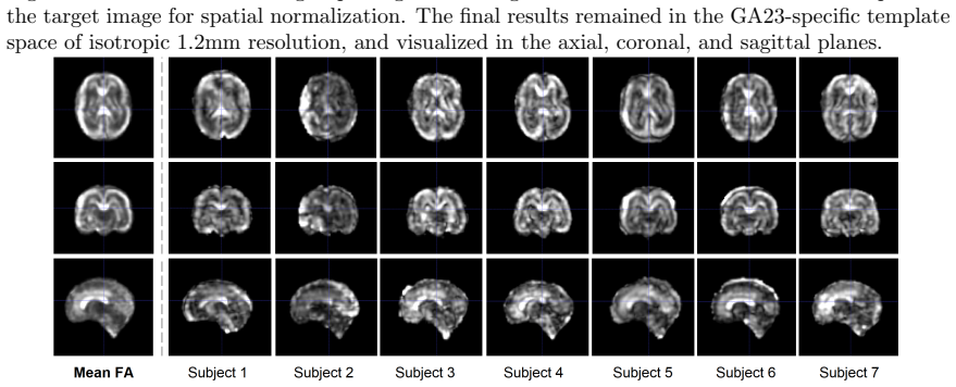
FetDTIAlign is presented as the first deep learning framework designed specifically for spatial normalization of fetal brain diffusion MRI. It estimates an affine transform and then a residual deformable field using two domain-specific encoders: one processes fractional anisotropy maps and the other the full diffusion tensor, with a recurrent multi-scale decoder and a differentiable tensor-reorientation layer that keeps fiber orientations anatomically consistent. The authors claim that, across gestational ages 23 to 36 weeks and across 60 white matter tracts, FetDTIAlign consistently achieves the highest tract-overlap score among the compared methods in both the affine and deformable stages, produces visually sharper mean fractional anisotropy images, and maintains more physically plausible deformations than a generic deep learning baseline, while also transferring to an external dataset with different acquisition protocols.
Load-bearing premise
The load-bearing premise is that the hand-verified white matter tract maps used for scoring are an impartial measure of anatomical correspondence, even though the learning-based methods were trained to maximize overlap with those same maps while the classical methods were not.
Editorial extensions
If this is right
- Spatial normalization of fetal diffusion MRI across a wide gestational range becomes a single network inference, making large-cohort voxel-wise and tract-based analyses practical.
- Because the learned transformation is applied to the original image only once, the pipeline reduces cumulative interpolation smoothing and helps preserve tract boundaries for downstream analysis.
- The same estimated transformations can be applied to other diffusion-derived maps, such as mean diffusivity, extending the framework beyond fractional anisotropy and tensor comparisons.
- Cross-protocol generalization to data acquired with different acquisition schemes suggests the method can be applied to multi-shell fetal diffusion data without retraining.
- If the tract-overlap results are accepted, tract-specific developmental trajectories across 23 to 36 weeks can be built from cross-sectional in-utero data, supporting detection of early deviations in neurodevelopment.
Reading between the lines
- A fairer head-to-head would evaluate tract overlap on independently prepared segmentations from the external dataset, where the training loss was not applied; this is feasible and would break the circularity of training on the same masks used for scoring.
- The dual fractional-anisotropy-plus-tensor input is partly redundant because fractional anisotropy is derived from the tensor; the paper's observation that adding the tensor reduced image correlation but improved deformation plausibility suggests the two channels play distinct roles, and replacing fractional anisotropy with other scalar maps could target structures such as the cortical plate.
- The recurrent inference with mean-squared-error-based model selection is effectively a per-subject test-time optimization; whether this stage should be regarded as unsupervised learning or as a hybrid optimizer is a question the paper does not resolve.
- Since the fetal white matter atlas provides tract masks, an immediate extension is to use the aligned space for atlas-based tract parcellation, enabling automatic tract-specific measurements in new fetuses without manual tractography.
Editorial analysis
A structured set of objections, weighed in public.
Referee Report
Summary. The paper introduces FetDTIAlign, a deep learning framework for affine and deformable registration of fetal brain diffusion MRI, with a dual-encoder architecture, iterative feature-based inference, and tensor reorientation. The method is validated on internal BCH data across gestational ages 23–36 weeks and on external dHCP data, using Dice overlap of 60 white-matter tract masks, FA cross-correlation, tensor cosine similarity, and deformation Jacobian measures. The authors claim consistent superiority over two classical baselines (FSL, DTI-TK) and one learning baseline (VoxelMorph), and generalizability to the external dataset.
Significance. If the claimed improvements are supported by unbiased evaluation, the work would be a valuable contribution: it addresses a genuinely difficult problem (fetal dMRI registration with low SNR, rapid development, and few landmarks), covers a broad gestational range and a large set of white-matter tracts, includes careful comparison with several baselines on internal data, and the code is publicly released. The extensive visual inspection and the explicit discussion of DTI-TK's role in template construction are also strengths. However, the central benchmark is compromised by overlap between the evaluation metric and the training objective, and the external validation does not include any competing method. These issues must be resolved before the headline claims can be accepted.
major comments (1)
- [§5.4] The 'reference similarity score' in §5.4 is computed by translating each subject image by one voxel and computing CC between the translated and original image. This is not a registration baseline; it measures the sensitivity of CC to a small global shift, not the achievable alignment of any method. The statement that deformable registration 'achieved more accurate alignment than this minor misalignment' (p = 6.6 × 10⁻⁴) is therefore not a meaningful comparison against alternative registration approaches. This analysis should be removed or replaced with a comparison to actual competing methods on the same external data.
minor comments (6)
- [Table 2 caption] The caption says '** denotes a p-value ≪ 0.01'; it should be 'p < 0.01' rather than the informal 'much less than' notation, and the exact test (paired t-test) should be stated in the caption.
- [§3.1] The sentence 'The features ... were aggregated to estimate the twelve affine parameters by aligning the mass centers of the feature embeddings using a least-squares fit' is unclear: aligning mass centers alone determines only three translation parameters, not the twelve parameters of a general affine transform. Please clarify how the least-squares fit yields the full affine matrix.
- [§4.4] The list of FSL TBSS configurations includes a custom pipeline (item 5) that combines FSL and DTI-TK; when reporting 'FSL' results in Table 2 and Figures 7, 11–15, the configuration used should be explicitly identified to avoid confusion.
- [§4.1] The text says 'Overlapping subjects exist across age groups' and then states the test age groups consist of 'completely non-overlapping subjects'; please reconcile these statements by specifying whether the non-overlap applies within each age group or across the three test age groups.
- [§5.4] The paired t-tests report p-values but the number of pairs used in each comparison is not stated; without this, the reader cannot assess the power or the appropriateness of the paired test.
- [§1 and §7] The claim of being the 'first deep learning framework' for fetal brain dMRI registration should be qualified against existing learning-based dMRI registration methods such as DDMReg [32] and Segis-Net [24]; if the novelty is specifically the fetal application, the related work should explicitly explain why these prior methods do not apply.
Circularity Check
The deformable-registration Dice result is partially circular: the same white-matter tract masks from [60] are used both as a training loss term (Ltract) for FetDTIAlign and VoxelMorph and as the primary Dice evaluation metric in Table 2.
-
fitted input called prediction
[Section 3.2 (deformable objective LϕD), Section 4.3/Table 2, Section 4.4 (VoxelMorph baseline)]
"Section 3.2: "the objective function included spatial correspondence of white matter tracts Ltract quantified with the multi-class average Dice coefficient between aligned tract masks within the brain region of the tract atlas ΩA." Section 4.3: "The quality of affine and deformable registration was evaluated using multiple metrics: the Dice coefficient for the warped white matter tract segmentations..." Section 4.4: "we included the Dice coefficient for available white matter tract segmentations, in addition to the original F A similarity measured using normalized cross-correlation...""
FetDTIAlign's deformable training minimizes LϕD, which contains Ltract: a multi-class Dice term over the [60] tract-atlas masks, and VoxelMorph is also trained with an auxiliary Dice loss on the same masks. The headline evidence of 'superior anatomical correspondence' is the average Dice on these exact [60] masks reported in Table 2. FSL and DTI-TK never optimize any objective involving these masks, so the Dice gap against the classical baselines partly reflects how well each learned model was fit to the evaluation label rather than an independent measure of anatomical accuracy. The affine Dice is less affected because Ltract is added only in the deformable stage, but the deformable Dice, which carries the main claim, is the same functional that was optimized during training.
full rationale
The paper contains no derivation that is equivalent to its inputs in the sense of Eq. X = Eq. Y, and the method is a genuine empirical deep-learning system rather than a construct that is defined by its evaluation. The main circularity concern is a partial one: the deformable registration networks are explicitly optimized on multi-class Dice over the authors' own [60] tract masks, and the same Dice measure is the primary metric used to support the claim of superior anatomical correspondence, especially against classical methods that never saw those masks. That is a fitted-objective-renamed-as-benchmark issue, not a fully circular derivation. Independent content remains: the affine Dice is not part of the affine training loss, the external dHCP evaluation uses a different protocol and shows generalizability, the authors openly disclose that the DTI-TK-built template favors DTI-TK, and additional metrics (FA cross-correlation, NJD, Tenengrad sharpness, visual inspection) go beyond the Dice objective. However, because the central comparative claim relies substantially on a metric that is also a training loss for the learning-based methods, the score is 4 rather than 0-2.
Assumptions & free parameters
free parameters (5)
- lambda_affine (loss weight for FA and DTI terms) =
10
- lambda_deformable (loss weight for FA, DTI, and tract terms) =
100
- gamma (deformation smoothness weight) =
100
- augmentation ranges for affine training =
scale 0.9 to 1.1, rotation -20 to 20 degrees, translation -4 to 4 voxels
- number of recurrent inference iterations i =
chosen per test image by lowest MSE on target
assumptions (4)
- domain assumption The GA-specific CRL fetal DTI and FA template is a valid reference space for spatial normalization across 23 to 36 weeks.
- domain assumption White matter tract masks from the authors' prior atlas are accurate and complete enough to serve as ground truth for Dice evaluation.
- standard math Tensor reorientation via Jacobian polar decomposition (SVD) correctly preserves diffusion orientation under affine and nonlinear warps.
- domain assumption dHCP data preprocessed by SHARD and resampled to 1.2 mm is directly comparable to BCH data without additional harmonization.
Cite this review
Pith. "Pith review of FetDTIAlign: A Deep Learning Framework for Affine and Deformable Registration of Fetal Brain dMRI." pith.science (2026). https://pith.science/paper/Q4Q6MJXU
@misc{pith2026250201057,
  author       = {Pith},
  title        = {Pith review of: FetDTIAlign: A Deep Learning Framework for Affine and Deformable Registration of Fetal Brain dMRI},
  year         = {2026},
  howpublished = {\url{https://pith.science/paper/Q4Q6MJXU}},
  note         = {Machine review of arXiv:2502.01057}
}
read the original abstract
Diffusion MRI (dMRI) provides unique insights into fetal brain microstructure in utero. Longitudinal and cross-sectional fetal dMRI studies can reveal crucial neurodevelopmental changes but require precise spatial alignment across scans and subjects. This is challenging due to low data quality, rapid brain development, and limited anatomical landmarks. Existing registration methods, designed for high-quality adult data, struggle with these complexities. To address this, we introduce FetDTIAlign, a deep learning approach for fetal brain dMRI registration, enabling accurate affine and deformable alignment. FetDTIAlign features a dual-encoder architecture and iterative feature-based inference, reducing the impact of noise and low resolution. It optimizes network configurations and domain-specific features at each registration stage, enhancing both robustness and accuracy. We validated FetDTIAlign on data from 23 to 36 weeks gestation, covering 60 white matter tracts. It consistently outperformed two classical optimization-based methods and a deep learning pipeline, achieving superior anatomical correspondence. Further validation on external data from the Developing Human Connectome Project confirmed its generalizability across acquisition protocols. Our results demonstrate the feasibility of deep learning for fetal brain dMRI registration, providing a more accurate and reliable alternative to classical techniques. By enabling precise cross-subject and tract-specific analyses, FetDTIAlign supports new discoveries in early brain development.
Figures


Figures from the paper (16 more)




Reference graph
Works this paper leans on
-
[60]
A detailed spatiotemporal atlas of the white matter tracts for the fetal brain
Camilo Calixto, Matheus Dorigatti Soldatelli, Camilo Jaimes, Lana Pierotich, Simon K Warfield, Ali Gholipour, and Davood Karimi. A detailed spatiotemporal atlas of the white matter tracts for the fetal brain. Proceedings of the National Academy of Sciences, 122(1):e2410341121, 2025
work page 2025
-
[1]
Quantitative magnetic resonance imaging of brain development in premature and mature newborns
Petra S H¨ uppi, Simon Warfield, Ron Kikinis, Patrick D Barnes, Gary P Zientara, Ferenc A Jolesz, Miles K Tsuji, and Joseph J Volpe. Quantitative magnetic resonance imaging of brain development in premature and mature newborns. Annals of Neurology: Official Journal of the American Neurological Association and the Child Neurology Society , 43(2):224–235, 1998
work page 1998
-
[2]
As- sessment of cortical gyrus and sulcus formation using mr images in normal fetuses
Seiji Abe, Kenjiro Takagi, Tatsuo Yamamoto, Yoshitaka Okuhata, and Toshinori Kato. As- sessment of cortical gyrus and sulcus formation using mr images in normal fetuses. Prena- tal Diagnosis: Published in Affiliation With the International Society for Prenatal Diagnosis , 23(3):225–231, 2003
work page 2003
-
[3]
Brugger, Gerda Ricken, Gerlinde M
Christian Mitter, Andr´ as Jakab, Peter C. Brugger, Gerda Ricken, Gerlinde M. Gruber, Dieter Bettelheim, Anke Scharrer, Georg Langs, Johannes A. Hainfellner, Daniela Prayer, and Gregor Kasprian. Validation of in utero tractography of human fetal commissural and internal capsule fibers with histological structure tensor analysis. Frontiers in neuroanatomy,...
work page 2015
-
[4]
Tract-specific group analysis in fetal cohorts using in utero diffusion tensor imaging
Shadab Khan, Caitlin K Rollins, Cynthia M Ortinau, Onur Afacan, Simon K Warfield, and Ali Gholipour. Tract-specific group analysis in fetal cohorts using in utero diffusion tensor imaging. In International Conference on Medical Image Computing and Computer-Assisted Intervention , pages 28–35. Springer, 2018
work page 2018
-
[5]
A.K. Prohl, B. Scherrer, X. Tomas-Fernandez, P.E. Davis, R. Filip-Dhima, S.P. Prabhu, J.M. Peters, E.M. Bebin, D.A. Krueger, H. Northrup, and J.Y. Wu. Early white matter develop- ment is abnormal in tuberous sclerosis complex patients who develop autism spectrum disorder. Journal of neurodevelopmental disorders , 2019
work page 2019
-
[6]
S. Wilson, M. Pietsch, L. Cordero-Grande, A.N. Price, J. Hutter, J. Xiao, L. McCabe, M.A. Rutherford, E.J. Hughes, S.J. Counsell, and J.D. Tournier. Development of human white matter pathways in utero over the second and third trimester. Proceedings of the National Academy of Sciences, 2021
work page 2021
-
[7]
J. Ashburner and K.J. Friston. Voxel-based morphometry—the methods. Neuroimage, 2000
work page 2000
Show all 75 references
-
[8]
Smith, M
S.M. Smith, M. Jenkinson, H. Johansen-Berg, D. Rueckert, T.E. Nichols, C.E. Mackay, K.E. Watkins, O. Ciccarelli, M.Z. Cader, P.M. Matthews, and T.E. Behrens. Tract-based spatial statistics: voxelwise analysis of multi-subject diffusion data. Neuroimage, 2006
2006
-
[9]
Smith, M
S.M. Smith, M. Jenkinson, M.W. Woolrich, C.F. Beckmann, T.E. Behrens, H. Johansen-Berg, P.R. Bannister, M. De Luca, I. Drobnjak, D.E. Flitney, and R.K. Niazy. Advances in functional and structural MR image analysis and implementation as FSL. Neuroimage, 2004
2004
-
[10]
Jones, M.R
D.K. Jones, M.R. Symms, M. Cercignani, and R.J. Howard. The effect of filter size on VBM analyses of DT-MRI data. Neuroimage, 2005
2005
-
[11]
Smith, G
S.M. Smith, G. Kindlmann, and S. Jbabdi. In Diffusion MRI, chapter Cross-subject comparison of local diffusion MRI parameters. Academic Press, 2014
2014
-
[12]
A. Zalesky. Moderating registration misalignment in voxelwise comparisons of DTI data: a performance evaluation of skeleton projection. Magnetic resonance imaging , 2011
2011
-
[13]
Bach, F.B
M. Bach, F.B. Laun, A. Leemans, C.M. Tax, G.J. Biessels, B. Stieltjes, and K.H. Maier-Hein. Methodological considerations on tract-based spatial statistics (TBSS). Neuroimage, 2014
2014
-
[14]
G. Gong, T. Jiang, C. Zhu, Y. Zang, F. Wang, S. Xie, J. Xiao, and X. Guo. Asymmetry analysis of cingulum based on scale-invariant parameterization by diffusion tensor imaging. Human brain mapping, 2005
2005
-
[15]
Corouge, P.T
I. Corouge, P.T. Fletcher, S. Joshi, S. Gouttard, and G. Gerig. Fiber tract-oriented statistics for quantitative diffusion tensor MRI analysis. Medical image analysis , 2006
2006
-
[16]
O’Donnell, C.F
L.J. O’Donnell, C.F. Westin, and A.J. Golby. Tract-based morphometry for white matter group analysis. Neuroimage, 2009
2009
-
[17]
Yushkevich, H
P.A. Yushkevich, H. Zhang, T.J. Simon, and J.C. Gee. Structure-specific statistical mapping of white matter tracts. Neuroimage, 2009
2009
-
[18]
Pecheva, P
D. Pecheva, P. Yushkevich, D. Batalle, E. Hughes, P. Aljabar, J. Wurie, J.V. Hajnal, A.D. Edwards, D.C. Alexander, S.J. Counsell, and H. Zhang. A tract-specific approach to assessing white matter in preterm infants. Neuroimage, 2017
2017
-
[19]
Nonrigid registration of 3D tensor medical data
Juan Ruiz-Alzola, C-F Westin, Simon K Warfield, C Alberola, S Maier, and Ron Kikinis. Nonrigid registration of 3D tensor medical data. Medical image analysis , 6(2):143–161, 2002
2002
-
[20]
Ball, S.J
G. Ball, S.J. Counsell, M. Anjari, N. Merchant, T. Arichi, V. Doria, M.A. Rutherford, A.D. Edwards, D. Rueckert, and J.P. Boardman. An optimised tract-based spatial statistics protocol for neonates: applications to prematurity and chronic lung disease. Neuroimage, 2010
2010
-
[21]
de Groot, M.W
M. de Groot, M.W. Vernooij, S. Klein, M.A. Ikram, F.M. Vos, S.M. Smith, W.J. Niessen, and J.L. Andersson. Improving alignment in tract-based spatial statistics: evaluation and optimiza- tion of image registration. Neuroimage, 2013
2013
-
[22]
Schwarz, R.I
C.G. Schwarz, R.I. Reid, J.L. Gunter, M.L. Senjem, S.A. Przybelski, S.M. Zuk, J.L. Whitwell, P. Vemuri, K.A. Josephs, K. Kantarci, and P.M. Thompson. Improved DTI registration allows voxel-based analysis that outperforms tract-based spatial statistics. Neuroimage, 2014
2014
-
[23]
Diffusion tensor image regis- tration using hybrid connectivity and tensor features
Qian Wang, Pew-Thian Yap, Guorong Wu, and Dinggang Shen. Diffusion tensor image regis- tration using hybrid connectivity and tensor features. Human brain mapping , 35(7):3529–3546, 2014
2014
-
[24]
B. Li, W.J. Niessen, S. Klein, M. de Groot, M.A. Ikram, M.W. Vernooij, and E.E. Bron. Longitudinal diffusion MRI analysis using Segis-Net: a single-step deep-learning framework for simultaneous segmentation and registration. Neuroimage, 2021
2021
-
[25]
Elastic matching of diffusion tensor images
Daniel C Alexander and James C Gee. Elastic matching of diffusion tensor images. Computer Vision and Image Understanding , 77(2):233–250, 2000
2000
-
[26]
Spatial transforma- tions of diffusion tensor magnetic resonance images
Daniel C Alexander, Carlo Pierpaoli, Peter J Basser, and James C Gee. Spatial transforma- tions of diffusion tensor magnetic resonance images. IEEE transactions on medical imaging , 20(11):1131–1139, 2001
2001
-
[27]
Zhang, B.B
H. Zhang, B.B. Avants, P.A. Yushkevich, J.H. Woo, S. Wang, L.F. McCluskey, L.B. Elman, E.R. Melhem, and J.C. Gee. High-dimensional spatial normalization of diffusion tensor images improves the detection of white matter differences: an example study using amyotrophic lateral sc...
2007
-
[28]
Irfanoglu, A
M.O. Irfanoglu, A. Nayak, J. Jenkins, E.B. Hutchinson, N. Sadeghi, C.P. Thomas, and C. Pier- paoli. DR-TAMAS: diffeomorphic registration for tensor accurate alignment of anatomical struc- tures. Neuroimage, 2016
2016
-
[29]
A continuous staple for scalar, vector, and tensor images: an application to dti analysis
Olivier Commowick and Simon K Warfield. A continuous staple for scalar, vector, and tensor images: an application to dti analysis. IEEE transactions on medical imaging , 28(6):838–846, 2008
2008
-
[30]
Unbiased white matter atlas construction using diffusion tensor images
Hui Zhang, Paul A Yushkevich, Daniel Rueckert, and James C Gee. Unbiased white matter atlas construction using diffusion tensor images. In International conference on medical image computing and computer-assisted intervention , pages 211–218. Springer, 2007
2007
-
[31]
Raffelt, J.D
D. Raffelt, J.D. Tournier, J. Fripp, S. Crozier, A. Connelly, and O. Salvado. Symmetric diffeo- morphic registration of fibre orientation distributions. Neuroimage, 2011
2011
-
[32]
Zhang, W.M
F. Zhang, W.M. Wells, and L.J. O’Donnell. Deep diffusion MRI registration (DDMReg): a deep learning method for diffusion MRI registration. IEEE transactions on medical imaging , 2021
2021
-
[33]
Commowick, A
O. Commowick, A. Maarouf, J.C. Ferr´ e, J.P. Ranjeva, G. Edan, and C. Barillot. Diffusion MRI abnormalities detection with orientation distribution functions: A multiple sclerosis longitudinal study. medical image analysis. Medical image analysis , 2015
2015
-
[34]
Karimi, H
D. Karimi, H. Kebiri, and A. Gholipour. TBSS++: A novel computational method for tract- based spatial statistics. bioRxiv
-
[35]
Garyfallidis, O
E. Garyfallidis, O. Ocegueda, D. Wassermann, and M. Descoteaux. Robust and efficient linear registration of white-matter fascicles in the space of streamlines. NeuroImage, 2015
2015
-
[36]
Garyfallidis, M.A
E. Garyfallidis, M.A. Cˆ ot´ e, F. Rheault, J. Sidhu, J. Hau, L. Petit, D. Fortin, S. Cunanne, and M. Descoteaux. Recognition of white matter bundles using local and global streamline-based registration and clustering. NeuroImage, 2018
2018
-
[37]
Unbi- ased groupwise registration of white matter tractography
Lauren J O’Donnell, William M Wells, Alexandra J Golby, and Carl-Fredrik Westin. Unbi- ased groupwise registration of white matter tractography. In Medical Image Computing and Computer-Assisted Intervention–MICCAI 2012: 15th International Conference, Nice, France, October 1-5,...
2012
-
[38]
Taquet, B
M. Taquet, B. Scherrer, O. Commowick, J.M. Peters, M. Sahin, B. Macq, and S.K. Warfield. A mathematical framework for the registration and analysis of multi-fascicle models for population studies of the brain microstructure. IEEE transactions on medical imaging , 2013
2013
-
[39]
Jbabdi, T.E
S. Jbabdi, T.E. Behrens, and S.M. Smith. Crossing fibres in tract-based spatial statistics. Neuroimage, 2010
2010
-
[40]
Keihaninejad, N.S
S. Keihaninejad, N.S. Ryan, I.B. Malone, M. Modat, D. Cash, G.R. Ridgway, H. Zhang, N.C. Fox, and S. Ourselin. The importance of group-wise registration in tract based spatial statistics study of neurodegeneration: a simulation study in Alzheimer’s disease. PloS one , 2012
2012
-
[41]
B. Li, W.J. Niessen, S. Klein, M.A. Ikram, M.W. Vernooij, and E.E. Bron. Learning unbiased group-wise registration (LUGR) and joint segmentation: evaluation on longitudinal diffusion MRI. In Medical Imaging 2021: Image Processing , 2021
2021
-
[42]
Longitudinal reliability of tract-based spatial statistics in diffusion tensor imaging
Tara Madhyastha, Susan Merillat, Sarah Hirsiger, Ladina Bezzola, Franziskus Liem, Thomas Grabowski, and Lutz J¨ ancke. Longitudinal reliability of tract-based spatial statistics in diffusion tensor imaging. Human brain mapping , 35(9):4544–4555, 2014
2014
-
[43]
Toward a quantitative assessment of diffusion anisotropy
Carlo Pierpaoli and Peter J Basser. Toward a quantitative assessment of diffusion anisotropy. Magnetic resonance in Medicine , 36(6):893–906, 1996
1996
-
[44]
Estimation of the effective self-diffusion tensor from the nmr spin echo
Peter J Basser, James Mattiello, and Denis LeBihan. Estimation of the effective self-diffusion tensor from the nmr spin echo. Journal of Magnetic Resonance, Series B , 103(3):247–254, 1994
1994
-
[45]
Non-linear registration, aka spatial normalisation fmrib technical report tr07ja2
Jesper LR Andersson, Mark Jenkinson, Stephen Smith, et al. Non-linear registration, aka spatial normalisation fmrib technical report tr07ja2. FMRIB Analysis Group of the University of Oxford, 2(1):1–22, 2007
2007
-
[46]
Accurate and robust brain image alignment using boundary- based registration
Douglas N Greve and Bruce Fischl. Accurate and robust brain image alignment using boundary- based registration. Neuroimage, 48(1):63–72, 2009
2009
-
[47]
Brian C. Hall. Lie Groups, Lie Algebras, and Representations: An Elementary Introduction, Graduate Texts in Mathematics , volume 222 (2nd ed.). Springer, 2015
2015
-
[48]
Zhang, P
H. Zhang, P. A. Yushkevich, D. C. Alexander, and J. C. Gee. Deformable registration of diffusion tensor MR images with explicit orientation optimization. Medical image analysis , 2006
2006
-
[49]
Detailed anatomic segmentations of a fetal brain diffusion tensor imaging atlas between 23 and 30 weeks of gestation
Camilo Calixto, Fedel Machado-Rivas, Davood Karimi, Maria C Cortes-Albornoz, Lina M Acosta-Buitrago, Sebastian Gallo-Bernal, Onur Afacan, Simon K Warfield, Ali Gholipour, and Camilo Jaimes. Detailed anatomic segmentations of a fetal brain diffusion tensor imaging atlas between...
2023
-
[50]
Elastix: a toolbox for intensity-based medical image registration
Stefan Klein, Marius Staring, Keelin Murphy, Max A Viergever, and Josien PW Pluim. Elastix: a toolbox for intensity-based medical image registration. IEEE transactions on medical imaging, 29(1):196–205, 2009
2009
-
[51]
Rectifier nonlinearities improve neural network acoustic models
Andrew L Maas, Awni Y Hannun, Andrew Y Ng, et al. Rectifier nonlinearities improve neural network acoustic models. In Proc. icml, volume 30, page 3. Atlanta, GA, 2013
2013
-
[52]
Gradient-based learning applied to document recognition
Yann LeCun, L´ eon Bottou, Yoshua Bengio, and Patrick Haffner. Gradient-based learning applied to document recognition. Proceedings of the IEEE, 86(11):2278–2324, 1998
1998
-
[53]
Anatomy-aware and acquisition-agnostic joint registration with synthmorph
Malte Hoffmann, Andrew Hoopes, Douglas N Greve, Bruce Fischl, and Adrian V Dalca. Anatomy-aware and acquisition-agnostic joint registration with synthmorph. Imaging Neu- roscience, 2024
2024
-
[54]
Brainmorph: A foundational keypoint model for robust and flexible brain MRI registration
Alan Q Wang, Rachit Saluja, Heejong Kim, Xinzi He, Adrian Dalca, and Mert R Sabuncu. Brainmorph: A foundational keypoint model for robust and flexible brain MRI registration. arXiv preprint arXiv:2405.14019 , 2024
2024 arXiv
-
[55]
Mathematical methods and algorithms for signal processing
Todd K Moon and Wynn C Stirling. Mathematical methods and algorithms for signal processing. (No Title) , 2000
2000
-
[56]
Hammer: hierarchical attribute matching mechanism for elastic registration
Dinggang Shen and Christos Davatzikos. Hammer: hierarchical attribute matching mechanism for elastic registration. IEEE transactions on medical imaging , 21(11):1421–1439, 2002
2002
-
[57]
Correlation-aware coarse-to-fine mlps for deformable medical image registration
Mingyuan Meng, Dagan Feng, Lei Bi, and Jinman Kim. Correlation-aware coarse-to-fine mlps for deformable medical image registration. In Proceedings of the IEEE/CVF Conference on Computer Vision and Pattern Recognition , pages 9645–9654, 2024
2024
-
[58]
Fetal diffusion mri acquisition and analysis in the developing human connectome project
Daan Christiaens, Lucilio Cordero-Grande, Anthony N Price, Jana Hutter, Emer Hughes, Ser- ena J Counsell, J-Donald Tournier, and Joseph V Hajnal. Fetal diffusion mri acquisition and analysis in the developing human connectome project. In ISMRM Annual Meeting & Exhibition, page...
2019
-
[59]
Motion-robust diffusion-weighted brain MRI reconstruction through slice-level registration-based motion tracking
Bahram Marami, Benoit Scherrer, Onur Afacan, Burak Erem, Simon K Warfield, and Ali Gholipour. Motion-robust diffusion-weighted brain MRI reconstruction through slice-level registration-based motion tracking. IEEE transactions on medical imaging , 35(10):2258–2269, 2016
2016
-
[61]
Anatomically constrained tractography of the fetal brain
Camilo Calixto, Camilo Jaimes, Matheus D Soldatelli, Simon K Warfield, Ali Gholipour, and Davood Karimi. Anatomically constrained tractography of the fetal brain. NeuroImage, 297:120723, 2024
2024
-
[62]
The white matter query language: a novel approach for describ- ing human white matter anatomy
Demian Wassermann, Nikos Makris, Yogesh Rathi, Martha Shenton, Ron Kikinis, Marek Ku- bicki, and Carl-Fredrik Westin. The white matter query language: a novel approach for describ- ing human white matter anatomy. Brain Structure and Function , 221(9):4705–4721, 2016
2016
-
[63]
Scat- tered slice shard reconstruction for motion correction in multi-shell diffusion mri
Daan Christiaens, Lucilio Cordero-Grande, Maximilian Pietsch, Jana Hutter, Anthony N Price, Emer J Hughes, Katy Vecchiato, Maria Deprez, A David Edwards, Joseph V Hajnal, et al. Scat- tered slice shard reconstruction for motion correction in multi-shell diffusion mri. Neuroima...
2021
-
[64]
A unifying theoretical and algorithmic framework for least squares methods of estimation in diffusion tensor imaging
Cheng Guan Koay, Lin-Ching Chang, John D Carew, Carlo Pierpaoli, and Peter J Basser. A unifying theoretical and algorithmic framework for least squares methods of estimation in diffusion tensor imaging. Journal of magnetic resonance , 182(1):115–125, 2006
2006
-
[65]
Itk-snap: An interactive tool for semi- automatic segmentation of multi-modality biomedical images
Paul A Yushkevich, Yang Gao, and Guido Gerig. Itk-snap: An interactive tool for semi- automatic segmentation of multi-modality biomedical images. In 2016 38th annual international conference of the IEEE engineering in medicine and biology society (EMBC) , pages 3342–3345. IEEE, 2016
2016
-
[66]
Fetal-bet: Brain extraction tool for fetal mri
Razieh Faghihpirayesh, Davood Karimi, Deniz Erdo˘ gmu¸ s, and Ali Gholipour. Fetal-bet: Brain extraction tool for fetal mri. IEEE Open Journal of Engineering in Medicine and Biology , 2024
2024
-
[67]
Haitch: A framework for distortion and motion correction in fetal multi-shell diffusion-weighted mri
Haykel Snoussi, Davood Karimi, Onur Afacan, Mustafa Utkur, and Ali Gholipour. Haitch: A framework for distortion and motion correction in fetal multi-shell diffusion-weighted mri. ArXiv, pages arXiv–2406, 2024
2024
-
[68]
Analysis of focus measure operators for shape-from-focus
Said Pertuz, Domenec Puig, and Miguel Angel Garcia. Analysis of focus measure operators for shape-from-focus. Pattern Recognition, 46(5):1415–1432, 2013
2013
-
[69]
Voxel- morph: a learning framework for deformable medical image registration
Guha Balakrishnan, Amy Zhao, Mert R Sabuncu, John Guttag, and Adrian V Dalca. Voxel- morph: a learning framework for deformable medical image registration. IEEE transactions on medical imaging, 38(8):1788–1800, 2019
2019
-
[70]
S. Khan, L. Vasung, B. Marami, C.K. Rollins, O. Afacan, C.M. Ortinau, E. Yang, S.K. Warfield, and A. Gholipour. Fetal brain growth portrayed by a spatiotemporal diffusion tensor MRI atlas computed from in utero images. Neuroimage, 2019
2019
-
[71]
Adam: A method for stochastic optimization
Diederik P Kingma. Adam: A method for stochastic optimization. arXiv preprint arXiv:1412.6980, 2014
2014 arXiv
-
[72]
Repro- ducibility of diffusion MRI-based tractography in the fetal brain.Journal of Magnetic Resonance Imaging, 2024
Jiaxin Xiao, Cong Sun, Ruike Chen, Zhiyong Zhao, Guangbin Wang, and Dan Wu. Repro- ducibility of diffusion MRI-based tractography in the fetal brain.Journal of Magnetic Resonance Imaging, 2024
2024
-
[73]
Radial organization of developing preterm human cerebral cortex revealed by non-invasive water diffu- sion anisotropy MRI
Robert C McKinstry, Amit Mathur, Jeffrey H Miller, Alpay Ozcan, Abraham Z Snyder, Geor- gia L Schefft, C Robert Almli, Shelly I Shiran, Thomas E Conturo, and Jeffrey J Neil. Radial organization of developing preterm human cerebral cortex revealed by non-invasive water diffu- s...
2002
-
[74]
Transmorph: Transformer for unsupervised medical image registration
Junyu Chen, Eric C Frey, Yufan He, William P Segars, Ye Li, and Yong Du. Transmorph: Transformer for unsupervised medical image registration. Medical image analysis , 82:102615, 2022
2022
-
[75]
Affine medical image registration with coarse-to-fine vision transformer
Tony CW Mok and Albert Chung. Affine medical image registration with coarse-to-fine vision transformer. In Proceedings of the IEEE/CVF Conference on Computer Vision and Pattern Recognition, pages 20835–20844, 2022
2022
Reviewed August 9, 2026 · model on record in the stance chip above.
Discussion (0). Continue with ORCID to comment.