REVIEW 4 major objections 5 minor 54 references
QMaxViT-Unet+: A Query-Based MaxViT-Unet with Edge Enhancement for Scribble-Supervised Segmentation of Medical Images
T0 review · 4 major / 5 minor · reviewed 2026-08-07 · deepseek-v4-flash
Pith's one-line read QMaxViT-Unet+ claims that scribble-supervised medical segmentation can rival fully supervised accuracy by adding explicit edge supervision and a query-based transformer decoder to a MaxViT-backed U-Net.
desk verdict The paper's empirical SOTA claims are unsupported because the baseline tables contain rows copied across datasets; the architecture is plausible but the reported numbers are not trustworthy. read the letter →
The pith
A machine-rendered reading of the paper's core claim, the machinery that carries it, and where it could break.
The reading
What carries the argument
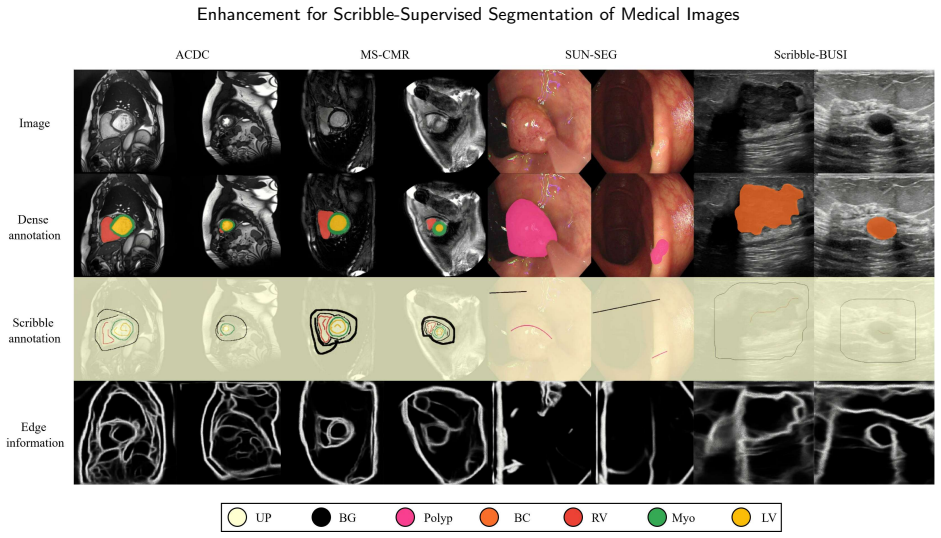
The load-bearing machinery is a U-Net whose encoder and decoder blocks are replaced by MaxViT stages, which combine MBConv layers with block and grid attention so that local and global features are learned at linear complexity. Three additions carry the argument: an edge-enhancement module that turns early-encoder features into edge predictions and attention maps, supervised by edge masks from a pretrained edge detector; a query-based transformer decoder with a query enhancer that refines bottleneck features and produces an auxiliary segmentation output; and a loss that mixes the two decoder outputs into dynamic pseudo-labels while weighting the edge-supervised regression loss at 0.2. The edge module is what the paper credits for the boundary-quality gains; the query decoder is what it credits for the feature-refinement and Dice gains.
What would settle it
Train the same model on a dataset with dense ground-truth segmentations, once with the pretrained edge detector's masks and once with human-traced boundaries or a second independent edge extractor, and compare HD95 against the no-edge baseline; if the edge module's boundary improvement disappears or reverses with the alternative edge source, the reported gains are specific to that detector rather than to edge information in general.
Extended reading notes
Core claim
On its own terms, the paper's central discovery is that the combination of a pre-trained MaxViT backbone in a U-Net layout, a query-based transformer decoder, and an edge-enhancement module supervised by automatically generated edge masks recovers most of the performance gap between scribble supervision and full supervision. The abstract and results tables claim state-of-the-art Dice and HD95 among scribble-supervised methods on all four benchmarks, and the cross-dataset experiment shows smaller degradation than prior hybrid models when moving between cardiac MRI protocols. The ablation attributes the boundary improvement mainly to the edge-enhancement module: on MS-CMRSeg, adding it to the dual-decoder setup lowers average HD95 from 6.749 mm to 2.508 mm while the query decoder contributes most of the remaining Dice gain.
Load-bearing premise
The boundary gains rest on treating the outputs of a pretrained edge detector as correct edge ground truth; if those masks are systematically biased, the model may learn the detector's errors rather than true organ boundaries.
Editorial extensions
If this is right
- On ACDC, MS-CMRSeg, SUN-SEG, and BUSI, the reported Dice scores of 89.1%, 88.4%, 71.4%, and 69.4% beat the scribble-supervised baselines compared, meaning less expensive scribble labels can replace dense labels in these tasks without the expected accuracy penalty.
- In the ablation, adding the edge-enhancement module to the dual-decoder model cuts HD95 on MS-CMRSeg from 6.749 mm to 2.508 mm, while the query decoder adds more Dice than boundary accuracy; the paper concludes the two components complement each other.
- In the cross-dataset test, training on one cardiac MRI protocol and evaluating on another drops Dice by 16.6% and 18.1% for this model, far less than the 25.8% to 61.2% drops reported for prior hybrid models; the paper takes this as evidence of better generalization across imaging domains.
- At 109.01 million parameters the model is large, but at 39.10 GMACs it has lower computational cost than the hybrid baselines it is compared with, supporting the paper's efficiency claim.
Reading between the lines
- Editorial inference: the boundary-quality gains may be partly tied to the specific pretrained edge detector used to generate supervision, so on a new image modality the HD95 advantage could shrink if that detector's edge maps are biased.
- Editorial inference: the cross-dataset robustness the paper attributes to MaxViT could instead come largely from large-scale natural-image pretraining; training the same architecture from random initialization would separate the two effects.
- Editorial inference: the same recipe — a pretrained transformer backbone plus a pseudo-ground-truth boundary signal and a query decoder — should transfer to other weak labels such as points or bounding boxes; the paper does not test this.
Editorial analysis
A structured set of objections, weighed in public.
Referee Report
Summary. The manuscript proposes QMaxViT-Unet+, a U-Net-like architecture with MaxViT encoder/decoder blocks, a query-based Transformer decoder, and an edge enhancement module, trained with a combination of scribble, pseudo-label, and edge losses. It reports state-of-the-art results on four scribble-supervised medical image segmentation benchmarks (ACDC, MS-CMRSeg, SUN-SEG, BUSI) and includes ablations, a cross-dataset generalization experiment, and a complexity comparison.
Significance. If the empirical claims were valid, this would be a meaningful contribution to scribble-supervised medical image segmentation. The architectural components are reasonable, the use of pre-trained MaxViT blocks for transfer learning is a sensible direction, and the paper provides detailed ablations and a cross-dataset study. However, the comparison tables contain internal inconsistencies that invalidate the state-of-the-art claim as printed. The empirical evidence for the central contribution does not currently stand, notwithstanding the plausible design and the availability of code.
major comments (4)
- [Table 3 and Section 3.2] The ScribbleVC row in Table 3 (MS-CMRSeg) reports DSC .868/.829/.904 (avg .867) and HD95 4.503/10.049/13.785 (avg 9.446), which are numerically identical to the DMPLS row in Table 2 for the ACDC dataset. Because MS-CMRSeg and ACDC are different datasets with different splits and patient populations, equal values to three decimals are not plausible. The claimed margin over ScribbleVC on MS-CMRSeg (1.9% DSC and 7.22 mm HD95, Section 3.2) is therefore computed against a baseline that appears to be the ACDC DMPLS result, so the comparison is unsupported. Please provide corrected, dataset-specific baseline numbers.
- [Table 4] The Unet_pce entry in Table 4 (SUN-SEG) is .375 DSC and 237.816 mm HD95, which is identical to the MS-CMRSeg Unet_pce average in Table 3. This again indicates a copied baseline row rather than a valid measurement on the SUN-SEG test set. The text claims that QMaxViT-Unet+ surpasses S2ME by 4% DSC on SUN-SEG, but the baseline table cannot substantiate that comparison in its current form.
- [Table 9 and Section 4] The paper states that QMaxViT-Unet+ 'has the lowest computational complexity with 39.10 GMACs' (Section 4, Model Complexity Comparison), but Table 9 reports SwinUnet at 5.92 GMACs, TransUnet at 25.37 GMACs, and UNet at 2.97 GMACs. The efficiency claim, which is one of the explicit contributions listed in the introduction, is contradicted by the paper's own measurements.
- [Section 2.3, Eq. (3) and Table 7] The edge enhancement module is supervised by edge masks generated by the pre-trained edge detector [34], and the Limitations section correctly notes that these masks may contain errors. Table 7 shows that the edge component is the main driver of the HD95 improvement in the dual-decoder setting (from 6.749 mm to 2.508 mm). Without an analysis that separates genuine boundary quality from fitting the detector's idiosyncratic errors, the central claim of improved boundary segmentation is not established. Please provide a boundary-quality evaluation against dense annotations or an ablation using an alternative edge source.
minor comments (5)
- [Section 2.2] The statement that 'the first two E-blocks typically capture low-level features such as edges' is presented as an observation, but no supporting evidence is shown; it should be framed as a design hypothesis or backed by feature visualizations.
- [Table 7] The Query enhancer's contribution is not isolated in the ablation study; the paper acknowledges this, but the limitation should also be stated explicitly in the experimental design section.
- [Section 3.2] The omission of data augmentation baselines such as Puzzle Mix, Cutout, MixUp, and CycleMix is justified by 'slower training convergence and inferior performance,' but no experimental evidence is provided for this claim.
- [Data and code availability] The abstract states that code is available at a GitHub URL, while the Data and code availability section says datasets and codes are available 'upon reasonable request.' Please align these statements.
- [Table 9] Inference times are reported from a 'free version virtual machine on Lightning Studios' with unspecified hardware and software versions; please specify the exact environment for reproducibility.
Circularity Check
No significant circularity: the central claims are benchmark comparisons against external baselines, and no predicted quantity reduces to a fitted input by construction.
full rationale
QMaxViT-Unet+ is an empirical architecture paper, not a derivation paper. The central claim is that the proposed model outperforms existing methods on four public benchmarks, and that claim is supported by comparisons against external baselines and by ablations measured with DSC/HD95 against independent dense ground truth. The edge enhancement module is supervised by edge masks from an external pre-trained detector [34], and the authors explicitly acknowledge these masks may be imperfect; this is a training-supervision quality issue, not a circular reduction, because the final evaluation metric is not the edge-mask output and the ground-truth segmentations are independent of the training signal. The only self-citation by a co-author (reference [7], a related U-Net/attention/transformer paper) appears in a general related-work list and is not load-bearing for any architectural choice or result. No uniqueness theorem from the authors is invoked, no fitted parameter is renamed as a prediction, and no known method is repackaged as a derivation. Separately, the baseline tables contain internally inconsistent copied rows across datasets and the complexity table contradicts the 'lowest computational complexity' wording, but those are correctness and integrity concerns outside the circularity definition and do not change the circularity score.
Assumptions & free parameters
free parameters (2)
- Loss weights (lambda1, lambda2, lambda3) =
(1, 0.5, 0.2)
- Number of MaxViT blocks per stage =
not reported
assumptions (4)
- domain assumption ImageNet-1k pre-trained MaxViT weights transfer to medical images across MRI, ultrasound, and endoscopy
- ad hoc to paper The first two E-blocks capture low-level edge features
- domain assumption Pseudo-labels generated by dynamically mixing predictions y1 and y2 are sufficiently reliable for Dice supervision
- domain assumption The pre-trained edge detector's output can be used as ground truth for edge supervision
Cite this review
Pith. "Pith review of QMaxViT-Unet+: A Query-Based MaxViT-Unet with Edge Enhancement for Scribble-Supervised Segmentation of Medical Images." pith.science (2026). https://pith.science/paper/WCZPGZVC
@misc{pith2026250210294,
  author       = {Pith},
  title        = {Pith review of: QMaxViT-Unet+: A Query-Based MaxViT-Unet with Edge Enhancement for Scribble-Supervised Segmentation of Medical Images},
  year         = {2026},
  howpublished = {\url{https://pith.science/paper/WCZPGZVC}},
  note         = {Machine review of arXiv:2502.10294}
}
read the original abstract
The deployment of advanced deep learning models for medical image segmentation is often constrained by the requirement for extensively annotated datasets. Weakly-supervised learning, which allows less precise labels, has become a promising solution to this challenge. Building on this approach, we propose QMaxViT-Unet+, a novel framework for scribble-supervised medical image segmentation. This framework is built on the U-Net architecture, with the encoder and decoder replaced by Multi-Axis Vision Transformer (MaxViT) blocks. These blocks enhance the model's ability to learn local and global features efficiently. Additionally, our approach integrates a query-based Transformer decoder to refine features and an edge enhancement module to compensate for the limited boundary information in the scribble label. We evaluate the proposed QMaxViT-Unet+ on four public datasets focused on cardiac structures, colorectal polyps, and breast cancer: ACDC, MS-CMRSeg, SUN-SEG, and BUSI. Evaluation metrics include the Dice similarity coefficient (DSC) and the 95th percentile of Hausdorff distance (HD95). Experimental results show that QMaxViT-Unet+ achieves 89.1\% DSC and 1.316mm HD95 on ACDC, 88.4\% DSC and 2.226mm HD95 on MS-CMRSeg, 71.4\% DSC and 4.996mm HD95 on SUN-SEG, and 69.4\% DSC and 50.122mm HD95 on BUSI. These results demonstrate that our method outperforms existing approaches in terms of accuracy, robustness, and efficiency while remaining competitive with fully-supervised learning approaches. This makes it ideal for medical image analysis, where high-quality annotations are often scarce and require significant effort and expense. The code is available at: https://github.com/anpc849/QMaxViT-Unet
Figures


Figures from the paper (4 more)


Reference graph
Works this paper leans on
-
[34]
The treasure beneath multiple annotations: An uncertainty-aware edge detector
Caixia Zhou, Yaping Huang, Mengyang Pu, Qingji Guan, Li Huang, and Haibin Ling. The treasure beneath multiple annotations: An uncertainty-aware edge detector. InProceedings of the IEEE/CVF Conference on Computer Vision and Pattern Recognition, pages 15507– 15517, 2023
work page 2023
-
[1]
U-net: Convolutional networks for biomedical image segmentation
Olaf Ronneberger, Philipp Fischer, and Thomas Brox. U-net: Convolutional networks for biomedical image segmentation. InMedical image computing and computer-assisted intervention–MICCAI 2015: 18th international conference, Munich, Germany, October 5-9, 2015, proceedings, part III 18, pages 234–241. Springer, 2015
2015
-
[2]
Attention u-net: Learning where to look for the pancreas.arXiv preprint arXiv:1804.03999, 2018
Ozan Oktay, Jo Schlemper, Loic Le Folgoc, Matthew Lee, Mattias Heinrich, Kazunari Misawa, Kensaku Mori, Steven McDonagh, Nils Y Hammerla, Bernhard Kainz, et al. Attention u-net: Learning where to look for the pancreas.arXiv preprint arXiv:1804.03999, 2018
arXiv 2018
-
[3]
Unet++: A nested u-net architecture for medical imagesegmentation
Zongwei Zhou, Md Mahfuzur Rahman Siddiquee, Nima Tajbakhsh, and Jianming Liang. Unet++: A nested u-net architecture for medical imagesegmentation. InDeepLearninginMedicalImageAnalysisandMultimodalLearningforClinicalDecisionSupport:4thInternational Workshop, DLMIA 2018, and 8th International Workshop, ML-CDS 2018, Held in Conjunction with MICCAI 2018, Gran...
work page 2018
-
[4]
FabianIsensee,JensPetersen,AndreKlein,DavidZimmerer,PaulFJaeger,SimonKohl,JakobWasserthal,GregorKoehler,TobiasNorajitra, Sebastian Wirkert, et al. nnu-net: Self-adapting framework for u-net-based medical image segmentation.arXiv preprint arXiv:1809.10486, 2018
arXiv 2018
-
[5]
Bo Li, Sikai Liu, Fei Wu, GuangHui Li, Meiling Zhong, and Xiaohui Guan. Rt-unet: an advanced network based on residual network and transformer for medical image segmentation.International Journal of Intelligent Systems, 37(11):8565–8582, 2022
work page 2022
-
[6]
Shen Jiang and Jinjiang Li. Transcunet: Unet cross fused transformer for medical image segmentation.Computers in Biology and Medicine, 150:106207, 2022
work page 2022
-
[7]
Thien B. Nguyen-Tat, Thien-Qua T. Nguyen, Hieu-Nghia Nguyen, and Vuong M. Ngo. Enhancing brain tumor segmentation in mri images: A hybrid approach using unet, attention mechanisms, and transformers.Egyptian Informatics Journal, 27:100528, 2024
work page 2024
Show all 54 references
-
[8]
An image is worth 16x16 words: Transformers for image recognition at scale.arXiv preprint arXiv:2010.11929, 2020
AlexeyDosovitskiy,LucasBeyer,AlexanderKolesnikov,DirkWeissenborn,XiaohuaZhai,ThomasUnterthiner,MostafaDehghani,Matthias Minderer, Georg Heigold, Sylvain Gelly, et al. An image is worth 16x16 words: Transformers for image recognition at scale.arXiv preprint arXiv:2010.11929, 2020
2010 arXiv
-
[9]
Transunet:Transformers make strong encoders for medical image segmentation.arXiv preprint arXiv:2102.04306, 2021
JienengChen,YongyiLu,QihangYu,XiangdeLuo,EhsanAdeli,YanWang,LeLu,AlanLYuille,andYuyinZhou. Transunet:Transformers make strong encoders for medical image segmentation.arXiv preprint arXiv:2102.04306, 2021
2021 arXiv
-
[10]
nnformer:Interleavedtransformerforvolumetric segmentation
Hong-YuZhou,JiansenGuo,YinghaoZhang,LequanYu,LianshengWang,andYizhouYu. nnformer:Interleavedtransformerforvolumetric segmentation. arXiv preprint arXiv:2109.03201, 2021
2021 arXiv
-
[11]
Swin unetr: Swin transformers for semantic segmentation of brain tumors in mri images
Ali Hatamizadeh, Vishwesh Nath, Yucheng Tang, Dong Yang, Holger R Roth, and Daguang Xu. Swin unetr: Swin transformers for semantic segmentation of brain tumors in mri images. InInternational MICCAI brainlesion workshop, pages 272–284. Springer, 2021
2021
-
[12]
Unetr: Transformers for 3d medical image segmentation
AliHatamizadeh,YuchengTang,VishweshNath,DongYang,AndriyMyronenko,BennettLandman,HolgerRRoth,andDaguangXu. Unetr: Transformers for 3d medical image segmentation. InProceedings of the IEEE/CVF winter conference on applications of computer vision, pages 574–584, 2022
2022
-
[13]
A robust volumetric transformer for accurate 3d tumor segmentation
Himashi Peiris, Munawar Hayat, Zhaolin Chen, Gary Egan, and Mehrtash Harandi. A robust volumetric transformer for accurate 3d tumor segmentation. InInternational conference on medical image computing and computer-assisted intervention, pages 162–172. Springer, 2022
2022
-
[14]
Levit-unet:Makefasterencoderswithtransformerformedicalimagesegmentation
GuopingXu,XuanZhang,XinweiHe,andXinglongWu. Levit-unet:Makefasterencoderswithtransformerformedicalimagesegmentation. In Chinese Conference on Pattern Recognition and Computer Vision (PRCV), pages 42–53. Springer, 2023. Hoang-An Vo et al.: Page 16 of 17 Enhancement for Scribble...
2023
-
[15]
Stu- net: Scalable and transferable medical image segmentation models empowered by large-scale supervised pre-training.arXiv preprint arXiv:2304.06716, 2023
Ziyan Huang, Haoyu Wang, Zhongying Deng, Jin Ye, Yanzhou Su, Hui Sun, Junjun He, Yun Gu, Lixu Gu, Shaoting Zhang, et al. Stu- net: Scalable and transferable medical image segmentation models empowered by large-scale supervised pre-training.arXiv preprint arXiv:2304.06716, 2023
2023 arXiv
-
[16]
Scribble-based hierarchical weakly supervised learning for brain tumor segmentation
Zhanghexuan Ji, Yan Shen, Chunwei Ma, and Mingchen Gao. Scribble-based hierarchical weakly supervised learning for brain tumor segmentation. InMedical Image Computing and Computer Assisted Intervention–MICCAI 2019: 22nd International Conference, Shenzhen, China, October 13–17,...
2019
-
[17]
Scribble-supervised medical image segmentation via dual-branch network and dynamically mixed pseudo labels supervision
Xiangde Luo, Minhao Hu, Wenjun Liao, Shuwei Zhai, Tao Song, Guotai Wang, and Shaoting Zhang. Scribble-supervised medical image segmentation via dual-branch network and dynamically mixed pseudo labels supervision. InInternational Conference on Medical Image Computing and Comput...
2022
-
[18]
Scribblevc: Scribble-supervised medical image segmentation with vision-class embedding
Zihan Li, Yuan Zheng, Xiangde Luo, Dandan Shan, and Qingqi Hong. Scribblevc: Scribble-supervised medical image segmentation with vision-class embedding. InProceedings of the 31st ACM International Conference on Multimedia, pages 3384–3393, 2023
2023
-
[19]
Scribformer: Transformer makes cnn work better for scribble-based medical image segmentation.IEEE Transactions on Medical Imaging, 2024
Zihan Li, Yuan Zheng, Dandan Shan, Shuzhou Yang, Qingde Li, Beizhan Wang, Yuanting Zhang, Qingqi Hong, and Dinggang Shen. Scribformer: Transformer makes cnn work better for scribble-based medical image segmentation.IEEE Transactions on Medical Imaging, 2024
2024
-
[20]
Weakly-supervisedsalientobjectdetectionviascribbleannotations
JingZhang,XinYu,AixuanLi,PeipeiSong,BowenLiu,andYuchaoDai. Weakly-supervisedsalientobjectdetectionviascribbleannotations. In Proceedings of the IEEE/CVF conference on computer vision and pattern recognition, pages 12546–12555, 2020
2020
-
[21]
Cyclemix: A holistic strategy for medical image segmentation from scribble supervision
Ke Zhang and Xiahai Zhuang. Cyclemix: A holistic strategy for medical image segmentation from scribble supervision. InProceedings of the IEEE/CVF Conference on Computer Vision and Pattern Recognition, pages 11656–11665, 2022
2022
-
[22]
S 2 me: Spatial-spectral mutual teaching and ensemble learning for scribble-supervised polyp segmentation
An Wang, Mengya Xu, Yang Zhang, Mobarakol Islam, and Hongliang Ren. S 2 me: Spatial-spectral mutual teaching and ensemble learning for scribble-supervised polyp segmentation. InInternational Conference on Medical Image Computing and Computer-Assisted Intervention, pages 35–45....
2023
-
[23]
In European conference on computer vision, pages 459–479
ZhengzhongTu,HosseinTalebi,HanZhang,FengYang,PeymanMilanfar,AlanBovik,andYinxiaoLi.Maxvit:Multi-axisvisiontransformer. In European conference on computer vision, pages 459–479. Springer, 2022
2022
-
[24]
Deep learning techniques for automatic mri cardiac multi-structures segmentation and diagnosis: is the problem solved?IEEE transactions on medical imaging, 37(11):2514–2525, 2018
Olivier Bernard, Alain Lalande, Clement Zotti, Frederick Cervenansky, Xin Yang, Pheng-Ann Heng, Irem Cetin, Karim Lekadir, Oscar Camara, Miguel Angel Gonzalez Ballester, et al. Deep learning techniques for automatic mri cardiac multi-structures segmentation and diagnosis: is t...
2018
-
[25]
Learning to segment from scribbles using multi-scale adversarial attention gates
Gabriele Valvano, Andrea Leo, and Sotirios A Tsaftaris. Learning to segment from scribbles using multi-scale adversarial attention gates. IEEE Transactions on Medical Imaging, 40(8):1990–2001, 2021
1990
-
[26]
Multivariatemixturemodelforcardiacsegmentationfrommulti-sequencemri
XiahaiZhuang. Multivariatemixturemodelforcardiacsegmentationfrommulti-sequencemri. InSebastienOurselin,LeoJoskowicz,MertR. Sabuncu, Gozde Unal, and William Wells, editors,Medical Image Computing and Computer-Assisted Intervention – MICCAI 2016, pages 581–588, Cham, 2016. Sprin...
2016
-
[27]
Multivariate mixture model for myocardial segmentation combining multi-source images.IEEE Transactions on Pattern Analysis and Machine Intelligence, 41(12):2933–2946, 2019
Xiahai Zhuang. Multivariate mixture model for myocardial segmentation combining multi-source images.IEEE Transactions on Pattern Analysis and Machine Intelligence, 41(12):2933–2946, 2019
2019
-
[28]
Shapepu: A new pu learning framework regularized by global consistency for scribble supervised cardiac segmentation
Ke Zhang and Xiahai Zhuang. Shapepu: A new pu learning framework regularized by global consistency for scribble supervised cardiac segmentation. In International Conference on Medical Image Computing and Computer-Assisted Intervention, pages 162–172. Springer, 2022
2022
-
[29]
Video polyp segmentation: A deep learning perspective.Machine Intelligence Research, 19(6):531–549, 2022
Ge-Peng Ji, Guobao Xiao, Yu-Cheng Chou, Deng-Ping Fan, Kai Zhao, Geng Chen, and Luc Van Gool. Video polyp segmentation: A deep learning perspective.Machine Intelligence Research, 19(6):531–549, 2022
2022
-
[30]
Progressively normalized self-attention networkforvideopolypsegmentation
Ge-Peng Ji, Yu-Cheng Chou, Deng-Ping Fan, Geng Chen, Huazhu Fu, Debesh Jha, and Ling Shao. Progressively normalized self-attention networkforvideopolypsegmentation. In InternationalConferenceonMedicalImageComputingandComputer-AssistedIntervention ,pages 142–152. Springer, 2021
2021
-
[31]
Pranet: Parallel reverse attention network for polyp segmentation
Deng-Ping Fan, Ge-Peng Ji, Tao Zhou, Geng Chen, Huazhu Fu, Jianbing Shen, and Ling Shao. Pranet: Parallel reverse attention network for polyp segmentation. InInternational conference on medical image computing and computer-assisted intervention, pages 263–273. Springer, 2020
2020
-
[32]
MasashiMisawa,Shin-eiKudo,YuichiMori,KinichiHotta,KazuoOhtsuka,TakahisaMatsuda,ShoichiSaito,ToyokiKudo,ToshiyukiBaba, Fumio Ishida, et al. Development of a computer-aided detection system for colonoscopy and a publicly accessible large colonoscopy video database (with video).G...
2021
-
[33]
Dataset of breast ultrasound images.Data in brief, 28:104863, 2020
Walid Al-Dhabyani, Mohammed Gomaa, Hussien Khaled, and Aly Fahmy. Dataset of breast ultrasound images.Data in brief, 28:104863, 2020
2020
-
[35]
Masked-attention mask transformer for universal image segmentation
Bowen Cheng, Ishan Misra, Alexander G Schwing, Alexander Kirillov, and Rohit Girdhar. Masked-attention mask transformer for universal image segmentation. InProceedings of the IEEE/CVF conference on computer vision and pattern recognition, pages 1290–1299, 2022
2022
-
[36]
Mask2former with improved query for semantic segmentation in remote-sensing images.Mathematics, 12(5):765, 2024
Shichen Guo, Qi Yang, Shiming Xiang, Shuwen Wang, and Xuezhi Wang. Mask2former with improved query for semantic segmentation in remote-sensing images.Mathematics, 12(5):765, 2024
2024
-
[37]
Maxvit-unet: Multi-axis attention for medical image segmentation
Abdul Rehman Khan and Asifullah Khan. Maxvit-unet: Multi-axis attention for medical image segmentation. arXiv preprint arXiv:2305.08396, 2023
2023 arXiv
-
[38]
Imagenet: A large-scale hierarchical image database
Jia Deng, Wei Dong, Richard Socher, Li-Jia Li, Kai Li, and Li Fei-Fei. Imagenet: A large-scale hierarchical image database. In2009 IEEE conference on computer vision and pattern recognition, pages 248–255. Ieee, 2009
2009
-
[39]
Batch normalization: Accelerating deep network training by reducing internal covariate shift
Sergey Ioffe and Christian Szegedy. Batch normalization: Accelerating deep network training by reducing internal covariate shift. In International conference on machine learning, pages 448–456. pmlr, 2015
2015
-
[40]
Deep learning using rectified linear units (relu).arXiv preprint arXiv:1803.08375, 2018
AF Agarap. Deep learning using rectified linear units (relu).arXiv preprint arXiv:1803.08375, 2018. Hoang-An Vo et al.: Page 17 of 17 Enhancement for Scribble-Supervised Segmentation of Medical Images
2018 arXiv
-
[41]
Et-net: A generic edge-attention guidance network for medical image segmentation
Zhijie Zhang, Huazhu Fu, Hang Dai, Jianbing Shen, Yanwei Pang, and Ling Shao. Et-net: A generic edge-attention guidance network for medical image segmentation. In Medical Image Computing and Computer Assisted Intervention–MICCAI 2019: 22nd International Conference, Shenzhen, C...
2019
-
[42]
Per-pixel classification is not all you need for semantic segmentation.Advances in neural information processing systems, 34:17864–17875, 2021
Bowen Cheng, Alex Schwing, and Alexander Kirillov. Per-pixel classification is not all you need for semantic segmentation.Advances in neural information processing systems, 34:17864–17875, 2021
2021
-
[43]
Query-guided generalizable medical image segmentation.Pattern Recognition Letters, 184:52–58, 2024
Zhiyi Yang, Zhou Zhao, Yuliang Gu, and Yongchao Xu. Query-guided generalizable medical image segmentation.Pattern Recognition Letters, 184:52–58, 2024
2024
-
[44]
Segment anything
AlexanderKirillov,EricMintun,NikhilaRavi,HanziMao,ChloeRolland,LauraGustafson,TeteXiao,SpencerWhitehead,AlexanderCBerg, Wan-Yen Lo, et al. Segment anything. InProceedings of the IEEE/CVF International Conference on Computer Vision, pages 4015–4026, 2023
2023
-
[45]
Sparse instance activation for real-time instance segmentation
Tianheng Cheng, Xinggang Wang, Shaoyu Chen, Wenqiang Zhang, Qian Zhang, Chang Huang, Zhaoxiang Zhang, and Wenyu Liu. Sparse instance activation for real-time instance segmentation. InProceedings of the IEEE/CVF Conference on Computer Vision and Pattern Recognition, pages 4433–...
2022
-
[46]
In Proceedings of the IEEE conference on computer vision and pattern recognition, pages 2117–2125, 2017
Tsung-YiLin,PiotrDollár,RossGirshick,KaimingHe,BharathHariharan,andSergeBelongie.Featurepyramidnetworksforobjectdetection. In Proceedings of the IEEE conference on computer vision and pattern recognition, pages 2117–2125, 2017
2017
-
[47]
Pyramid scene parsing network
Hengshuang Zhao, Jianping Shi, Xiaojuan Qi, Xiaogang Wang, and Jiaya Jia. Pyramid scene parsing network. InProceedings of the IEEE conference on computer vision and pattern recognition, pages 2881–2890, 2017
2017
-
[48]
Puzzle mix: Exploiting saliency and local statistics for optimal mixup
Jang-Hyun Kim, Wonho Choo, and Hyun Oh Song. Puzzle mix: Exploiting saliency and local statistics for optimal mixup. InInternational conference on machine learning, pages 5275–5285. PMLR, 2020
2020
-
[49]
Improved regularization of convolutional neural networks with cutout
Terrance DeVries and Graham W Taylor. Improved regularization of convolutional neural networks with cutout. arXiv preprint arXiv:1708.04552, 2017
2017 arXiv
-
[50]
mixup: Beyond empirical risk minimization.arXiv preprint arXiv:1710.09412, 2017
Hongyi Zhang, Moustapha Cisse, Yann N Dauphin, and David Lopez-Paz. mixup: Beyond empirical risk minimization.arXiv preprint arXiv:1710.09412, 2017
2017 arXiv
-
[51]
Scribblesup:Scribble-supervisedconvolutionalnetworksforsemanticsegmentation
DiLin,JifengDai,JiayaJia,KaimingHe,andJianSun. Scribblesup:Scribble-supervisedconvolutionalnetworksforsemanticsegmentation. In Proceedings of the IEEE conference on computer vision and pattern recognition, pages 3159–3167, 2016
2016
-
[52]
Semi-supervisedlearningbyentropyminimization
YvesGrandvaletandYoshuaBengio. Semi-supervisedlearningbyentropyminimization. Advancesinneuralinformationprocessingsystems , 17, 2004
2004
-
[53]
Weakly supervised segmentation of covid19 infection with scribble annotation on ct images.Pattern recognition, 122:108341, 2022
Xiaoming Liu, Quan Yuan, Yaozong Gao, Kelei He, Shuo Wang, Xiao Tang, Jinshan Tang, and Dinggang Shen. Weakly supervised segmentation of covid19 infection with scribble annotation on ct images.Pattern recognition, 122:108341, 2022
2022
-
[54]
Semi-supervised semantic segmentation with cross pseudo supervision
Xiaokang Chen, Yuhui Yuan, Gang Zeng, and Jingdong Wang. Semi-supervised semantic segmentation with cross pseudo supervision. In Proceedings of the IEEE/CVF conference on computer vision and pattern recognition, pages 2613–2622, 2021. Hoang-An Vo et al.: Page 18 of 17
2021
Reviewed August 7, 2026 · model on record in the stance chip above.
Discussion (0). Sign in to comment.